

Article Figures & Data
Figures
- Figure 1.
CREB is phosphorylated at a specific development stage of adult hippocampal neurogenesis. a , The phosphorylated form of CREB (pCREB, red) is present in many but not all DCX-expressing (green) immature neurons in the subgranular zone of the hippocampal dentate gyrus. DAPI in blue. White arrowheads indicate DCX-positive immature neurons, which are pCREB negative. Scale bars (from left), 50 and 25 μm, respectively. b , Time course of phosphorylation of CREB (red) and DCX expression (blue) in newborn hippocampal neurons using a retroviral birth-dating paradigm. Confocal images of GFP-transduced cells at 3, 14, and 28 dpi. Top, Low magnification; lower rows are higher magnifications of indicated boxes. At 3 dpi, a fraction of GFP-positive cells becomes DCX positive. Most of these cells are pCREB negative (white arrowhead). Some transduced cells are negative for DCX and pCREB (yellow arrowhead). At 14 dpi the vast majority of GFP-transduced cells are DCX positive and pCREB positive (red arrowheads). At 28 dpi a large fraction of transduced cells display the morphology of mature granule neurons and are DCX and pCREB negative (yellow arrowheads). Scale bar, 25 μm. c , Analysis of GFP-transduced cells for pCREB expression and expression of markers corresponding to different stages of adult neuronal development at different time points after injection. Phosphorylation of CREB is preceded by DCX and NeuroD expression. Loss of CREB phosphorylation is paralleled by the loss of DCX and NeuroD expression and the initiation of calbindin expression.
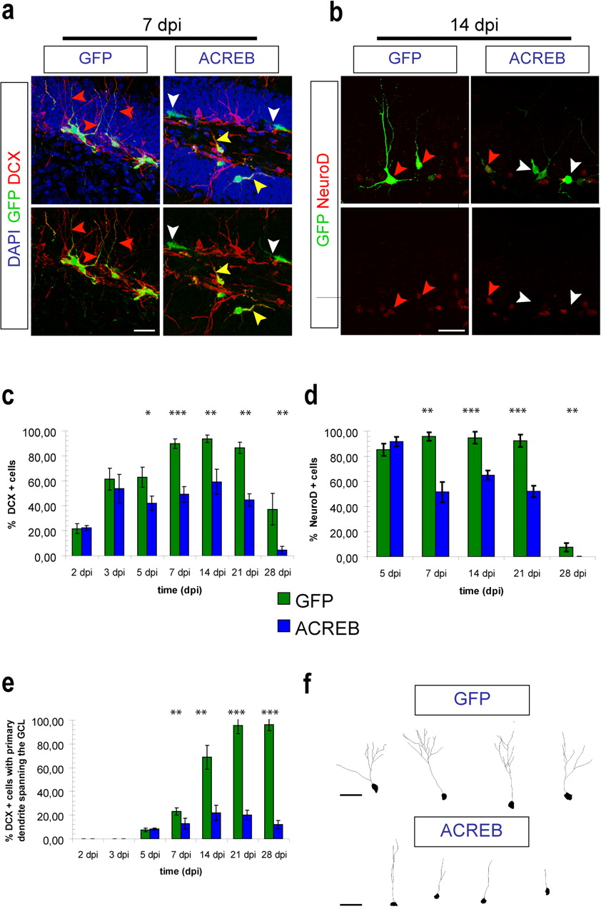
- Figure 2.
Loss of CREB function affects the differentiation and morphological maturation of newborn dentate granule cells. a , Analysis of DCX expression and morphology of newborn cells transduced with GFP-retrovirus (GFP; green) or ACREB-retrovirus (ACREB; green) at 7 dpi. GFP-transduced cells express DCX (red) and have extended an apical dendrite toward the molecular layer (red arrowhead). In contrast, a large number of ACREB-transduced cells are negative for DCX (white arrowhead). ACREB-transduced DCX+ neurons frequently failed to extend an apical dendrite toward the molecular layer (yellow arrowhead). DAPI in blue. Scale bar, 25 μm. b , NeuroD expression in newborn cells transduced with GFP-retrovirus (GFP; green) or ACREB-retrovirus (ACREB; green) at 14 dpi. The majority of GFP-transduced cells are NeuroD positive (red) (red arrowheads). In contrast, ACREB-transduced cells are frequently negative for NeuroD expression (white arrowheads). Scale bar, 25 μm. c , Quantification of the percentage of retrovirally transduced cells in the granule cell layer expressing DCX. Expression of DCX is significantly reduced among ACREB-transduced cells starting at 5 dpi (*p < 0.05,**p < 0.01, ***p < 0.001). d , Quantification of the percentage of retrovirally transduced cells in the granule cell layer expressing NeuroD. Expression of NeuroD is significantly reduced among ACREB-transduced cells after 5 dpi (**p < 0.01, ***p < 0.001). e , Morphological analysis of retrovirally transduced immature neurons. The percentage of ACREB-transduced DCX+ immature neurons, which extend an apical dendrite through the granule cell layer toward the molecular layer, is significantly reduced (**p < 0.01, ***p < 0.001). f , Confocal three-dimensional reconstruction of dendrites of GFP- and ACREB-transduced immature neurons, which had developed an apical dendrite spanning the granule cell layer at 14 dpi. Scale bar, 25 μm.
- Figure 3.
Loss of CREB function affects the survival rate of newborn dentate granule cells. a , Confocal images of transduced cells in control animals (GFP/RFP) and ACREB-injected animals (ACREB/RFP) at 5, 14, and 28 dpi. The percentage of double-transduced cells (yellow) in ACREB-injected animals is decreased compared with controls at 14 and 28 dpi. RFP-only transduced cells appear in red. DAPI in blue. Scale bar, 25 μm. b , Significant reduction in the survival rate of newborn cells, in which CREB signaling has been ablated starting at 7 dpi (***p < 0.001).
- Figure 4.
GABA-mediated depolarization regulates CREB phosphorylation, differentiation, and morphological maturation of new dentate granule cells during the first 2 weeks after their birth. a , Analysis of CREB phosphorylation and DCX expression after shRNA-mediated knockdown of NKCC1. Confocal images of control shRNA-transduced cells (shCTR) (green) and shNKCC1-transduced cells (green) at 14 dpi. Bottom, Magnifications of indicated boxes. A large fraction of shNKCC1-transduced DCX+ (blue) immature neurons is negative for pCREB (red) (white arrowhead). Moreover, many shNKCC1-transduced cells do not express DCX (yellow arrowhead). pCREB+-transduced immature neurons (red arrowheads). Scale bars, 25 μm. b , The percentage of pCREB-positive immature neurons (identified by DCX expression) among the shNKCC1-transduced immature neurons is significantly lowered (***p < 0.001). c , Expression of DCX is significantly reduced among shNKCC1-transduced cells (***p < 0.001). d , Analysis of NeuroD expression (red) in newborn cells after transduction with shCTR (green) or shNKCC1 (green) at 14 dpi. Scale bar, 25 μm. A large percentage of shNKCC1-transduced cells do not express NeuroD (white arrowheads). Red arrowheads indicate transduced cells, which express NeuroD. e , Quantification of NeuroD expression in transduced cells. Expression of NeuroD is significantly reduced among the shNKCC1-transduced cells (**p < 0.01). f , The percentage of shNKCC1-transduced immature neurons (DCX+), which extend an apical dendrite through the granule cell layer toward the molecular layer, is significantly reduced (**p < 0.01).
- Figure 5.
NKCC1 knockdown affects the survival rate of newborn dentate granule cells. a , Confocal images of transduced cells in control animals (shCTR/TagRFP injected) and shNKCC1/TagRFP-injected animals at 7 and 14 dpi. The percentage of double-transduced cells (yellow) in shNKCC1/TagRFP animals is decreased. shCTR- and shNKCC1-transduced cells in green, TagRFP in red. Scale bars, 25 μm. b , Quantification of the survival rate. There is a significant reduction in the fraction of double cells in the shNKCC1/TagRFP group between 7 and 14 dpi (**p < 0.01), indicating that NKCC1 knockdown affects survival of newborn neurons.
- Figure 6.
NKCC1-knockdown phenotype can be compensated by enhanced activation of CREB signaling. Analysis of neuronal protein expression and cellular morphology of animals injected with shNKCC1- and RFP-encoding retroviruses and animals injected with shNKCC1- and CREBYF-encoding retroviruses. a , Confocal images from animals at 7 dpi. Analysis of DCX expression and of the morphology of newborn cells. shNKCC1 (green)- and RFP (red)-injected control animals in left column. shNKCC1 (green)- and CREBYF (red)-injected animals in right column. Many shNKCC1/RFP-cotransduced cells are negative for DCX (blue) (white arrowhead). Yellow arrowheads indicate RFP-only transduced cells expressing DCX. Right column, shNKCC1/CREBFY-cotransduced cells expressing DCX and sending an apical dendrite toward the molecular cell layer (red arrowheads). Yellow arrowhead indicates CREBYF-only transduced immature neuron. Scale bar, 25 μm. b , Confocal images from animals at 7 dpi. Analysis of NeuroD expression and of the morphology of newborn cells. shNKCC1 (green)- and RFP (red)-injected control animals in left column. shNKCC1 (green)- and CREBYF (red)-injected animals in right column. The majority of shNKCC1/CREBFY double-transduced cells express NeuroD (blue) (red arrowheads). White arrowheads indicate shNKCC1-transduced cells which have lost NeuroD expression. Yellow arrowheads indicate RFP- or CREBYF-transduced cells which are NeuroD+. Scale bar, 25 μm. c , Quantification of single-transduced (shNKCC1, RFP, or CREBYF) and double-transduced (shNKCC1/RFP or shNKCC1/CREBYF) cells expressing DCX. CREBYF expression significantly increases the fraction of DCX+ immature neurons among shNKCC1-transduced cells (***p < 0.001). d , Quantification of single- and double-transduced cells expressing NeuroD. CREBYF expression significantly increases the fraction of NeuroD+ cells among shNKCC1-transduced cells (**p < 0.01) e , Quantification of single-transduced and double-transduced immature neurons, which extended an apical dendrite spanning the granule cell layer. CREBYF expression significantly increases the fraction of immature neurons, which have developed a dendrite spanning the granule cell layer among single-transduced cells and shNKCC1-transduced cells (***p < 0.001).


Additional Files
Supplemental Data
Files in this Data Supplement:
- supplemental material - Supplemental Legend
- supplemental material - Supplemental Figure 1
- supplemental material - Supplemental Figure 2
- supplemental material - Supplemental Figure 3
- supplemental material - Supplemental Figure 4
- supplemental material - Supplemental Figure 5
- supplemental material - Supplemental Figure 6


