

Article Figures & Data
Figures
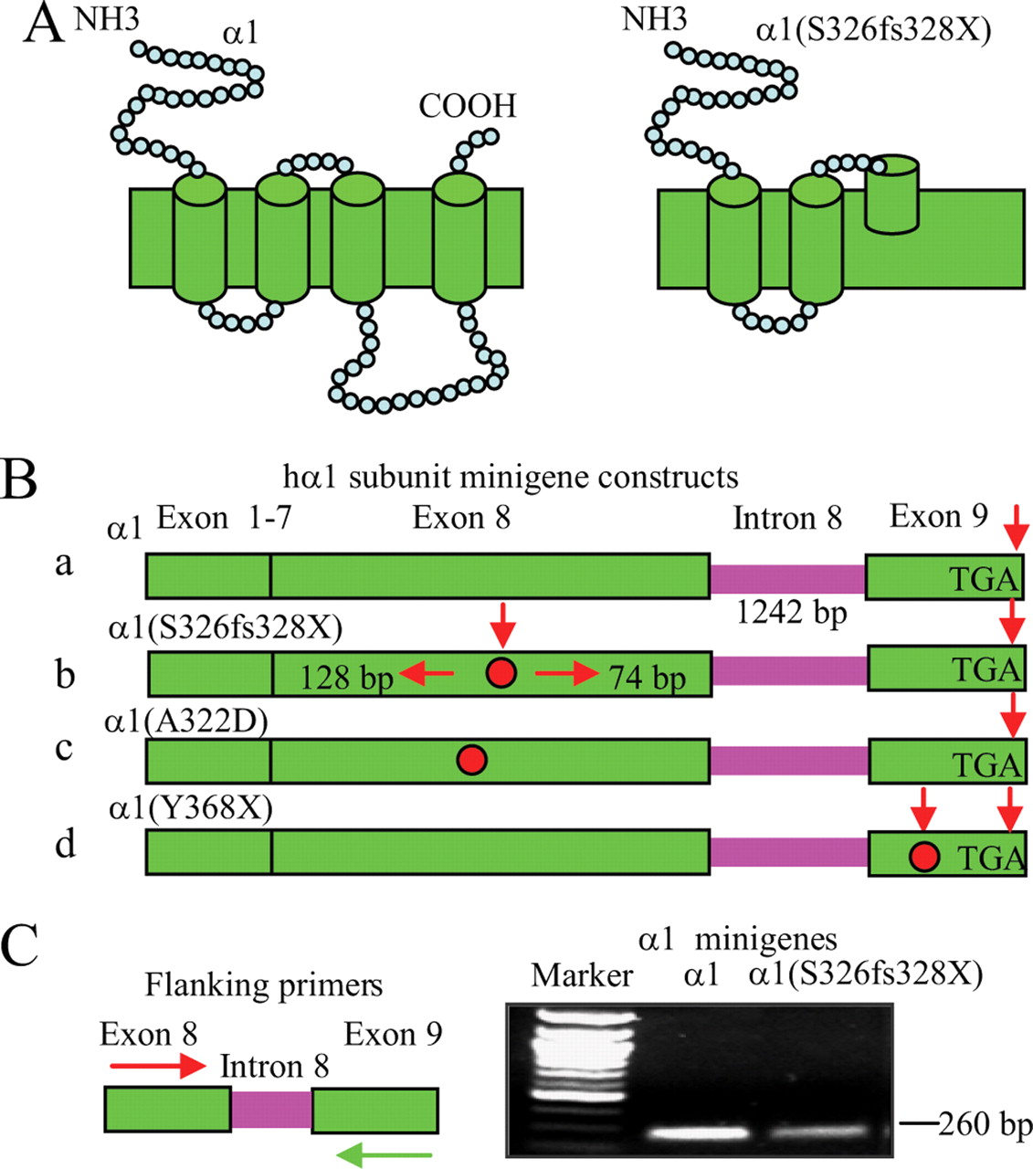
- Figure 1.
A minigene strategy was used to study the α1 subunit mutation, 975delC, S326fs328X. A, Schematic topologies of GABAA receptor wild-type (left) and mutant α1(S326fs328X) subunit (right). B, The α1 subunit gene, GABRA1, has 9 exons and 8 introns. Wild-type and mutant α1 subunit minigenes were constructed by including intron 8 (1242 bp). The stop codons of the wild-type minigene (α1) (Ba) and α1(A322D) (red dot) (Bc) are located at the end of exon 9, and the nonsense stop codon caused by the α1 subunit mutation, 975delC, S326fs328X, is located at the middle of exon 8 (Bb). An NMD-incompetent control minigene had a nonsense stop codon inserted in the last (ninth) exon of the α1 subunit gene α1(Y368X) (Bd). C, With primers in exon 8 and 9 that flank intron 8, both wild-type and mutant α1 subunit minigenes displayed a band at 260 bp, suggesting correct splicing in the reverse transcribed cDNA products from RNAs of α1 and α1(S326fs328X) subunit minigenes.
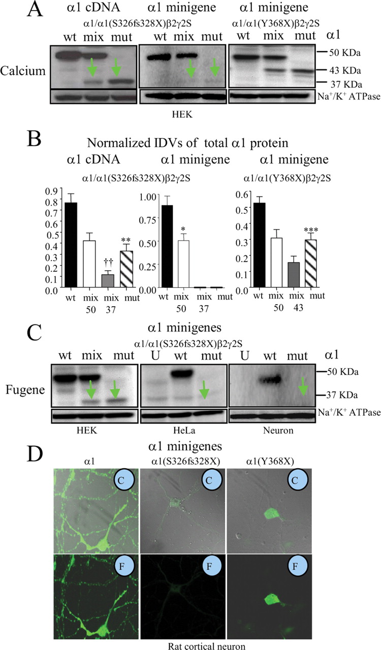
- Figure 2.
Expression of the NMD competent α1(S326fs328X) subunit minigene produced minimal mutant protein. A, Total lysates from HEK 293T cells cotransfected with β2 and γ2S subunit cDNAs and α1 subunit cDNAs (left) or minigenes (middle and right) with wild-type (wt) subunits (ratio of 1:1:1), mixed (mix) α1/α1(S326fs328X) or α1/α1(Y368X) subunits (0.5:0.5:1:1) and mutant (mut) α1(S326fs328X) or α1/α1(Y368X) subunits (1:1:1) with the Calcium phosphate precipitation method (Calcium). The total lysates were analyzed by immunoblot. Na+/K+ ATPase was used for a loading control. Wild-type α1 subunits migrated at ∼50 KD, α1(S326fs328X) subunits migrated at 37 KD, and α1(Y368X) subunits migrated at 43 KD. B, The relative amount of total wt, mix and mut α1 subunit protein from A was plotted. Mix 50 stands for the wild-type subunit level detected at 50 KDa in the mixed transfection condition, mix 37 or 43 stands for the mutant truncated subunits detected at 37 or 43 KDa in the mixed condition (**p < 0.01, ***p < 0.001 vs wild-type, ††p < 0.01 vs mix 50). C, HEK 293T cells, HeLa cells and rat cortical neurons were cotransfected with β2 and γ2S subunit cDNAs and wild-type α1, mixed α1/α1(S326fs328X) or mutant α1(S326fs328X) subunit minigenes (1:1:1) with Fugene transfection. In A and C, the green arrows indicate the mutant subunit. In C, U stands for untransfected. wt, mix, and mut are as described in A. D, Rat cortical neurons were transfected with β2 and γ2S subunit cDNAs and the wild-type α1 subunit minigene (left), the mutant α1 subunit minigene (S326fs328X) (middle) or the mutant α1(Y368X) subunit minigene (right) at a cDNA ratio of 1:1:1. After 6 d in culture, the neurons were permeabilized and stained with anti-monoclonal human α1 subunit antibody conjugated with fluorophore Alexa-647. In the insets, C refers to combined transmitted and fluorescent images, and F refers to fluorescent images. In C and D, neurons were transfected by nucleofection.
- Figure 3.
Mixed α1/α1(S326fs328X)β2γ2S receptors had reduced peak current amplitudes and reduced α1 subunit surface expression. A–D, Human GABAA receptor currents were obtained from HEK 293T cells cotransfected with β2 and γ2S subunit cDNAs and wild-type α1 subunit minigenes for wild-type (wt α1:β2:γ2S 1:1:1 cDNA ratio, black) or for haploinsufficiency control (0.5:1:1, hc, silver) or with mixed α1/α1(S326fs328X) minigenes (0.5:0.5:1:1, mix, green) and mutant (mut) α1(S326fs328X) minigene (1:1:1, mut, gray) with 1 mm GABA applied for 6 s (A) or 28 s (C). In A, arrows indicate the peak of each trace. B, The mean peak amplitude of each group was plotted (n = 7 for wt, n = 8 for hc, n = 7 for mix). C, The peak haploinsufficiency control and mixed receptor currents were normalized to the wild-type receptor current. D, The ratios of steady-state current/peak amplitude of both haploinsufficiency control and mixed receptor currents were similar to wild-type current (n = 7 for each group). In A and C, lifted whole cells were voltage-clamped at −50 mV. E, Equal amounts of membrane-bound protein were biotinylated and immunoblotted with anti-α1 antibody. The green arrows indicate the mutant protein. F, The relative amounts of surface wild-type, haploinsufficiency control, mixed and mutant α1 subunit protein were plotted (n = 6). In B and F, ***p < 0.001 versus wild-type.
- Figure 4.
The truncated mutant α1(S326fs328X) subunit protein was subject to ERAD. A, Total lysates of HEK 293T cells coexpressing β2 and γ2S subunit cDNAs and wild-type α1 subunit minigenes or mutant α1(S326fs328X) subunit cDNAs were undigested (U) or digested with Endo-H (H) or PNGase-F (F). The relatively high level of nonspecific signal with mutant expression of the mutant subunit was due to overexposing the gel to detect the very low level of mutant subunit protein that was present when the mutant subunit minigene was used. B, Total lysates of HEK 293T cells expressing β2 and γ2S subunit cDNAs and wild-type α1 subunit minigene (wt) or half amount of wild-type α1 subunit minigene (haploinsufficiency control; hc), mixed α1/α1(S326fs328X) subunit minigenes (mix) and mutant α1(S326fs328X) subunit minigene (mut) with or without lactacystin (Lac,10 μm) treatment for 6 h were analyzed by immunoblot. The arrows indicate the mutant protein. Calcium stands for Calcium phosphate precipitation transfection while Fugene stands for Fugene transfection. C, The total α1 subunit protein in each condition before lactacystin was quantified and normalized to the total α1 subunit expression of wild-type receptors (n = 8, ***p < 0.001 vs wild-type, Calcium; n = 4, *p < 0.05, ***p < 0.001 vs wild-type, Fugene). D, The histogram displays the ratio of increased α1 subunit protein from Fugene-transfected HEK 293T cells with lactacystin (10 μm) treatment over α1 subunit protein without treatment (**p < 0.01 vs wild-type, n = 4). E, HEK 293T or HeLa cells cotransfected with β2 and γ2S subunit cDNAs and α1 subunit cDNAs were treated with or without lactacystin, and the cell lysates were analyzed by immunoblot. F, The graph was plotted with the ratios of the mean IDVs ± SEM obtained with lactacystin treatment divided by results obtained without treatment (n = 7; *p < 0.05, ***p < 0.001 vs wild-type).
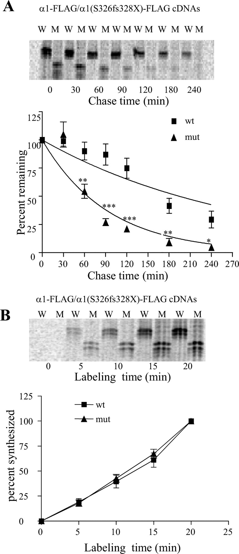
- Figure 5.
Mutant α1(S326fs328X) subunit protein had a reduced half-life. A, HEK 293T cells containing pulse-chase 35S methionine radio-labeled wild-type (W), and mutant α1 (S326fs328X) (M) subunits were lysed, immunopurified and analyzed by 12.5% SDS-PAGE. After 20 min labeling, the cells were chased for the indicated times (n = 5). The same amount of total protein (800 μg) from each sample was used for immunopurification. The percentage of radioactivity remaining is plotted versus the amount of radioactivity measured at time 0 for either the wild-type or mutant subunits (*p < 0.05, **p < 0.01, ***p < 0.001 vs wild-type). B, HEK 293T cells expressing wild-type (W) and mutant α1 (S326fs328X) (M) were pulse-labeled for 0, 5, 10, 15, and 20 min and lysed for immunopurification and SDS-PAGE (n = 4). The percentage of radioactivity incorporated during subunit synthesis was plotted by normalizing to the value obtained at 20 min for either the wild-type or mutant subunits.
- Figure 6.
Mutant α1(S326fs328X) subunit protein had increased association with the ER resident chaperone Bip. HEK 293T cells expressing wild-type and mutant FLAG-tagged α1 cDNA subunits were lysed and immunopurified with FLAG antibody conjugated beads. The immunoblots show the expression level of the Flag tagged α1 subunit and its association with the chaperone Bip.
- Figure 7.
Mutant α1(S326fs328X) subunit mRNA was reduced and reversed by ribosome inhibition or by silencing the NMD essential factor hUpf-1. A, The relative amount of wild-type α1 and α1(S326fs328X) subunit mRNAs were measured by quantitative RT-PCR in different cells expressing α1 and α1(S326fs328X) minigenes (**p < 0.01, ***p < 0.001 vs wt; n = 15 for HEK, n = 7 for neuron, n = 11 for HeLa). B, HeLa cells expressing α1 and α1(S326fs328X) subunit minigenes were either treated or untreated with cycloheximide (CHX) (100 μg/ml) for 6 h before the total RNA extraction. The mRNAs were measured by RT-PCR and were normalized to wild-type mRNAs without CHX treatment (***p < 0.001 vs mutant α1(S326fs328X) subunit without CHX; n = 7). C, Total cell lysates of HEK 293T or HeLa cells untreated (U) or treated with negative control siRNA (100 nm, neg) or transfected with hUpf-1 siRNA (10 nm, 50 and 100 nm) for 2–4 d were immunoblotted with hUpf-1 antibody. D, Wild-type α1 or mutant α1(A322D) minigenes with β2 and γ2S subunits or wild-type and α1(S326fs328X) minigenes alone were transfected into sister cultures of the HeLa cells from the experiment in C 2 d later after siRNA treatment. The total proteins were harvested 48–56 h later with or without 6 h of lactacystin (10 μm) (Lac) treatment and then analyzed. Equal amounts of protein (90 μg) were loaded, and α1 subunits (3 μg) were expressed to better visualize the α1(S326fs328X) protein. E, The total mutant α1 subunit protein IDVs with different treatments were normalized to their internal control and then normalized to the mock treated group (**p < 0.01 vs neg, †p < 0.05 vs lac; n = 4 for A322D, n = 8–11 for S326fs328X). F, Sister cultures from the experiment in E were harvested and analyzed for RT-PCR. The mRNA levels from mock, negative control or Upf-1 were normalized to the results from wild-type cells treated with the same conditions (**p < 0.01 vs neg; n = 6). In D–F, mock stands for siRNA mock transfection, neg for negative control siRNA transfection, Upf for human Upf-1 specific siRNA, Upf+lac for both human Upf-1 specific siRNA and Lactacystin treated and lac for only Lactacystin treated.
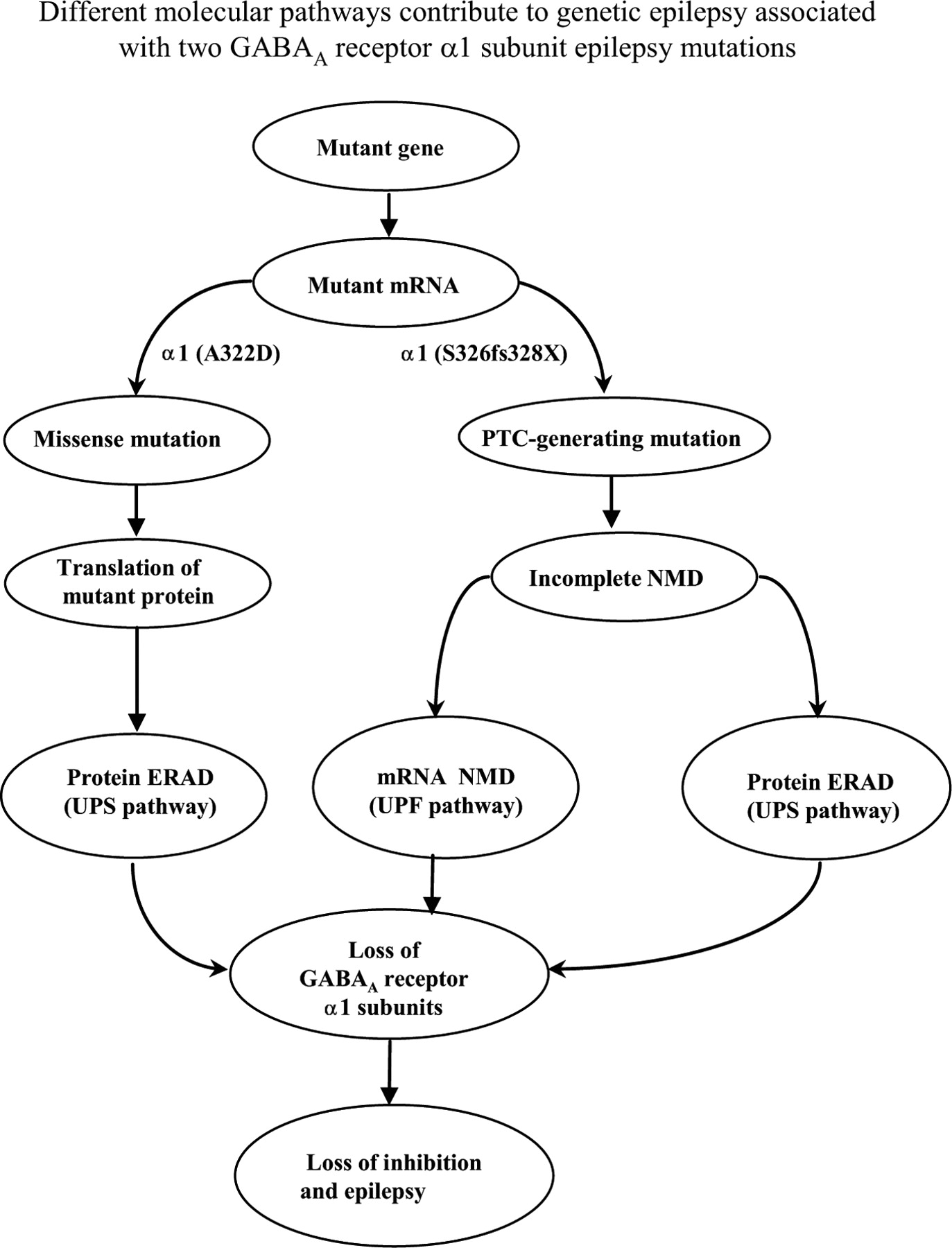
- Figure 8.
Different molecular pathways contribute to epileptogenesis of a missense and a frameshift PTC-generating epilepsy GABRA1 gene mutation. Although GABAA receptor α1 subunit mutations, 975delC, S326fs328X and A322D, are located in the same transmembrane domain (TM3), the two mutations caused haploinsufficiency through different molecular pathways. The α1 subunit mutation, A322D, caused mutant protein degradation through proteasome-ubiquitin pathway (ERAD). The α1 subunit mutation, 975delC, S326fs328X, activated NMD, resulting in loss of majority mutant mRNAs and the portion of mutant mRNA that escaped NMD translated mutant truncated protein that was subject to rapid degradation through the ubiquitin-proteasome pathway.


Additional Files
Supplemental Data
Files in this Data Supplement:
- supplemental material - Supplemental Figure 1
- supplemental material - Supplemental Figure 2
- supplemental material - Supplemental Figure 3
- supplemental material - Supplemental Figure 4


