

Article Figures & Data
Figures
- Figure 1.
A, Schematic representation of two experimental apparatuses. (1) Electrical stimulation of the buccal motor branch (b) of the seventh nerve (FMN) (cuff electrode) induced whisker protraction and passive retraction. (2) Juxtacellular stimulation of motoneurons in facial nucleus evoked small whisker movement (Herfst and Brecht, 2008). B, Transformation of MN spikes into whisker movements. The two separate processes involved were as follows: (1) transformation of spikes into muscle force Fm(t) and (2) translation of muscle force into whisker movements via the biomechanics of the pad. C, Top view of a rat head and whisker. The whisker resting angle, θ0, is measured between the whisker shaft and the tangent to the pad. The angle θ is the instantaneous protraction angle. D, Block diagram of the transformation of MN spikes (Harish and Golomb, 2010) into muscle force. The model consists of four concatenated processes: the process r (Eq. 1), intracellular [Ca2+]i accumulation (Eqs. 2–4), Ca2+-dependent muscle force development, FC (Eq. 5), and the force–length curve, FL, applicable only for large protraction angles (Eqs. 6–8). The output is the muscle force Fm. E, Biomechanical model of the plant: row of whiskers connected only by intrinsic muscles. Springs and dampers, which model the viscoelastic properties of the tissue, connect each whisker to stationary local sites of the mystacial pad, sites that are represented by anchor points. Elements that belong to one whisking motor unit (WMU), which consists of two whiskers connected by single intrinsic muscle, are plotted in red. θ0i and θi are the resting angle and the protraction angle of the ith whisker, respectively, and ld is the length between the skin and anchor point where the sling intrinsic muscle is attached to the follicle. The dot (black or red) on each whisker shaft represents the center of mass of the whisker (Xicm, Yicm), which is located at a distance −C along the follicle from the skin.
- Figure 2.
Electrical whisking. A, Sample trace of a whisker protraction angle, θ(t) (top panel), and of translation of the pad along the x-axis at the whisker base near the skin, xσ(t) (bottom panel), during 1 s trials (5 cycles), during two-pulse FMN stimulation (Δt = 15 ms, 200 ms cycles). B, Traces (black) of whisker dynamics in the θ–xσ plane during the protraction phase (100 ms, n = 9 whiskers). Colored solid lines [adapted from Hill et al. (2008)] represent the outcomes of stimulating different muscle types in anesthetized rats: intrinsic muscles (blue), the protracting extrinsic muscle (m. nasalis; red), and the retracting extrinsic muscles (m. nasolabialis and m. maxillolabialis; green).
- Figure 3.
Nonlinear temporal summation of whisker movements. A, Whisker trajectories upon single-pulse (dash blue) and two-pulse (solid blue, Δt = 17 ms) stimulation. The trajectory expected from a linear summation of two single-pulse responses delayed by Δt is also plotted (black). Results were computed by averaging 10 stimulations of the same whisker. B, Whisker trajectories upon single-pulse stimulation (dash blue) and three-pulse stimulation (solid blue, Δt = 17 ms), and of the calculated linear sum (black). C, Linearity indices LI2 (blue) and LI3 (brown) resulting from two-pulse and three-pulse stimulation as functions of the interpulse interval Δt. Solid lines denote the means, averaged over nine tracked whiskers for Δt values above 5 ms. For Δt values between 1 and 4 ms, means were calculated over three whiskers. Dashed lines denote the means ± SEM. ΔtM is the value Δt for which LI2 is maximal, and ΔtL is minimal Δt for linear summation (∼35 ms). Inset, Linearity indices LI2 and LI3 computed for the single whisker presented in A and B. D, Peak linearity indices:
 (LI3) versus (LI2) (n = 9; asterisks); the dashed line denotes the diagonal.
 - Figure 4.
Temporal summation properties of whisker protraction responses to spikes fired by one MN stimulated juxtacellularly [based on data from Herfst and Brecht (2008) (blue lines and solid circles)], assuming linearity based on the response to one spike (black line) and computed from our model (green lines). Protraction angles are small. The model consists of a row of five whiskers, and the movement of the middle whisker is presented. Only one muscle, connecting to the top of the second follicle and the bottom of the third follicle (denoted by red in Fig. 1E), was activated. Parameters, ld = 4 mm, θ0 = 65°, A = 1.33. A, Whisker trajectory upon single-spike stimulation. B, Whisker trajectory upon two-spike stimulation (interspike interval, Δt = 19 ms). C, Linearity index LI2 as a function of ISI Δt, calculated from two-spike events. Data from each of four MNs are depicted by circles of different colors (blue, brown, red, and black).
- Figure 5.
Temporal summation properties of whisker protraction responses to one, two, and three pulses stimulating the FMN as measured experimentally (blue lines) and computed from our model (green lines). Protraction angles are not small. The model consists of a row of five whiskers, and the movement of the middle whisker is presented. For this computation, all five muscles were activated. Muscle parameters different from the reference parameter set are as follows: A = 1.087, r0 = 2.55, τc = 7.4 ms, zl = 0.33, kσ,x = kπ,x = 0.2 mg/ms2, ζσ,x = ζπ,x = 2.2 mg/ms, kπ,y = 0.6 mg/ms2, ζπ,y = 6 mg/ms, ld = 3.5 mm, θ0 = 75°. A, Whisker protraction in response to single-pulse stimulation of the FMN. B, C, Whisker protraction in response to two- (B) and three- (C) pulse stimulations of the FMN with Δt = 15 ms. D, E, Linearity indices LI2 (D) and LI3 (E) versus Δt. Vertical bars denote SEM. Experimental data are the same as for the inset of Figure 3C.
- Figure 6.
Computed responses of two adjacent whiskers in our model to weak stimulation of the intrinsic muscle connecting them. Parameter, A = 1.33. The resting angle θ0 is 70° for A–C and 95° for D and E. Symbols and lines related to the posterior and anterior whiskers are plotted in black and red, respectively. A, D, Schemes of the WMU. B, E, Time traces of the protraction angles of the whiskers, θ. C, Time traces of the coordinates of the centers of mass of the whiskers, (x, y). F, Peak amplitudes of whisker protraction angles, θposteriorpeak and θanteriorpeak, as functions of θ0. Arrows indicate θ0,pr, where θposteriorpeak switches from negative to positive values; θ0,ar, where θanteriorpeak switches from positive to negative values; θ0,eq, where θposteriorpeak = θanteriorpeak; and θ0 values for the simulations in B, E, G, and H. G, H, Time traces of the protraction angles of the whiskers, θ, for θ0 = 45° (G) and for θ = 120° (H).
- Figure 7.
Dependencies of the peak amplitude ratio R (θanteriorpeak/θposteriorpeak) on θ0 for the architecture of A and D are shown when model parameters, such as the ld/lf, spring constants, and damping constants are varied. Values of R for the parameters of Figure 7 (A = 1.33) are denoted by black lines in each panel. Other colors denote R when a parameter is varied. A, The length ld/lf is varied: 0.4 (green), 0.8 (black), and 1 (blue). B, The ratio kπ,y/kπ,x of the vertical and horizontal spring constants is varied: 0 (green), 1 (blue), 3.3 (black). This ratio is varied while keeping kπ,x and ζπ,x constant and the ratio ζπ,y/ζπ,x equal to kπ,y/kπ,x. C, The ratio kπ,x/kσ,x of the plate and skin spring constants is varied: 1 (black), 1.5 (green), and 2 (blue). This ratio is varied while keeping kσ,x and ζσ,x constant and ζπ,x/ζσ,x equal to kπ,x/kσ,x. Graphs on the right side present the same curves with wider ranges of θ0 and R.
- Figure 8.
Maximum amplitudes of the protraction angles of two adjacent (posterior and anterior) whiskers, θposteriormax (black) and θanteriormax (red), for whisker resting angles θ0 (70° (solid line), and 95° (dash line), as functions of the dynamic properties of the whisker. A, Effects of the absolute value of the whisker center of mass (|C|), normalized to lf, the follicle length, on the maximum amplitudes. B, C, Effects of the whisker moment of inertia (I) and whisker mass (M) on the maximum amplitudes. Reference parameter set values are denoted by arrows labeled “Ref.”
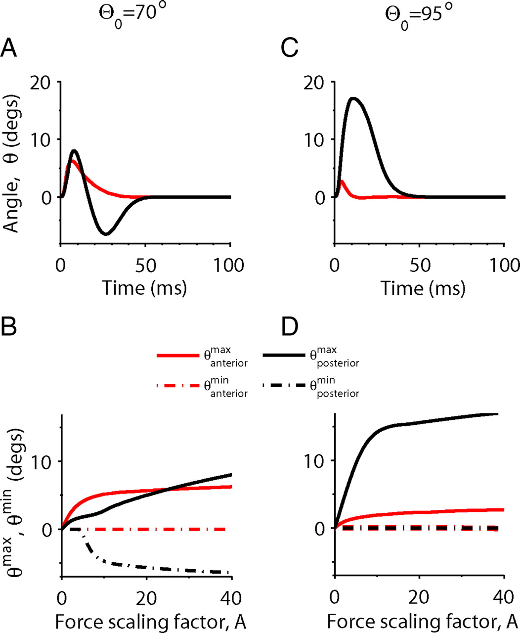
- Figure 9.
Responses of two adjacent whiskers to stimulation that is not weak of their connecting intrinsic muscle, computed from the model with the reference parameter set. A, C, Time traces of the protraction angles of the whiskers, θ, for A = 40. B, D, The maximal and minimal protraction amplitudes θanteriormax, θanteriormin, θposteriormax, and θposteriormin, as functions of the force amplitude A. The resting angles, θ0, are 70° (A, B) and 95° (C, D). Lines representing the movements of the posterior and anterior whiskers are plotted in black and red, respectively.
- Figure 10.
Experimental responses of two adjacent whiskers in a row to juxtacellular stimulation of a single FMN (Herfst and Brecht, 2008). A, Time traces of the protraction angles, θ, of the whiskers C2 and C3. B, The ratio R, as a function of the resting angle, θ0, averaged over the two whiskers, as extracted from stimulations of three MNs. The pairs of whiskers that were moved are as follows: C2 and C3 (blue asterisk, shown in A), C2 and C3 (black cross), and C3 and C4 (red diamond). The solid green line is computed from simulating the model with the reference parameter set, ld = 2.8 mm, and A = 1.
- Figure 11.
Spatiotemporal summation of protraction angles in response to stimulation of two adjacent intrinsic muscles. A, Scheme of the biomechanical model. The middle follicle is innervated by two intrinsic muscles, the anterior (red) and the posterior (black). The arrows indicate stimuli to the posterior and anterior muscles (denoted by “Stim Post” and “Stim Ant,” respectively). B, Maximal amplitudes of protraction angles of the middle whisker as functions of the muscle force scaling factor, A, in response to the innervations of the posterior (θpost,musclemax, black), anterior (θant,musclemax, red), or both (θpost+ant,musclemax, green) muscles. The maximum value of the linear sum of the separate responses to each muscle is also plotted (
(θpost,muscle(t) + θant,muscle(t)), blue). The whisker resting angle is θ0 = 70° and the two muscles are stimulated simultaneously (Δt = 0). C, The maximal amplitudes of protraction angles described in B as functions of Δt for θ0 = 70°, A = 10. Lines are as in B. D, The “spatiotemporal linearity index” [S(A, Δt), Eq. 16] for A = 10 as a function of θ0 and Δt. Inset, S(A, 0) as a function of θ0 for A = 10. E, F, Peak amplitudes of whisker protraction angles, θposteriorpeak (black), θmiddlepeak (blue), and θanteriorpeak (red), as functions of θ0. Parameters, A = 1.33 (E, same as in Fig. 6F) and A = 10 (F); Δt = 0. Note that the θpeak values of the posterior and anterior whiskers among the three (black and red lines) when A is small (F) are equal to the θpeak values of the posterior and anterior whiskers in Figure 6F, where they are adjacent. - Figure C1.
Scheme of the WMU, which consists of two whiskers connected by a single intrinsic muscle. Symbols are defined as in Figure 1, and ϕ is the angle between the muscle and the x-axis.


Tables
Superscript Definition Forces s Spring d Damper m Muscle Attachment points σ Skin π Plate μ Muscle Direction p Posterior a Anterior b Bone Other cm Center of mass Parameter Units Reference value Description Source NW 5 Number of whiskers in a row Dörfl (1982) M mg 10.5 Mass of whisker Hill et al. (2008) I mg · mm2 112 Moment of inertia of whisker Hill et al. (2008) lf mm 4 Length of follicle Hill et al. (2008) C mm −1.43 Position of whisker center of mass Hill et al. (2008) s mm 2 Distance between vibrissae at rest Hill et al. (2008) w mm 20 Length of mystacial pad Hill et al. (2008) ld mm 3 Muscle attachment point ϴ0 ° 75 Whisker rest angle kσ,x, kπ,x mg/ms2 0.3 Spring constants in the x-direction ζσ,x, ζπ,x mg/ms 3 Damper constants in the x-direction kπ,y mg/ms2 1 Spring constant in the y-direction ζπ,y mg/ms 10 Damper constant in the y-direction
Supplemental Data
Files in this Data Supplement:
- supplemental material - Supplemental Legend
- supplemental material - Supplemental Figure
- supplemental material - Supplemental Movie 1
- supplemental material - Supplemental Movie 2
- supplemental material - Supplemental Movie 3
- supplemental material - Supplemental Movie 4
In this issue


Jump to section
- Article
- Abstract
- Introduction
- Materials and Methods
- Results
- Discussion
- Appendix A: Model of the Biomechanical Plant for Rat Whiskers
- Appendix B: For Small Protraction Angles, the Linearity Index Depends Only on the Muscle Activity
- Appendix C: Steady-State Whisker Mechanics for Small Protraction Angles
- Footnotes
- References
- Figures & Data
- Info & Metrics
- eLetters