

Article Figures & Data
Figures
- Figure 1.
The optomotor maze paradigm. A, Maze. Arrow, Grating direction and velocity. Black trace, Filmed path of an individual fly. B, Each individual fly path is quantified for turning behavior. The number of successive turns in the same direction (+, with the moving grating; −, against the grating) is tallied per fly. Reversals of direction were also counted (rev). C, The normalized frequency of consecutive turn categories is plotted as a histogram for wild-type flies (n = 40 flies; weighted average ± SEM). The average value for either direction is indicated. D, Histogram for data created by a random model (50% turn probability at each choice level), with corresponding consecutive turn averages. E, Histogram and turn averages for radish1 mutants (n = 40 flies). F, Average ± SEM distribution of flies among the nine collection tubes at the end of the maze (n = 8 mazes of 25–30 flies for wild type and radish1) compared with a theoretical distribution for the random model.
- Figure 2.
radish1 optomotor responsiveness and distractibility. A, Optomotor responsiveness (means ± SEM; n = 8 maze runs for each point) for wild type and radish1 for different grating velocities. radish mutants do not respond to gratings moving 3 Hz or faster but do respond to more slowly moving gratings (1 and 2 Hz, p < 0.05 by t test against zero). *p < 0.05, significantly different from wild type by t test of means. B, Optomotor responsiveness (means ± SEM; n = 8 maze runs for each point) to a moving grating for wild type (at 3 Hz) and radish1 (at 1 Hz; see above) in the presence of a visual distracter. A vertical white bar (5 × 25 cm) on a red background, positioned opposite of the direction of grating movement, was varied in intensity (percentage white; a schema is shown below the graph) to quantitatively test the distractibility of flies responding to the moving grating. *p < 0.05, significantly different from wild type by t test. C, Phototaxic responses of radish1 and wild type in the maze to the distracter alone (the same schema shown above the graph).
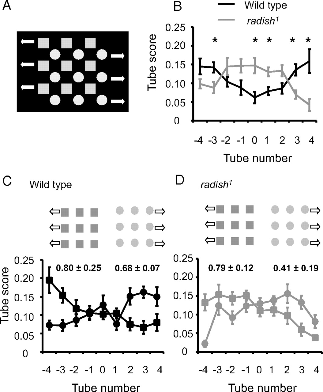
- Figure 3.
Optomotor responsiveness to competing objects. A, A field of blue squares (1 cm wide) moving right to left at 1 Hz is intercalated with a field of green circles (1 cm diameter) moving left to right at 1 Hz (see Materials and Methods). B, Average ± SEM distribution in the maze (n = 8 maze runs) for wild type and radish1 in response to the visual in A. *p < 0.05, significantly different proportion of flies by t test. C, Wild-type responses to the moving squares or circles presented individually, with average ± SEM optomotor indices (n = 8 maze runs). D, radish1 responses to the moving squares or circles presented individually, with average ± SEM optomotor indices (n = 8 maze runs).
- Figure 4.
Tethered flight. A, Average ± SEM power spectra between 0 and 5 Hz for wild-type (black line; n = 25) and radish1 (gray line; n = 24) torque behavior in 6 min closed-loop flights with two distinct visual objects (see Materials and Methods). B, Average power spectra between 0 and 5 Hz for wild-type (black line; n = 25) and radish1 (gray line; n = 21) torque behavior in 6 min open-loop flights without any visual landmarks.
- Figure 5.
radish1 brain recordings. A, Arena setup. Visual objects rotate around the fly clockwise with a period of 3 s. B, Average ± SEM power spectrum of wild-type and radish1 brain activity between 0 and 5 Hz (n = 14 flies for both genotypes). The large peaks below 1 Hz (off scale) represents responses to the visual objects rotating around the fly at 0.33 Hz. C, Flies were exposed for 100 s to two identical squares before one of the squares changed to a cross. The amplitude (or power) of brain LFPs bandpass filtered at 20–30 Hz was correlated with the position of either competing object as it swept in front of the fly following a novelty transition. Data for a sample wild-type fly are shown. The black vertical line indicates when the square changed to a cross. D, Sample 20–30 and 1–2 Hz bandpass-filtered wild-type data for several rotations of the visual panorama before and after a novelty transition. Circled epochs indicate successive 20–30 Hz responses to the novel cross. E, Enlarged section of the novelty response in D. F, Sample 20–30 and 1–2 Hz bandpass-filtered radish1 data for several rotations of the visual panorama before and after a novelty transition. G, Enlarged section of the novelty response in F. The response to the novel cross (circled) does not recur selectively, as it does in wild type.
- Figure 6.
Average responses to visual novelty in radish1 and wild type. A, radish1. Left, Average LFP activity for the 10 s after a novelty transition for three frequency domains (10–20, black Hz; 20–30 Hz, dark gray, 30–40 Hz, light gray) for radish mutants (n = 14 flies). Right, The same 20–30 Hz radish1 data as summarized in the left but partitioned into successive 3 s epochs after a novelty transition (*p < 0.05, significant response) (supplemental Methods 1, available at www.jneurosci.org as supplemental material). B, Wild type. Left, Average LFP activity for the 10 s after a novelty transition was calculated for three frequency domains; n = 6 flies. The direction of panorama flow is indicated. Right, The same 20–30 Hz wild-type data as summarized in the left but partitioned into successive 3 s epochs after a novelty transition [*p < 0.05, significant response (data are from van Swinderen, 2007)]. C, Average LFP responses to each of the two visual objects presented individually to radish mutants (n = 14), for the three frequency domains indicated. Average wild-type 20–30 Hz responses to the same individual objects are shown for comparison (van Swinderen, 2007) (n = 6).
- Figure 7.
Attention-like bias. A, Opposing visual stimuli (a square and a cross, 180° apart) rotate around the fly at 3 s per cycle. Each object is thus in front of the fly for 1.5 s, for which summed 20–30 Hz activity is separately calculated (during the epochs symbolized by the black and gray bars). B, Log ratio of summed 20–30 Hz activity plotted for successive cycles of image rotation in a sample wild-type fly. AT, Alteration time, or the duration (in cycles) when the ratio is biased in succession for one of the objects before alternating, indicated numerically above the graph. C, Successive AT values plotted as a time series histogram in the same sample wild-type fly. The size of five contiguous AT groupings (supplemental Methods 2, available at www.jneurosci.org as supplemental material) is tallied in a column on the right (Σ AT). D, Shuffled data from the same wild-type fly, replotted as a time series histogram with five tallied AT groups (Σ AT) shown on the right. E, The same analysis performed on data from the radish1 mutant.
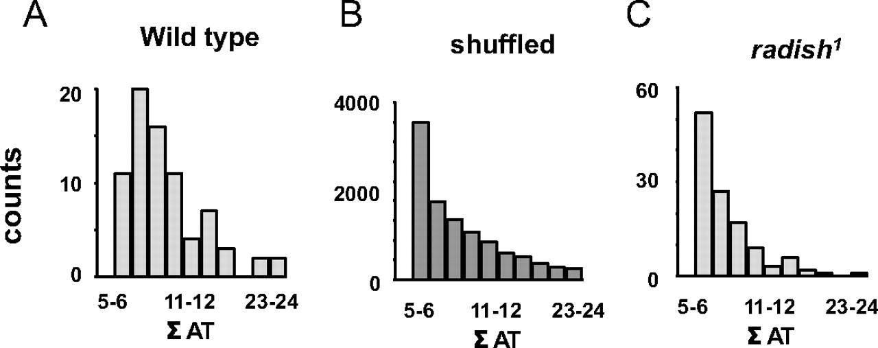
- Figure 8.
Distribution of attention-like bias. A, Frequency distribution of AT data for eight wild-type flies exposed to the two competing objects, as in Figure 7. Σ AT, A measure of attention-like bias, is the sum of AT values within each grouping (as indicated in Fig. 7C–E). B, Frequency distribution for eight sets of shuffled wild-type data. C, Frequency distribution for summed AT data from 14 radish flies.
- Figure 9.
MPH treatment. A, Optomotor responsiveness in wild-type flies treated with 0.5 mg/ml MPH (red histograms). Starved flies were transferred to drug-laced food and allowed to feed for 10 min, 3 h, or 24 h. Control flies were similarly transferred but to food without drug and tested 10 min later (Control1) or the next day (Control2). Flies chronically exposed to drug for 24 h were transferred back to normal food for 1–2 h and tested (Recovery). *p < 0.05, significantly different from controls by t test. n = 8 runs of 25–30 flies for each experiment. B, MPH (0.5 mg/ml) was administered acutely (2–4 h feeding on drug-laced food) to a panel of learning and memory mutants, as well as to flies with altered dopamine function [Th–Gal4 (Friggi-Grelin et al., 2003) × UAS–tnt, wherein dopaminergic neurons are silenced, or × UAS–eagΔ932, in which dopaminergic neurons are activated]. *p < 0.05, significantly different from controls (red vs blue bars) by t test. n = 8 runs of 25–30 flies for each experiment. C, Spectral analysis (0–5 Hz) of LFP activity in the brains of radish mutants treated to acute MPH (red line). Blue line, The same flies before treatment. Data are averages of z-scored spectrograms (n = 5 flies). *p < 0.05, significantly different within 0.3 Hz band range surrounding the peak, by t test. D, The effect of MPH on 20–30 Hz responsiveness (±SEM) to visual novelty in radish1 and wild-type flies. MPH feeding (red box) resulted in a significant novelty response in radish mutants (n = 5 flies), whereas radish mutants fed without the drug (blue box) show no response to novelty (n = 14). Wild-type responses to visual novelty were similar with and without MPH (n = 4 and 6 flies, respectively). *p < 0.05 by t test of means for either competing object. A visual explanation of how these data were calculated is in supplemental Methods 1 (available at www.jneurosci.org as supplemental material).


Additional Files
Supplemental Data
Files in this Data Supplement:
- supplemental material - Supplemental Material


