

Article Figures & Data
Figures
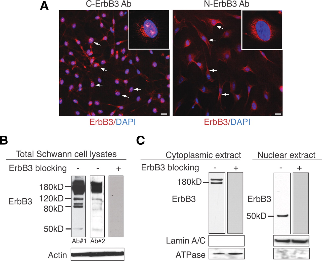
- Figure 1.
A 50 kDa fragment of the cytoplasmic domain of ErbB3 is expressed in the nucleus of Schwann cells. A, Immunostaining of purified rat primary Schwann cells with ErbB3 antibodies specific for the intracellular domain (C-ErbB3) or the extracellular domain (N-ErbB3) of ErbB3. Both pictures represent median images at the level of the nucleus from z-stacks (12 μm total with 2 μm increments) at 20× magnification. C-ErbB3 displayed staining in Schwann cell cytoplasm, membrane, perinuclear region, and nucleus (inset shows distinct pattern of speckled nuclear staining at 60×), whereas N-ErbB3 staining was restricted to the membrane, cytoplasm, and perinuclear region (inset shows lack of nuclear staining at 60×). B, Total protein lysates of rat primary Schwann cells blotted with two antibodies against separate epitopes of the cytoplasmic domain of ErbB3. This reveals the full-length ErbB3 band at 180 kDa and additional bands at 120, 80, and 50 kDa. Incubation with an ErbB3 blocking peptide abrogates all bands, suggesting that these are ErbB3 specific. Densitometric analysis from three independent experiments shows that the 50 kDa band corresponds to ∼1/50th of the density of the full-length ErbB3 band. β-Actin was used a loading control antibody. C, Protein lysates of Schwann cells were separated into cytoplasmic and nuclear fractions and blotted with an antibody that recognizes the cytoplasmic portion of ErbB3. This revealed the existence of the full-length ErbB3 receptor (∼180 kDa band) in the cytoplasmic fraction (left panel) and the ∼50 kDa band in the nuclear fraction (right panel). The purity of the cytoplasmic and nuclear fractions was confirmed by the expression of ATPase and Lamin A/C in the corresponding lysates. Specificity of the ErbB3 antibody is verified with preincubation with an ErbB3 blocking peptide, which eliminates antibody binding in both the cytoplasmic and the nuclear lysates.
- Figure 2.
ErbB3 nuclear expression in rat primary Schwann cells does not depend on ligand-induced protease cleavage. A, Sequence comparison analysis of rat ErbB3 with various isoforms of ErbB4. ErbB3 clearly resembles the JM-b isoform that lacks the cleavage recognition site for metalloproteases. B, Immunostaining for ErbB3 expression using a cytoplasmic-specific ErbB3 antibody showed no difference in nuclear ErbB3 expression after addition of β1-heregulin in the presence of the following inhibitors: GM6001 (metalloprotease inhibitor), ZLL2 (site-2 protease), IX (γ-secretase), DIC (Rhomboid). C, Ectopic reconstitution of functionally active ErbB2/ErbB3 heterodimers in Cos-7 cells using full-length ErbB2 and ErbB3 constructs. Addition of β1-heregulin in cotransfected Cos-7 cells induced transphosphorylation of ErbB3, suggesting that ErbB2 and ErbB3 form active heterodimers on the cell surface. D, Cos-7 cells cotransfected with ErbB2 and ErbB3 were immunostained with ErbB3 antibody with or without the addition of β1-heregulin. ErbB3 is localized on the membrane, in the cytoplasm, and at the perinuclear region, but not in the nucleus. Scale bars represent 20× magnification. Western blot of the cotransfected Cos-7 cells after addition of heregulin shows complete absence of the 50 kDa band that corresponds to nuc-ErbB3.
- Figure 3.
Identification of the nuclear variant of ErbB3 (nuc-ErbB3) in Schwann cells. A, Schematic representation of the genomic organization of the human ErbB3 nuclear variant AK125028 and the Schwann cell nuc-ErbB3 variant. Note that nuc-ErbB3 of the Schwann cell lacks the 50 nt unique region upstream of exon 23 and that its ATG codon is located at the start of exon 23 as opposed to the intronic ATG that AK125028 uses. B, Schematic showing the primer combinations used to amplify various regions of nuc-ErbB3 from Schwann cell mRNA and the respective PCR products. The nuc-ErbB3 PCR product and the nested PCR products correspond to processed mRNA excluding introns. C, Northern blotting of Schwann cell mRNA with an ErbB3 RNA probe that binds to both the full-length ErbB3 and the nuc-ErbB3 sequences reveals the existence of two (∼6 and ∼2.5 kb) discrete bands corresponding to the full-length ErbB3 sequence and the nuc-ErbB3, respectively. D, Tissue-specific expression of nuc-ErbB3. ErbB3 RNA probe cross-linked to biotin was used in Northern blotting performed on mouse mRNA from various tissues. Note that nuc-ErbB3 is expressed in whole embryos, thymus, testes, and ovaries, suggesting a broad distribution of expression. E, Transfection of Cos-7 cells that lack endogenous ErbB3 with nuc-ErbB3 construct showed that nuc-ErbB3 is localized to the nucleus. Control represents vector-transfected cells stained for ErbB3 expression. F, Western blotting on Cos-7 cell lysates transfected with the Schwann cell-derived nuc-ErbB3 construct reveals a 50 kDa protein that migrates equally with the endogenous Schwann cell nuc-ErbB3 band. G, Developmental profile of nuc-ErbB3 expression by RT-PCR using RNA from adult rat sciatic nerves and P2 rat sciatic nerves shows that nuc-ErbB3 depends on the differentiation state of Schwann cells and is abrogated in adult myelinated nerves in vivo. Actin was used as a housekeeping gene to normalize expression.
- Figure 4.
nuc-ErbB3 translocates to the nucleus because of an active NLS sequence. A, Computational analysis of the nuc-ErbB3 sequence revealed a classical NLS sequence between amino acids 296 and 305. B, nuc-ErbB3 variant from rat primary Schwann cells was cloned into pCDNA 3.1/TOPO and the NLS sequence was mutated using QuikChange mutagenesis kit (Stratagene) by substituting arginine at position 300 with alanine (R300A). Both wild-type and the mutant plasmids were transfected into Cos-7 cells and stained with C-ErbB3 antibody 72 h after transfection. This showed that point mutation R300A abolishes nuclear localization. The arrows indicate nuclear staining; arrowheads, perinuclear staining; and asterisks, membranous staining. C, nuc-ErbB3 associates with a range of gene promoters as revealed by genome-wide ChIP–chip array. In Schwann cells, nuc-ErbB3 associates with genomic regions that contain promoters of 63 expressed genes. Functional clustering of these 63 Schwann cell genes using DAVID reveals nine significant functional clusters (p < 0.005). D, nuc-ErbB3 directly regulates the promoter activities of ezrin and HMGB1 genes (p < 0.005) but not Frizzled-5 promoter as quantified by dual luciferase assays. The graphs represent the average of six independent experiments, and the luciferase units have been normalized to DNA content, protein content, and the expression of Renilla luciferase.
- Figure 5.
Translation of nuc-ErbB3 is regulated by neuregulin and siRNAs can inhibit nuc-ErbB3 expression in Schwann cells. A, Neuregulin increases the steady-state translation rate of nuc-ErbB3 and full-length ErbB3 in Schwann cells as shown by RT-PCR on eIF4E-bound mRNA. Actin RT-PCR on eiF4E precipitated mRNA was performed to determine the specificity of the neuregulin effect on nuc-ErbB3 translation. B, Transfection of nuc-ErbB3 siRNA into Schwann cells downregulates the expression of nuc-ErbB3 in nuclear extracts compared with control-siRNA (scrambled nuc-ErbB3 siRNA)-transfected cells but does not affect the expression of full-length ErbB3 in the cytoplasmic/membrane extracts. Actin and lamin A/C were used as loading controls for cytoplasmic and nuclear extracts, respectively. C, Densitometric analysis from three independent experiments shows that the nuc-ErbB3 siRNA does not affect the expression of full-length ErbB3 but induce a significant inhibition of nuc-ErbB3 expression (**p < 0.02) as quantified using a paired t test. The graphs are normalized to the average expression of full-length ErbB3 or nuc-ErbB3 in Schwann cell cultures treated with scrambled nuc-ErbB3 siRNAs. D, The nuc-ErbB3 siRNA does not affect the proliferation rate of Schwann cells as quantified by incorporation of BrdU. The graph shows the average of BrdU-positive cells as a percentage of DAPI-positive cells in the culture from three independent coverslips. E, Transfection of Schwann cells with nuc-ErbB3 siRNA does not alter their apoptotic rate. Apoptosis was quantified as the percentage of caspase-positive cells in the culture from three coverslips.
- Figure 6.
nuc-ErbB3 influences Schwann cell myelination. A, nuc-ErbB3 siRNA-transfected Schwann cell neuron cocultures produce significantly less compact myelin compared with scrambled nuc-ErbB3 siRNA-transfected cultures or control (nontransfected) cultures. Myelin is stained with anti-MBP antibodies (green), and DAPI shows the amount of cells in the field. B, Quantification of the number of myelin segments from three independent experiments (n = 10) shows that the reduction in the number of myelinated segments in nuc-Erbb3 siRNA-transfected cultures is significant (p < 0.05). C, Quantification of the number of DAPI-positive cells per field of the myelinated Schwann cell neuron cocultures shows that the amount of cells is equal between the control and the siRNA-transfected cultures.
- Figure 7.
Inhibition of nuc-ErbB3 expression affects the distribution of ezrin at the nodes of Ranvier. A, In scrambled nuc-ErbB3 siRNA-transfected cocultures, ezrin is expressed tightly at the nodal region (a, arrow). In nuc-ErbB3 siRNA cultures, ezrin is expressed at the nodes (b, arrow), in apposed heminodes (b, c, asterisk), and in single heminodes (b, d, arrowheads). B, Quantification of the number of ezrin-positive nodes reveals a significant (*p < 0.05) reduction of ezrin expression in tight nodes and an increase in apposed heminodes and single heminodes in nuc-ErbB3 siRNA-transfected cultures compared with scrambled nuc-ErbB3 siRNA-transfected cultures. The graph shows the average number of ezrin-positive nodes per coverslip ± SD. Significance was calculated using a paired Student t test from a total of six coverslips per condition.


Additional Files
Supplemental Material
Files in this Data Supplement:
- supplemental material - Supplemental Material


