

Article Figures & Data
Figures
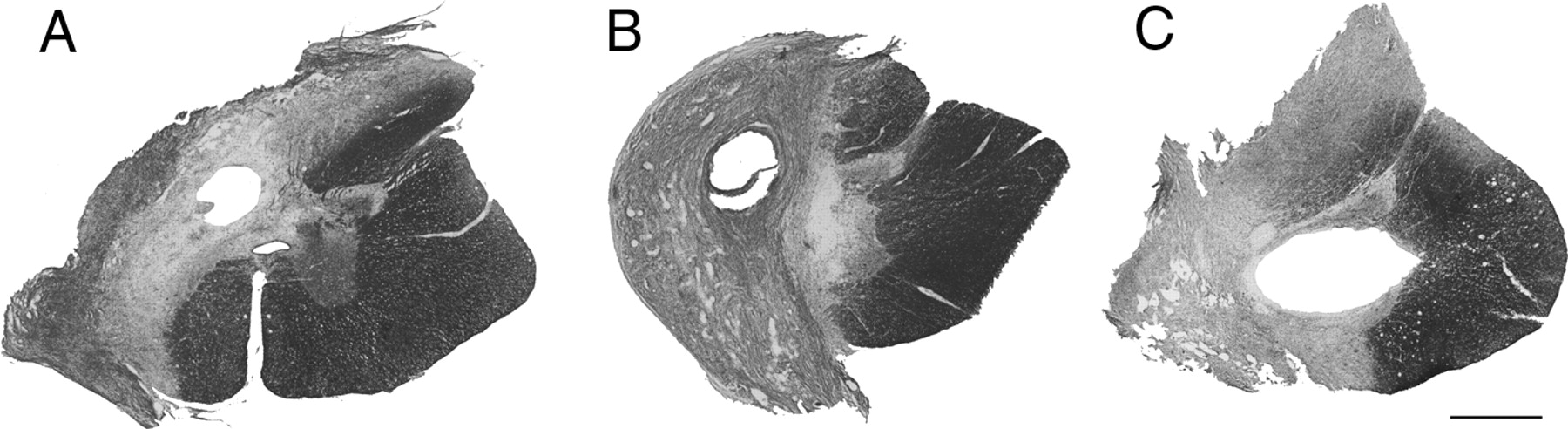
- Figure 1.
Spinal hemisections. A–C, Horizontal sections through the lesion epicenter stained with cresyl violet and myelin stains show that lesions ranged from an under-hemisection with ipsilateral ventromedial sparing (A), to hemisection lesions with interruption of all ipsilateral gray and white matter (B), to over-hemisections with interruption of contralateral gray and white matter (C). Scale bar, 1 mm.
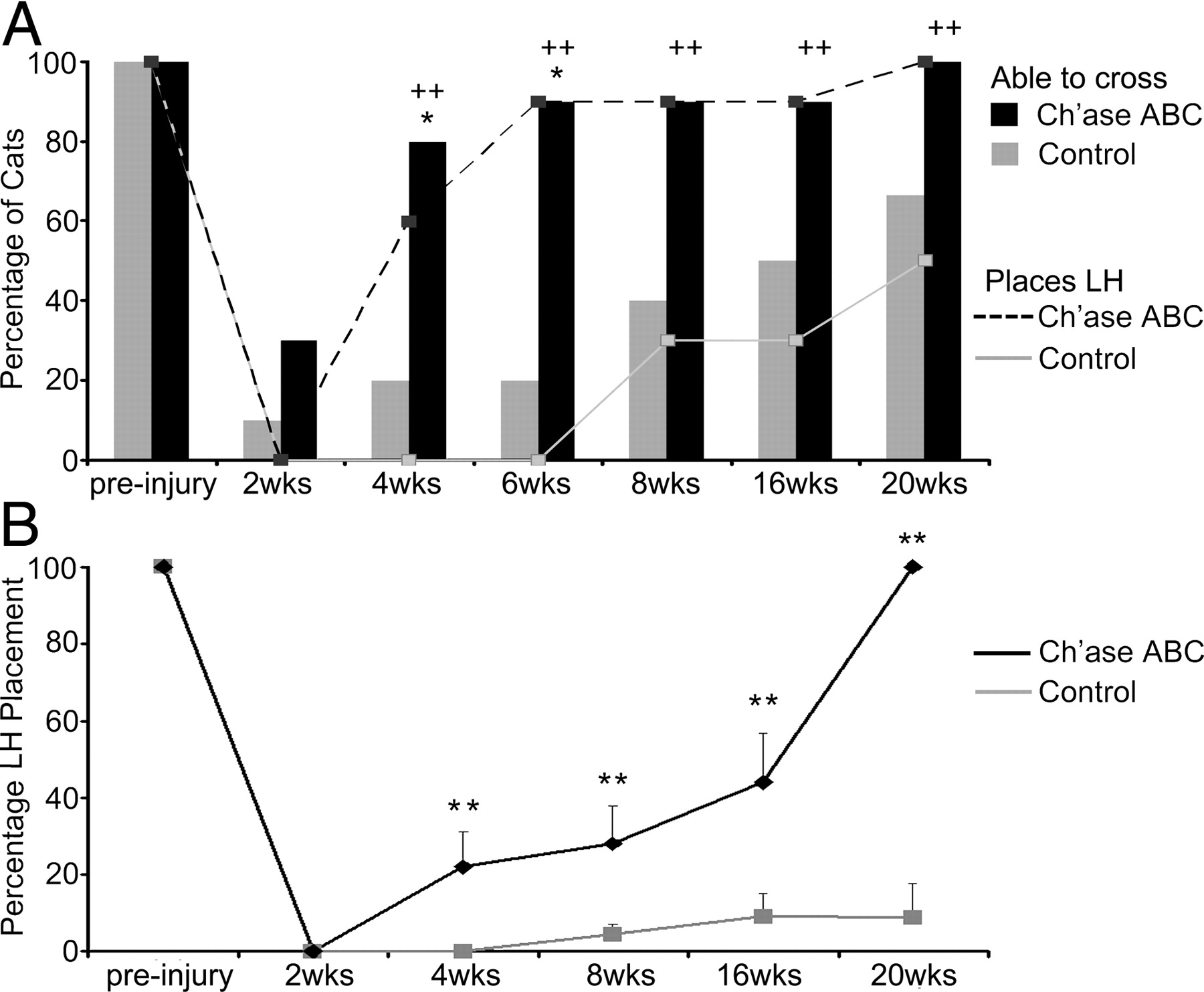
- Figure 2.
Peg walkway locomotor recovery is improved with Ch'ase ABC administration. A, The ability to cross the peg walkway (bar graphs) and place the LH on the pegs (line graphs) was assessed for Ch'ase ABC (black)-treated and control (gray)-treated cats pre- and post-injury. Pre-injury, all cats crossed the peg walkway and placed their LHs onto the pegs with 100% accuracy. Following injury, significantly more Ch'ase ABC-treated cats crossed independently at 4 (*p = 0.023) and 6 (*p = 0.005) weeks compared with controls. Significantly more Ch'ase ABC-treated cats also showed some placement of their LHs onto pegs at 4 (++p = 0.011), 6 (++p = 0.001), 8 (++p = 0.02), 16 (++p = 0.02), and 20 (++p = 0.033) weeks post-injury. B, LH placement onto the pegs was quantified. Ch'ase ABC-treated cats accurately placed their LHs a greater percentage of step cycles compared with controls at 4 (**p = 0.005), 8 (**p < 0.012), 16 (**p = 0.009), and 20 (**p = 0.005) weeks post-injury. Error bars denote SEM.
- Figure 3.
Interlimb coordination on the peg walkway. Pre-injury cats typically place their left limbs on the pegs on the left side of the board and the right limbs on the pegs on the right side. No two limbs occupy a peg at the same time (A). Sixteen weeks post-injury, control-treated animals typically cross on three limbs and do not place their LHs onto pegs (B). Ch'ase ABC-treated cats can place their LHs onto pegs (C), but do so using a novel pattern in which the LH is paired with the RF on a peg on the right side of the board (C, H). Footfall pattern diagrams also suggest differences in stance overlap and duration between pre-injury (D) and 16 weeks post-injury in control-treated (F) and Ch'ase ABC-treated (H) cats. Solid bars indicate when the limb is in stance, and open areas indicate when the limb is in swing. Time between vertical lines is one video frame (33.3 ms). Support pattern diagrams further illustrate the timing differences and the emergence of a single limb support period in the control-treated cats (G) and a quad support period in the Ch'ase ABC-treated cats (I), neither of which is seen pre-injury (E). The support formulas shown with the support diagrams indicate that each group has a different, but consistent pattern. Hindpaws are gray and forepaws black (A–C). Lines connecting limbs in the support pattern diagrams indicate pairing of the two limbs on the same peg. Two complete step cycles are shown in each example (D–I).
- Figure 4.
Hip and knee angular kinematics on the peg walkway. Pre-injury movement patterns (black; A, B) may vary some across cats but are consistent within each cat and used as a baseline for intra-animal pre–post comparisons. A, An example from 16 weeks post-injury in a control-treated cat shows that the ranges of hip and knee movements are much greater and more extended in the non-placing LH (gray) than were seen pre-injury. B, The angular excursions of the hip and knee also are much greater in the LHs of Ch'ase ABC-treated cats 16 weeks post-injury (gray) compared with pre-injury (black). However, as seen in this example, the coordination between the two joints appears similar to pre-injury (note crescent shapes) and the greater excursion is consistent with the skipping of every other peg by the LH. Smaller joint values on either axis represent increased flexion of the joint being assessed, and larger values represent increased joint extension. At each time point, representative step cycles are shown.
- Figure 5.
Increased pNF-H caudal to the lesion following Ch'ase ABC treatment. Nonbiased stereological quantification of the area fraction of pNF-H (A) was assessed in four regions of the spinal cord: ipsilateral gray matter (B, C), contralateral gray matter (D, E), ipsilateral white matter (F, G), and contralateral white matter (H, I). The pNF-H-positive area was significantly greater in Ch'ase ABC-treated animals compared with controls in the contralateral gray matter (A; D, E; *p = 0.003) and ipsilateral white matter (A; F, G; *p = 0.033). There was no significant difference between groups in the ipsilateral gray matter (A; B, C) or contralateral white matter (A; H, I). Error bars denote SEM. Photomicrograph examples shown are from one Ch'ase ABC and one control cat. The dorsal horn (DH) and ventral horn (VH) are shown in the ipsilateral and contralateral gray matter examples (B–E). The ipsilateral and contralateral white matter (F–I) examples are from the lateral funiculus. Scale bar, 0.1 mm.
- Figure 6.
Retrograde labeling of red nuclei neurons from below the lesion. A–C, FG retrogradely labeled neurons in control (left) (A) and experimental (axotomized) (B) red nuclei were counted. Significantly more neurons were labeled in the experimental nuclei of Ch'ase ABC-treated compared with control-treated cats (C). The number of neurons labeled in the experimental red nuclei are presented as a percentage of the number labeled in control nuclei, *p = 0.032. Error bars denote SEM. Scale bar, 0.1 mm. D–F, Retrogradely labeled neurons in the control nuclei (D) and experimental nuclei from control-treated (E) and Ch'ase ABC-treated (F) cats all showed costaining for the presynaptic terminal marker synaptophysin (purple), suggesting that these neurons (brown) were receiving input. Scale bar, 0.05 mm.


