

Article Figures & Data
Figures
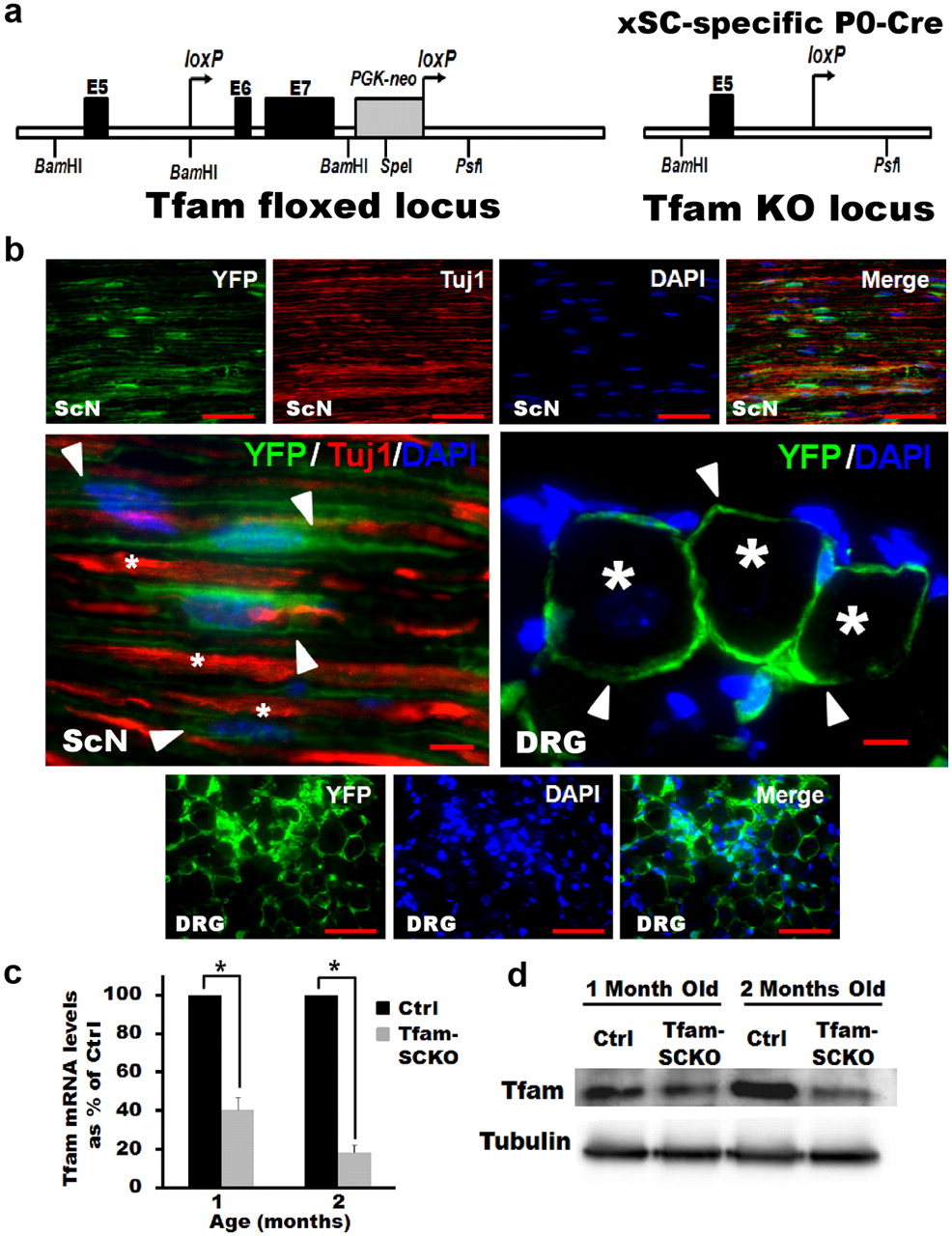
- Figure 1.
P0-Cre deletes Tfam from SCs specifically and efficiently in Tfam-SCKO mice. a, Diagram of the targeted Tfam locus in TfamloxP mice. When TfamloxP mice are crossed with mice that express Cre recombinase under the control of the P0 promoter, exons 6 and 7 of Tfam are excised to produce Tfam− mice. b, YFP fluorescence in SCs (arrowheads) of longitudinal sciatic nerve sections (ScN), as well as satellite SCs (arrowheads) in DRGs of Rosa26-YFP/P0-Cre mice. P0-Cre induces recombination specifically in SCs and excision-dependent YFP fluorescence is localized to regions of SC cytoplasm but not to axons (Tuj1 and stars; note non-overlapping Tuj1 and YFP staining) or DRG neuron cell bodies (stars). Scale bars: Top and bottom panels, 100 μm; middle magnified panel, 50 μm. c, qRT-PCR results showing efficient depletion of Tfam mRNA in Tfam-SCKO sciatic nerves. Tfam mRNA levels in the sciatic nerve of 1-month-old Tfam-SCKOs were decreased by 60% (p = 0.001, two-tailed Student's t test) compared with ctrl littermates. By 2 months of age Tfam mRNA levels were reduced by 85% (p = 0.002, two-tailed Student's t test) compared with ctrl littermates. Reported values are normalized to GAPDH. Error bars indicate SEM. n = 2 pools of 3 mice run in 3 independent experiments. d, Western blot showing efficient depletion of Tfam protein in the sciatic nerves of Tfam-SCKO mice compared with ctrl littermates by 2 months of age.
- Figure 2.
SC-specific excision of Tfam induces mtDNA and transcript depletion and respiratory dysfunction in Tfam-SCKOs. a, qRT-PCR results showing depletion of mtDNA content in the sciatic nerves of Tfam-SCKO mice compared with littermate ctrls. mtDNA content was significantly reduced by 50% (p = 0.01, two-tailed Student's t test) in 2-month-old Tfam-SCKO sciatic nerves compared with ctrl nerves. Reported values are normalized to nuclear DNA content. Error bars indicate SEM. n = 3 mice per genotype run in 2 independent experiments. b, qRT-PCR results showing depletion of mitochondrially encoded electron transport chain subunit transcripts (mt-ND2 and mt-Cox1) in Tfam-SCKO sciatic nerves. mRNA levels of these subunits were decreased 85% (p = 0.02, two-tailed Student's t test) in 2-month-old Tfam-SCKO sciatic nerves compared with ctrl littermates. In contrast, transcripts of nuclearly encoded electron transport chain subunits (nuclear SDHB) are not depleted in Tfam-SCKO sciatic nerves. Reported values are normalized to GAPDH. Error bars indicate SEM. n = 2 pools of 3 mice run in 3 independent experiments. c, Respiratory chain enzyme activity measured from mitochondria isolated from the sciatic nerves of 2-month-old Tfam-SCKO nerves and ctrl littermates. COX activity (complex IV), which contains critical mtDNA-encoded subunits, is reduced 65% (p < 0.01, two-tailed Student's t test) in Tfam-SCKO mice. Error bars indicate SEM. n = 3 pools of 2 mice per genotype. d, Mitochondrial respiration in 2-month-old Tfam-SCKO and littermate ctrl permeabilized sciatic nerves measured using high-resolution respirometry. Respiration induced by substrates delivering electrons to complex I (pyruvate plus malate) is reduced by 35% (p = 0.04, two-tailed Student's t test) in Tfam-SCKO sciatic nerves compared with littermate controls. Respiration induced by the convergent transport of electrons entering at complexes I and II (using pyruvate plus malate plus succinate as substrates) is decreased by 25% (p = 0.04, two-tailed Student's t test) in Tfam-SCKO sciatic nerves compared with littermate ctrls. Respiration induced by substrates delivering electrons to complex II alone (succinate), which is fully nuclearly encoded, was not significantly changed in Tfam-SCKO nerves compared with littermate ctrls. Error bars indicate SEM. n = 5 mice per genotype. e, COX enzymatic staining of 4-month-old ctrl and Tfam-SCKO sciatic nerves. The intense COX staining that localizes to SCs in ctrl nerves (arrowheads) is largely lost in Tfam-SCKO nerves, indicating mitochondrial dysfunction specifically in this cell type. f, Electron micrographs of Tfam-SCKO sciatic nerves show abundant abnormal enlarged mitochondria (arrowheads), often with distorted cristae. Aberrant mitochondria are mainly found in SCs, while axonal mitochondria show no morphological abnormalities (arrows). Pathological mitochondria are visible as early as 1 month in Tfam-SCKO nerves (top 3 panels) and become more abundant as these mice age (bottom panels from 4-month-old mice). Scale bars, 500 nm.
- Figure 3.
Tfam-SCKOs develop a progressive degenerative peripheral neuropathy. a, Photograph of end-stage Tfam-SCKO mouse (≈8 months of age). At this stage, Tfam-SCKOs display a characteristic swimming gait and are unable to support themselves on their hind legs. b, Typical nerve conduction trace from a 4-month-old Tfam-SCKO mouse showing marked temporal dispersion. Calibration: 1 ms, 1 mV. c, d, Electrophysiological studies of Tfam-SCKO mice show that, as early as 2 months of age, these mice have significantly reduced nerve conduction velocity (c) (p < 0.001, two-tailed Student's t test) compared with littermate ctrls. This deficit persists in 4-month-old animals (c) (p < 0.001, two-tailed Student's t test), which also have severe temporal dispersion as shown by an increased response duration (d) (p < 0.001, two-tailed Student's t test). These findings indicate segmental demyelination. Error bars indicate SEM. N = 4 mice per genotype at each age. e, Typical electromyography traces from a 4-month-old Tfam-SCKO mouse showing fibrillation (arrowheads) and fasciculations (arrows), common findings in diseases involving motor axon loss, indicative of muscle fiber denervation and motor unit degeneration/regeneration. These findings were never present in ctrl littermates. Calibration: 50 ms, 500 μV. f, Hematoxylin and eosin stain of the gastrocnemius muscle from a ctrl and a Tfam-SCKO mouse showing scattered and grouped muscle fiber atrophy (arrowheads). This is characteristic of motor axon degeneration. Scale bar, 50 μm.
- Figure 4.
Tfam-SCKOs display early preferential loss of small unmyelinated fibers: a–c, Toluidine blue-stained plastic sections of 1-month-old Tfam-SCKO sciatic nerve and littermate ctrls (a) show that at this age there are no differences in the number of myelinated fibers (b) or the extent of myelination (g-ratio, axon area/fiber area) (c). Scale bar, 25 μm. Error bars indicate SEM. n = 4 mice per genotype. d, Electron micrographs of 1-month-old ctrl and Tfam-SCKO sciatic nerves show that, while large myelinated fibers are mostly normal in Tfam-SCKO mice at this age (arrows), the structure of Remak bundles (arrowheads) is largely abnormal. Scale bar, 2 μm. e1–e4, Compared with ctrl littermates (e1), unmyelinated axons in Tfam-SCKO Remak bundles are found touching one another (e2, e4, stars) and interspersed by pathological SC processes known as bands of Bungner (e2, arrowheads). Free-floating excess basement membrane (e2, e3, arrows) enclosing SC cytoplasm (e3, star) with few or no axons left, indicative of extensive degeneration of unmyelinated axons, is also visible. By 2 months of age, most nonmyelinating SCs are associated with unstructured, degenerating Remak bundles, containing abnormal axons (e4, star) and filled with phagocytic vesicles and membranous debris (e4, arrowhead). Scale bar, 500 nm. f, Representative photographs of epidermal fiber innervation (arrowheads) in the footpads of 2-month-old Tfam-SCKO and littermate ctrl mice. Scale bar, 10 μm. g, Quantification of fiber density in the epidermis shows a 30% decrease in Tfam-SCKO by 2 months of age (p = 0.02, two-tailed Student's t test), indicating early degeneration of small unmyelinated C-fibers. Results reported are normalized to per unit area. Error bars indicate SEM. n = 3 mice per genotype at each age.
- Figure 5.
Degeneration of large-caliber myelinated fibers in older Tfam-SCKOs. a, b, Toluidine blue-stained plastic sections of Tfam-SCKO sciatic nerves and littermate ctrls at different ages (a) and quantification of total number of myelinated profiles per nerve (b). At 2 months, some initial signs of axonal degeneration are apparent (arrowhead) in Tfam-SCKO nerves, but at this age there are no differences in the number of myelinated fibers (b). By 4 months, there is prominent axonal degeneration, as shown by a significant decrease (p > 0.001, two-tailed Student's t test) in the total number of myelinated profiles in Tfam-SCKO nerves. Axonal degeneration worsens by 8 months of age (p > 0.001, two-tailed Student's t test). Scale bar, 25 μm. Error bars indicate SEM. n = 4 mice per genotype at each age. c1–c4, Electron micrographs of Tfam-SCKO nerves at different ages confirm that large myelinated fiber degeneration becomes prominent by 4 months of age (c2) and worsens by 8 months (c3), as shown by large portions of endoneurium completely devoid of axons (stars). By 4 months of age, actively demyelinating and degenerating axons filled with membranous debris (arrowheads) are also common in Tfam-SCKO nerves (c4). By this age, segmental demyelination as shown by large-caliber axons without myelin is also prominent (c2–c4, arrows). This type of pathology is not visible in ctrl nerves (c1). Scale bar, 2 μm. d, Fiber size distribution analysis in Tfam-SCKO and ctrl littermate nerves at different ages. No differences in fiber size distribution are evident despite severe axonal degeneration, indicating that myelinated fibers of all calibers are susceptible to degeneration. Error bars indicate SEM. n = 4 mice per group at each age. e, Osmicated teased fibers from 4-month-old Tfam-SCKO nerves confirm the presence of segmental demyelination. Scale bar, 25 μm.
- Figure 6.
Tfam-SCKOs have behavioral deficits consistent with early loss of unmyelinated fibers followed by extensive degeneration of myelinated axons. a, Two-month-old Tfam-SCKOs are less sensitive to an applied noxious heat stimulus than ctrl littermates (p < 0.01, two-tailed Student's t test), as shown by an increased withdrawal latency, which indicates loss of unmyelinated C-fiber nociceptors. This deficiency persists in 4-month-old Tfam-SCKO mice (p < 0.01, two-tailed Student's t test). b–d, At 2 months of age, consistent with the early preservation of large myelinated motor axons, Tfam-SCKOs do not display significant motor deficits as shown by the inverted screen test (b) or accelerating rotarod (c). Four-month-old Tfam-SCKOs, however, spend significantly less time on an inverted screen (p < 0.01, two-tailed Student's t test) (b) and accelerating rotarod (p < 0.01, two-tailed Student's t test) (d), motor deficits that indicate the loss of large myelinated fibers by this age. Error bars indicate SEM. N = 4 mice per genotype at each age.
- Figure 7.
SC mitochondrial dysfunction does not compromise SC survival. a, b, Nuclear staining (a, DAPI) and quantification (b) shows that there is no difference in the number of SC nuclei between Tfam-SCKO and ctrl sciatic nerves at different ages. a, c, Quantification of Rosa-YFP immunofluorescence (a, YFP/DAPI; c) in Tfam-SCKO/Rosa-YFP nerves at different ages shows that the majority of SCs are Tfam-deficient at all ages. a, d, Quantification of TUNEL immunohistochemistry (a, TUNEL/DAPI; d) of Tfam-SCKO and ctrl littermate shows no differences in the number of TUNEL-positive cells at different ages, indicating that SCs are able to survive despite the disruption in their mitochondria. Scale bar, 100 μm. Error bars indicate SEM. n = 3 mice per genotype at each age.
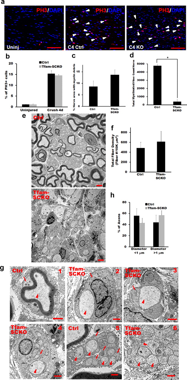
- Figure 8.
Tfam-deficient SCs support axonal regeneration but fail to remyelinate following injury. a, b, Phosphohistone 3 (PH3) immunostaining (a) and quantification (b) show that there is no difference in the number of proliferating SCs (arrowheads) between Tfam-SCKO and ctrl sciatic nerves 4 d after crush injury. Scale bar, 100 μm. Error bars indicate SEM. n = at least 3 mice per genotype. c, Quantification of the amount of myelin debris present in ctrl and Tfam-SCKO nerves 3 weeks after crush injury shows no impairment in the ability of Tfam-deficient SCs to break down existing myelin. Error bars indicate SEM. n = 3 mice per genotype. d, Quantification of the number of myelinated profiles in ctrl and Tfam-SCKO nerves 3 weeks after crush distal to the site of injury shows a significant decrease (p < 0.001, two-tailed Student's t test) in the number of myelinated profiles formed by Tfam-deficient SCs following injury. Error bars indicate SEM. n = 3 mice per genotype. e, Electron micrographs of ctrl and Tfam-SCKO nerves 3 weeks after crush depicting the inability of Tfam-deficient SCs to form mature myelin even though they are capable of supporting axonal regeneration. Scale bar, 2 μm. f, Quantification of the total number of fibers (myelinated and unmyelinated) distal to the site of injury 3 weeks after crush shows no differences between Tfam-SCKO and ctrl nerves, confirming that Tfam-deficient SCs are able to support axonal regeneration. Error bars indicate SEM. n = 3 mice per genotype. g, Electron micrographs of ctrl (g1) and Tfam-SCKO (g2–g4) large-caliber fibers distal to the site of injury 3 weeks after crush showing that, while Tfam-deficient SCs are capable of ensheathing axons and forming one-to-one associations with them (arrowheads, axons; arrows, SCs), they are not able to form mature myelin. Regenerating Remak bundles are present in ctrl nerves (g5) but are absent in Tfam-SCKO nerves (g6; arrowheads, axons; arrows, SCs). h, Proportion of axons >1 μm is similar between Tfam-SCKO and ctrl nerves following injury and cannot explain the absence of myelin in Tfam-SCKO nerves.


