

Article Figures & Data
Figures
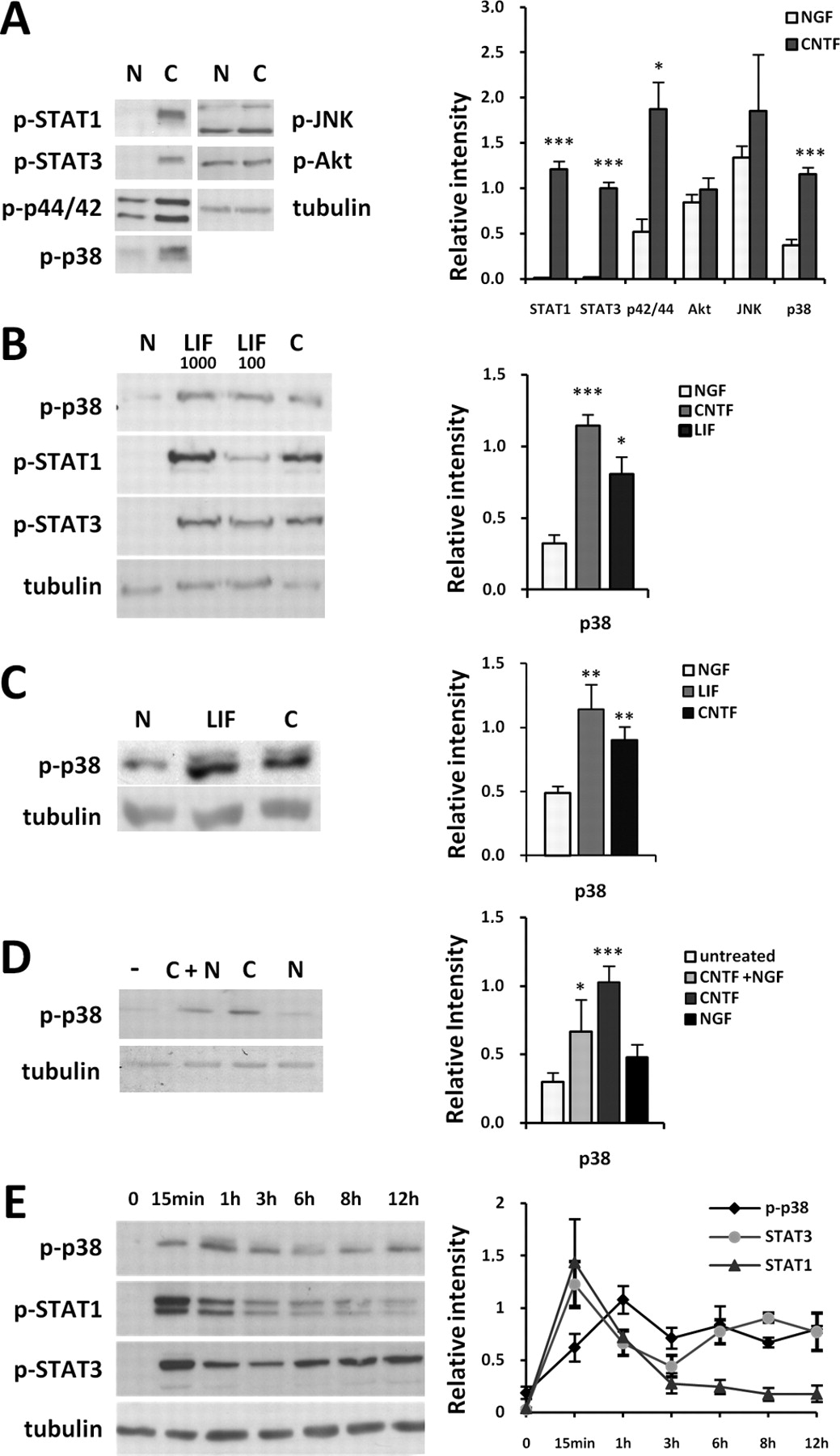
- Figure 1.
CNTF and LIF cause sustained hyperphosphorylation of p38 MAPK. A, Acute CNTF (C) treatment for 30 min leads to STAT1, STAT3, p42/p44 ERK, and p38 phosphorylation in SCG neurons compared to NGF (N)-treated cultures at DIV1 (n = 3). No difference is observed among levels of phosphorylated Akt and JNK. Tubulin was used as a loading control. Images were quantified with ImageJ. Values for phosphorylated proteins were calculated relative to tubulin levels and are represented as arbitrary units. B, CNTF and LIF (1000 or 100 U) treatment cause similar activation of STAT1, STAT3, and p38 (representative image of Western blot) in comparison with NGF treated cultures at DIV 1 (n = 3). C, p38 is also activated after acute CNTF or LIF stimulation of long-term cultures of SCG neurons (DIV4) (n = 3). D, Representative Western blot and quantified results showing the absence of p38 hyperphosphorylation after acute NGF treatment (30 min) compared to untreated controls. CNTF activates p38 in the absence and presence of NGF (n = 3). E, Quantification of Western blot results (representative image shown as left panel) reveals sustained p38 and STAT3 activation after CNTF treatment extending over at least 12 h. STAT1 is strongly activated only during the first 60 min (n = 3–7).
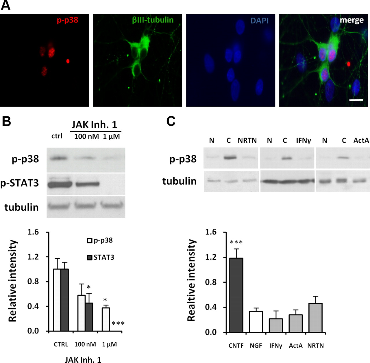
- Figure 2.
p38 activation occurs in neuropoietic cytokine-treated neurons. A, Primary SCG neurons after acute CNTF stimulation were costained for βIII-tubulin and phospho-p38. Nuclei were stained with DAPI. p-p38 staining is not detected in βIII-tubulin-negative cells (scale bar 20 μm). B, Immunoblots demonstrating a dose-dependent decrease in p38 and STAT3 phosphorylation after JAK Inhibitor 1 administration in comparison with neurons treated only with CNTF (Ctrl) (n = 3). Cells were lysed 4 h after CNTF treatment. C, No activation of p38 is detected after neurturin (NRTN), interferon γ (IFNγ), or activin A (ActA) treatment compared to NGF- and CNTF-treated cultures (n = 3).
- Figure 3.
p38 Inhibitors block cholinergic differentiation. A, Images of primary SCG neurons 16 h after plating in NGF- and CNTF-containing medium in the presence of 10 μm SB202190 compared to control cultures without inhibitor. No differences in cell morphology, neurite outgrowth or cell numbers were observed (scale bar 20 μm). B, Quantitative RT-PCR experiments demonstrate a complete block of upregulation of Chat Vacht and VIP by 10 μm SB202190 in CNTF-treated SCG neurons. Expression levels of the noradrenergic markers Th, Net, Dbh, Vmat2, and Gch were not changed in comparison with CNTF-treated control cultures (n = 3–6). C, In the absence of neuropoietic cytokine stimulation SB202190 had no effect on trace levels of Chat and Vacht present in noradrenergic neurons and significantly reduced VIP levels (n = 3). D, Effects of SB 202190 on Chat and VIP transcript levels in CNTF-treated SCG neurons are dose dependent. Comparison with CNTF-treated cultures as controls (n = 3). E, Administration of 10 μm SB202190 6 h after CNTF treatment still inhibits upregulation of Chat mRNA and does also affect VIP (p = 0.07) in comparison with control CNTF cultures (n = 4). F, Quantitative RT-PCR experiments demonstrating a significant decrease of Chat and VIP mRNA levels with p38 inhibitors PD169316 (10 μm) or SKF-86002 (50 μm), but not with the inactive compound SB202474 (10 μm) in comparison with CNTF-treated control cultures (n = 3–4).
- Figure 4.
p38 inhibitors do not affect STAT protein phosphorylation. A, Jak inhibitor 1 but not SB202190 (SB) reduces STAT3 tyrosine Y705 and STAT1 tyrosine Y701 phosphorylation after CNTF stimulation compared to control CNTF cultures and NGF-treated cultures (N). B, STAT3 serine S727 phosphorylation is also not affected by SB202190 compared with CNTF (C). C, Quantification of results shown in A and B (n = 3–4).
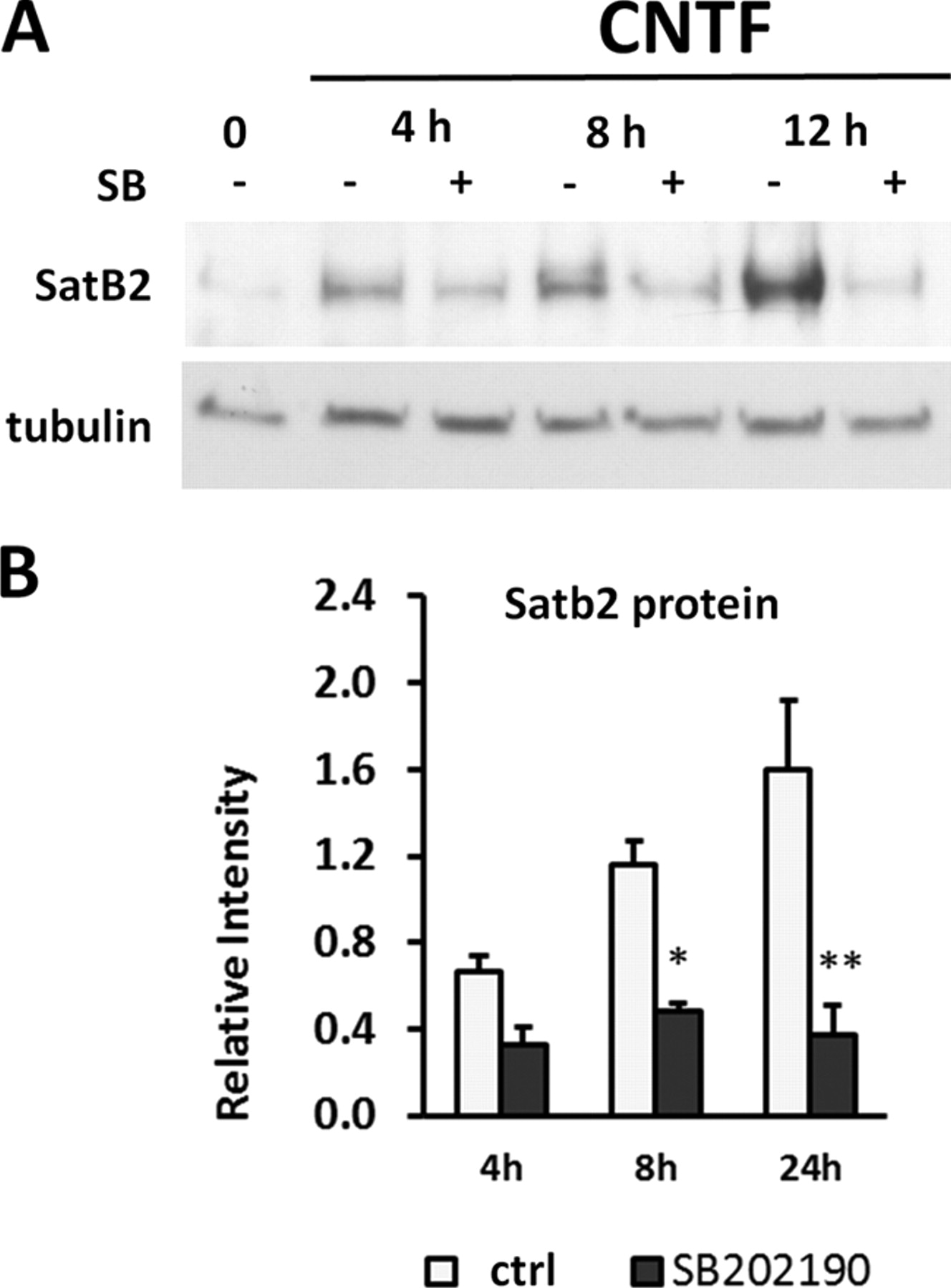
- Figure 5.
p38 inhibitor prevents Satb2 protein regulation. A, Representative Western blot of Satb2 protein in the absence (−) and presence (+) of SB202190 determined 4, 8, and 12 h after stimulation with CNTF. B, Quantification of the results. The upregulation of Satb2 protein in response to CNTF is effectively blocked by the p38 inhibitor at any time point tested.
- Figure 6.
Neurotransmitter markers are regulated after p38 overexpression. A, Quantitative RT-PCR experiments demonstrate a marked increase in Vacht and VIP transcript levels in NGF-treated SCG neurons coexpressing MKK6b[E] and p38 isoforms in the absence of neuropoietic cytokines. Stimulation of endogenous p38 by overexpression of the p38-MAPKK MKK6b[E] alone has no effect on Vacht levels but significantly increases VIP mRNA levels. B, The mRNA levels of Net and NF160 remain unaltered in all overexpression experiments. Approximately 80 GFP-positive nucleofected neurons were analyzed per experiment and compared with pCMV and pCDNA3-transfected neurons (Mock).
- Figure 7.
Cholinergic differentiation is impaired in p38β-deficient neurons in vitro and in vivo. A, Representative photograph of mouse SCGs at postnatal day 6 from either wild-type (WT) or p38β-deficient mice. B, Images of primary mouse SCG neurons from WT and p38β−/− mice 48 h after plating in the presence of CNTF and NGF. No differences in cell morphology, neurite outgrowth, or cell number was observed depending on the genotype (scale bar, 20 μm). C, Representative Western blot and quantified results showing a significant increase in p38 phosphorylation in CNTF-treated mouse SCG neurons in comparison to NGF-treated cultures. Tubulin was used as a loading control (n = 3). D, 38β deficiency in CNTF-treated mouse SCG neurons greatly reduces Chat, Vacht, and VIP transcript levels, without affecting Net, Tau, and p38α levels in comparison with CNTF-treated wild-type cultures after 48 h in culture (n = 4). E, Sections from postnatal day 60 mouse stellate ganglia were immunostained for Vacht in wild-type and p38β−/− mice. Vacht-positive cell somata are indicated by white arrows (scale bar 20 μm). Quantitative analysis of Vacht-immunoreactive neurons per ganglion demonstrates that the number of Vacht-positive cells is greatly reduced in p38β mutant ganglia. The percentage of Vacht-IR neurons per ganglion was calculated as Vacht/DAPI-positive neuron ratio in section where nuclei were stained with DAPI. Data are expressed as mean ± SEM (n = 5; 5 animals per group i.e., 10 ganglia were analyzed per genotype).


