

Article Figures & Data
Figures
- Figure 1.
Expression of TIP39 and PTH2R immunoreactivity in the preoptic hypothalamus. A, B, Low-magnification images show TIP39-ir (A) and PTH2R-ir (B) concentrated in hypothalamic preoptic areas. C, Schematic representation of the preoptic hypothalamus at the same level shows the location of β-gal-expressing cells in a PTH2R-β-gal knock-in mouse. Scale bar (in A) A, B, 500 μm. 3V, Third ventricle; ac, anterior commissure; LPO, lateral preoptic area; VP, ventral pallidus.
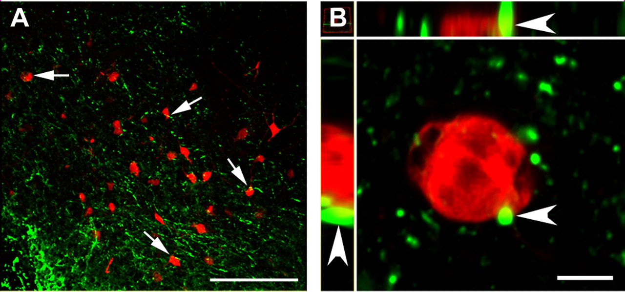
- Figure 2.
PTH2R and VGlut2 immunoreactivity overlap in the MnPO. PTH2R-ir (A, D, and G), GAD65-ir (B), MAP2-ir (E), and VGlut2-ir (H) are shown as single channels and as merged images (C, F, and I). There is no apparent colocalization of PTH2R-ir with GAD65-ir or MAP2-ir, a dendritic marker. A–F, Examples of puncta labeled only with the PTH2R antibody are indicated by arrowheads and ones labeled by the GAD65 or MAP2 antibodies are indicated by indented arrowheads. Images are confocal microscope single optical sections. G–I, The images are from a Z-series spanning ∼1.2 μm. The labeled structure indicated by the arrow is present in the selected X-Z (left of each panel) and Y-Z (top of each panel) planes. Arrowheads point to other puncta coexpressing the two markers (yellow). Scale bars, 10 μm.
- Figure 3.
Few MnPO PTH2R neurons project to the DMH. A, B, Neurons containing retrograde label (red) are shown in the MnPO following injection of FG into the DMH of a PTH2R-β-gal knock-in mouse. β-gal accumulates within vesicle-like structures in the cell bodies of PTH2R neurons (green dots indicated by arrows in A). The area indicated by the square in A is shown at higher magnification in B. One of three retrogradely labeled cells contains β-gal-ir. Scale bars: A, 200 μm; B, 10 μm.
- Figure 4.
MnPO neurons that project to the DMH are contacted by PTH2R-ir fibers. A, Arrows indicate MnPO neurons filled with FG (red) from a DMH injection and enmeshed in PTH2R-ir fibers (green). B, At higher magnification, PTH2-ir processes appear to contact DMH projecting MnPO neurons. The arrow indicates an area of apparent colocalization, which may be a PTH2R-containing fiber making a contact with an MnPO projection neuron. A is a single optical section, B is an XYZ projection of a confocal Z-series of 12 optical sections spanning 47.6 × 47.6 × 5.6 μm with X-Z images on top and Y-Z images at the left. Scale bars: A, 10 μm; B, 5 μm.
- Figure 5.
PRV infection of a PTH2R-ir cell in the MnPO. Five days following BAT injection, PRV-152-infected cells are observed in the preoptic hypothalamus (A–C). A PRV-infected cell with PTH2R-ir on its surface is indicated by the rectangle in C and shown at high magnification in D–G. Single optical sections are shown in D–F. The image in G is an XYZ plane representation of a Z-series of 12 optical sections spanning 47.6 × 47.6 × 3.4 μm. The X-Z (top) and Y-Z (left) views confirm the colocalization between the green and red voxels (yellow). Virally encoded GFP is pseudocolored red in all images and PTH2R-ir is green. Scale bars: A–C, 100 μm; D–F, 10 μm. In G, 5 μm (X) and 2 μm (Z) bars are shown.
- Figure 6.
PTH2R-ir processes appear to contact PRV-infected cells in the MnPO. A, Multiple cells infected with PRV-152 (red) are found in the MnPO following BAT injection of the virus. PTH2R-ir fibers are adjacent to many of the PRV-infected cells, some of which are indicated with arrows. The arrowhead in B indicate an area of PTH2R-ir in apparent contact with a PRV-152-infected cell. The image is presented as an XYZ plane from a confocal Z series covering 47.6 × 47.6 × 5.6 μm, with X-Z image on top and Y-Z image at the left of the panel. Scale bars: A, 100 μm; B, 5 μm.
- Figure 7.
MnPO glutamatergic and GABAergic neurons project to the DMH. Rhodamine latex beads were injected into the DMH. The general distribution of rhodamine bead-containing cells (A, G) overlaps the distribution of cells containing VGlut2 mRNA (B) and GAD67 mRNA (H) as detected by fluorescent in situ hybridization. Adjacent diagrams (C, I) indicate the approximate shape of the MnPO at the levels illustrated. DAPI (blue) counter staining helps to show the location of cell bodies containing rhodamine beads (red, D and J), VGlut2 mRNA (green, E) and GAD67 mRNA (green, K). F and L are merged images in which the arrows show double-labeled cells and the arrowheads single-labeled cells (VGlut2 mRNA in F and GAD67 mRNA in L). The fielding F also contains three cells containing VGlut2 mRNA but no rhodamine beads. Scale bars: A–C, 100 μm; D–F, 10 μm.
- Figure 8.
Distribution of neurons projecting from the MnPO to the DMH. A, Rhodamine latex bead injection sites in the four animals used for analysis are illustrated. B, The injection site in red is shown after counterstaining tissue with DAPI. C–F, The distribution of rhodamine-labeled neurons (red dots), VGlut2/Rhodamine (purple stars) and GAD67/Rhodamine (teal circles) coexpressing cells from this animal are illustrated at four levels (distance from bregma indicated on the figure). Scale bar (B), 1 mm. Re, Thalamic reuniens nucleus; zi, zona incerta; Arc, hypothalamic arcuate nucleus.
- Figure 9.
BAT-injected PRV infects preoptic glutamatergic neurons. A, B, PRV-614-infected cells (red) and VGlut2-GFP-expressing neurons (green) are shown at two levels of the preoptic hypothalamus (bregma +0.5 mm, left; +0.26 mm, right) after injection of the tracer virus into BAT of a VGlut2-GFP BAC transgenic mouse. C, D, The distribution of neurons containing GFP (green dots), PRV-614 (red dots), or both markers (blue stars) at these two levels is shown. High-magnification images taken from the outlined areas in A (E, G, I) and B (F, H, J) show VGlut2-GFP neurons (E, F) containing PRV-614 (G, H) in which the markers colocalize (I, J). In the high-magnification images (E–J), arrows indicate GFP/PRV-614 double-labeled cells. Scale bar (in A) A, B, 200 μm; (in I) E–J, 10 μm.
- Figure 10.
Body temperature following hypothalamic microinjection of TIP39. Vehicle or 50, 100, or 200 pmol of TIP39 was microinjected into the MnPO and body temperature measured hourly (A). Area under the curve measurements (B) indicate significant increases for the groups receiving 100 or 200 pmol of TIP39. TIP39 microinjected into the DMH did not have an effect on body temperature (C). *p < 0.05.
- Figure 11.
Body temperature following lateral ventricle administration of TIP39. Injection of 500 pmol of TIP39 into the lateral ventricle increased body temperature for several hours in wild-type animals but not in PTH2R-KO mice, seen as a time course (A), and in area under the curve measurement (B). *p < 0.05.
- Figure 12.
Peripheral β-adrenergic block prevents TIP39 elevation of body temperature. Pretreatment with nadolol (20 mg/kg, i.p.) 1 h before MnPO injection of TIP39 (100 pmol) or vehicle prevented the small temperature increase observed 1 h after intracranial injections and blocked the effect of TIP39 (A). Area under the curve measurements indicate that the temperature increase in animals receiving MnPO injection of TIP39 was significantly greater than either group receiving MnPO vehicle injection and the group that received MnPO TIP39 and peripheral nadolol (B). *p < 0.05, **p < 0.01.
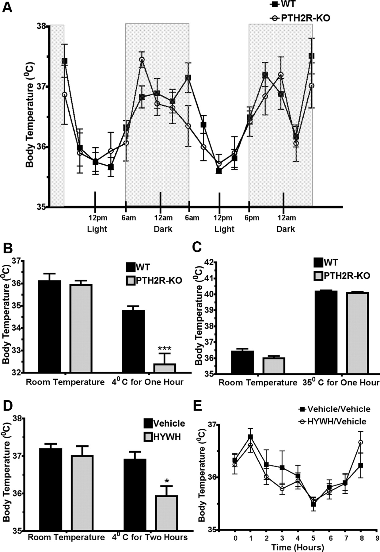
- Figure 13.
TIP39 signaling is required for an appropriate response to cold. The daily pattern of body temperature was not different between wild-type and PTH2R-KO mice (A). PTH2R-KO mice had a much larger decrease in body temperature than wild-type mice following 1 h cold (4°C) exposure (B), but no difference from wild-type when placed in a hot (35°C) environment for the same period (C). Injection of the PTH2R blocker HYWH into the lateral ventricle caused a significant decrease in body temperature in animals exposed to cold (D) but did not affect the temperature of animals that were kept at room temperature (E). *p < 0.05, ***p < 0.001.
- Figure 14.
Evidence for impaired BAT function in PTH2R-KO mice. Peripheral administration of norepinephrine (1 mg/kg, i.p.) did not change the body temperature of wild-type mice but decreased the temperature of PTH2R-KO mice for several hours shown as a time course (A) and cumulated area under the curve measurements (B). *p < 0.05.


