

Article Figures & Data
Figures
- Figure 1.
Faithful expression of GAD67-GFP in cortical GABAergic neurons. A–P, Immunostaining of GAD67+ brain sections with GABA antibody at the stages indicated. GAD67-GFP signals (A, D, G, J, M) were colocalized with those of GABA (B, E, H, K, N, single images; C, F, I, L, O, merged images). More than 93% of GAD67-GFP cells are immunopositive for GABA at the stages examined (P, n = 4, SEM as indicated). Q–U, Similar migratory patterns of GABAergic neurons in heterozygous GAD67-GFP and wild-type cortex. Sections of E14.5 (Q, R) and E17.5 embryonic brains (S, T) of wild-type (Q, S) and GAD67-GFP het (R, T) mice are stained with anti-GABA antibody. Overall, migratory patterns appear to be the same in both brains at both stages. GABA+ cells in each section have also been counted in the somatosensory cortex (U), and no significant differences in the numbers of GABA+ cells are found between genotypes. Scale bars: (in A) A–O, 50 μm; (in Q) Q–T, 0.5 mm.
- Figure 2.
Migration patterns of GAD67-GFP cells during development. A–I, Images of GAD67-GFP heterozygote mouse brains at indicated stages. A′–I′, High-magnification pictures of A–I at middle sections along the anterior/posterior and lateral/medial axes. GAD67+ cells begin to reach to the cortex tangentially at E13.5 (A, A′) over the two main migratory streams in the marginal zone (MZ) and subventricular zone (SVZ), and fill in those pathways at E14.5 (B, B′). Some cells enter the cortical plate (CP) and upper/lower intermediate zone (U-IZ and L-IZ) and migrate in these regions, but most cells use tangential streams (C–G, C′–G′). Entry into the cortical plate by GAD67+ cells becomes evident around E17.5–E18.5 (E′, F′) and most of these GABAergic cells have settled to their final position by P28 (H, I, H′, I′). J, Distribution of GAD67+ cells that are found in each of three cortical domains for each embryonic day. Ordinate is fraction of all GAD67+ cells (error bars indicate SEM) in marginal zone (MZ, open circles), in the cortical plate (CP, open squares), and in the lower subventricular zone and ventricular zone (SVZ + VZ, open diamonds), and abscissa is developmental age (days). Scale bars: (in A) A–G, 0.5 mm; H, 0.5 mm; I, 0.5 mm; (in A′) A′–I′, 50 μm.
- Figure 3.
Cortical cells stained with neuronal markers. A–G, Dissociated cells from embryonic mouse cortex at various stages from E10.5 to E18.5 are stained on a glass coverslip by TuJ1 (B) and anti-Map2 (C) antibodies. Representative images at E14.5 are shown. GFP signals are detected in dissociated cells prepared from GAD67-GFP mice (F). Merged images are shown in D and G. Cells are counterstained with DAPI (A, E) to label all cortical cells. Typical images of an E14.5 cortex are shown. Arrows indicate Tuj1+ cells and arrowheads Map2+ cells (A–D), or GAD67-GFP cells (F, G). Scale bar: (in A) A–G, 0.2 mm. H–K, Sections of GAD67-GFP postnatal brains at P28 were stained by NeuN antibodies (J). GFP signals indicate GAD67+ cells (I) that have been counterstained with TO-PRO3 to visualize all cell nuclei. Merged image in K. Scale bar: (in H) H–K, 50 μm. L–O, Faithful expression of NeuN in the cortical neurons. P28 sections after fluorescence in situ hybridization (N) and subsequent immunostaining with anti-NeuN antibody (M). Sections were counterstained with TOPO3 and the merged image is shown in O. Most, if not all, of NeuN+ cells are colocalized with Synapsin I mRNA. Arrows indicate double-positive cells for NeuN and Synapsin I and arrowheads negative for both (O), Scale bar: (in L) L–O, 50 μm.
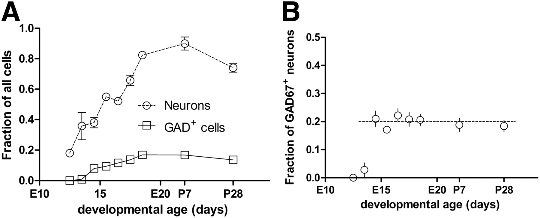
- Figure 4.
A, Fraction of all cells that are neurons and are GABAergic neurons during embryonic development and in the early postnatal period. Ordinate is the fraction of all neurons (open circles) and GAD67+ neurons (open squares) relative to all cells present in cortex, and the abscissa is developmental age (days). B, Fraction of all neurons that are GAD67+ for each developmental age. Ordinate is fraction of neurons that are GAD67+ and abscissa is developmental age (days). The dotted line is the fraction 1/5. Dissociated cells are counted from 3–4 independent experiments. Error bars indicate SEM.


