

Article Figures & Data
Figures
- Figure 1.
Generation and characterization of Rac1-CKO mice. A, DhhCre directed recombination in Rac1 mutant mice. Dhh activates Cre recombinase expression in SC from E12.5. After Cre recombination, exon1 of the Rac1 alleles between loxP sites are excised. B, Genotyping for DhhCre, Rac1-knock-out, and Rac1-flox alleles in samples of wild-type and Rac1-CKO nerves. C, Control and Rac1-CKO P30 mice are shown. Hindlimb paralysis in Rac1-CKO mice is present as noted by dragging their hindlimbs. D, Sciatic nerves from P30 Rac1-CKO and control mice are shown. Rac1-CKO sciatic nerves are thinner than wild-type nerves. E, Western blot analysis shows that Rac1 protein levels are decreased in P30 sciatic nerves of mutant mice. Residual Rac1 protein is attributed to axonal Rac1 because Rac1 protein is nearly absent in sciatic nerves after in vitro axon degeneration, confirmed by loss of neurofilament protein. F, Quantification from three independent experiments shows significant decrease of Rac1 protein in Rac1-CKO sciatic nerves. G, SCs cultured from control and Rac1-CKO sciatic nerves were immunostained using Rac-GTP (green) and phalloidin (F-actin cytoskeleton, red) antibodies. Nuclei were visualized by DAPI (blue). GTP-bound, active forms of Rac1 are eliminated in Rac1-CKO SCs (arrow) but remained in wild-type fibroblasts (arrowhead) from Rac1-CKO nerves. ***p < 0.001 by Student's t test. Error bars indicate ± SEM. Scale bars: G, 25 μm.
- Figure 2.
Myelination defects in Rac1-CKO mice. A, EM analysis of control and Rac1-CKO sciatic nerve cross-sections at P1, P30, P60, and P120. At P1, SCs begin to form a few myelin sheaths in control sciatic nerves. In contrast, no myelin sheaths are detectable in Rac1-CKO sciatic nerves. At P30, P60, and P120, SCs form myelin sheaths around large axons (myelinating SCs) in wild-type sciatic nerves. In contrast, SC myelination is essentially absent in Rac1-CKO sciatic nerves at all ages. SCs in Rac1-CKO nerves form one-to-one relationships with large axons without myelin sheath formation. Abnormal SCs protrusions (arrowheads) are found in Rac1-CKO nerves. B, There is a significant decrease in the number of myelinated axons in Rac1-CKO sciatic nerves. C, There is a significant increase in the number of one-to-one SC–axon relationships without myelination in Rac1-CKO sciatic nerves. D, There is no significant difference in axonal bundle size (axons/bundle) in Rac1-CKO sciatic nerves. E, SC ensheathing small axons as Remak bundles are shown (a, control; b–e, Rac1-CKO). SCs wrapping large axons (asterisk) form myelin sheath in wild-type nerve (a) but no myelin sheath in Rac1-CKO nerve (b–e). Abnormal SCs protrusions (arrowheads) are found in SCs in one-to-one relationships with large axons from Rac1-CKO nerves (c, d). One large axon is not completely sorted out from axonal bundle in Rac1-CKO nerves (d). Redundant Rac1-CKO SC wraps within the axonal bundles were observed (e, arrow). For B–D, n ≥ 15 fields from at least 3 animals per genotype per age were analyzed. *p < 0.05, **p < 0.01, ***p < 0.001 by Student's t test. Error bars indicate ± SEM. Scale bars: A, E, 1 μm.
- Figure 3.
Decreased PAK and merlin phosphorylation in Rac1-CKO sciatic nerves. A, Western blots analysis of Rac1-CKO sciatic nerves from P30 mice. Actin expression level is used as a protein loading control. Blots are representative of nerve protein extracts from at least four independent experiments for each genotype. B, Quantification from five independent experiments shows phosphorylation of PAK (P-Pak_Thr423/Thr402) and merlin (P-merlin_Ser518) are downregulated in Rac1-CKO sciatic nerves. Expression of P-Erk1/2, total Erk1/2, total PAK, and total merlin are not changed in Rac1-CKO sciatic nerves. ***p < 0.001 by Student's t test. Error bars indicate ± SEM.
- Figure 4.
Myelination deficiency in Rac1-CKO mice is rescued by NF2/merlin mutation. A, Sciatic nerves from 3-month-old control, Rac1-CKO, NF2-del, and Rac1-CKO&NF2-del mice are shown. Rac1-CKO sciatic nerves are thinner than control nerves. Rac1-CKO&NF2-del nerves are similar to the size of control nerves. B, EM analysis of control, Rac1-CKO, NF2-del, and Rac1-CKO&NF2-del sciatic nerve cross-sections at P120. More myelinated axons are present in Rac1-CKO&NF2-del sciatic nerves than in Rac1-CKO nerves. Irregular SC protrusions in Rac1-CKO sciatic nerves in vivo remained in Rac1-CKO&NF2-del sciatic nerves (arrowheads). C, The number of myelinated axons is significantly increased in Rac1-CKO&NF2-del mice compared with Rac1-CKO mice. D, The number of axons in one-to-one SC–axon profile without myelination is decreased in Rac1-CKO&NF2-del mice versus Rac1-CKO mice. E, Western blot analysis of Pmp22, a peripheral myelin protein, in control, Rac1-CKO, NF2-del, and Rac1-CKO&NF2-del sciatic nerves. Quantification from three independent experiments shows Pmp22 expression was significantly decreased in Rac1-CKO sciatic nerves. Reduced Pmp22 expression in Rac1-CKO sciatic nerves was rescued in Rac1-CKO&NF2-del sciatic nerves. F, EM analysis of control (a), Rac1-CKO (b), NF2-del (c), and Rac1-CKO&NF2-del (d) nerves. Small axons are normally segregated by SCs and form Remak bundles in Rac1-CKO nerves and Rac1-CKO&NF2-del nerves, although no myelin sheath forms in Rac1-CKO nerves. For C and D, n ≥ 25 fields from at least 5 animals per genotype were analyzed. *p < 0.05, **p < 0.01, ***p < 0.001 by ANOVA statistical analysis, followed by Tukey's test. Error bars indicate ± SEM. Scale bars: B, top row, F, 5 μm; B, bottom row, 2 μm.
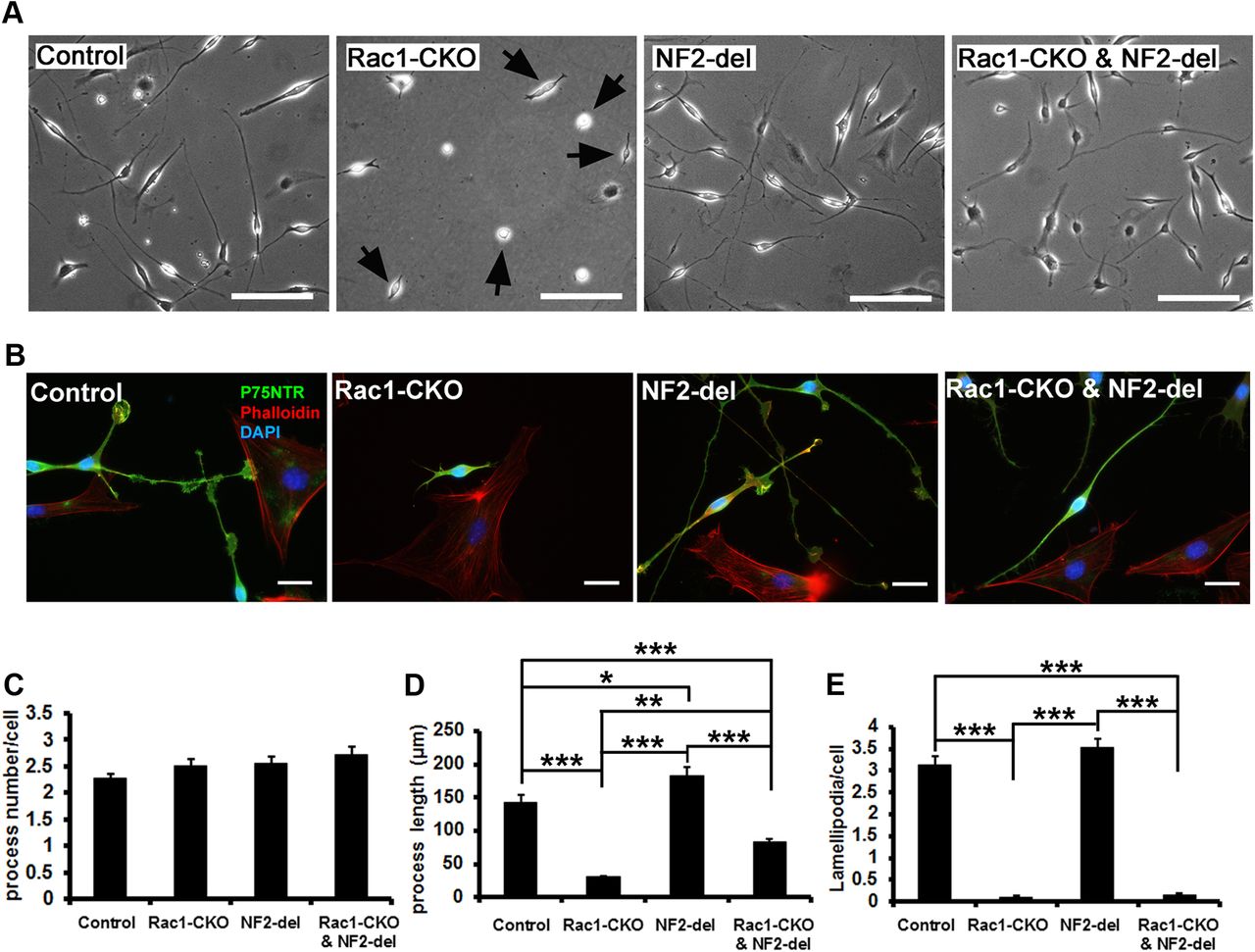
- Figure 5.
Impaired SC process elongation in Rac1-CKO SC is rescued by NF2/merlin mutation. A, SC cultures from control, Rac1-CKO, NF2-del, and Rac1-CKO&NF2-del sciatic nerves are shown. SC process length in Rac1-CKO culture (arrows) is much shorter than that in wild-type culture. B, SCs cultured from P30 control, Rac1-CKO, NF2-del, and Rac1-CKO&NF2-del sciatic nerves were immunostained using P75NTR (an SC marker, green) and phalloidin (F-actin cytoskeleton, red). Nuclei were visualized by DAPI (blue). Rac1-CKO SCs, but not Rac1-CKO&NF2-del SCs, have short processes. C, There is no significant change of SC process numbers per cell in Rac1-CKO or Rac1-CKO&NF2-del cultures. D, The average SC process length is significantly decreased in Rac1-CKO SCs. Process length in Rac1-CKO&NF2-del SCs is increased compared with Rac1-CKO SCs. E, Lamellipodia numbers per cell are significantly decreased in Rac1-CKO SCs and in Rac1-CKO&NF2-del SCs. For C–E, n > 50 cells from 3 independent experiments per genotype. *p < 0.05, **p < 0.01, ***p < 0.001 by ANOVA statistical analysis, followed by Tukey's test. Error bars indicate ± SEM. Scale bars: A, 100 μm; B, 25 μm.
- Figure 6.
Rac1 and NF2/merlin regulate cAMP in SCs. A, Western blots analysis of E-cadherin and P-NF2/merlin in control, Rac1-CKO, NF2-del, and Rac1-CKO&NF2-del sciatic nerves. B, Quantification from four independent experiments shows that E-cadherin expression and NF2/merlin phosphorylation were significantly decreased in Rac1-CKO sciatic nerves. Reduced E-cadherin expression in Rac1-CKO sciatic nerves was rescued in Rac1-CKO&NF2-del sciatic nerves. C, Forskolin was added in control or Rac1-CKO nerve grafts in vitro for 48 h and then E-cadherin expression and NF2/merlin phosphorylation were detected by Western blots. D, Quantification from three independent experiments shows that decreased E-cadherin expression and NF2/merlin phosphorylation in Rac1-CKO nerve grafts was rescued by forskolin treatment in vitro. E, cAMP levels were measured in sciatic nerves of control (n = 8), Rac1-CKO (n = 8), NF2-del (n = 7), and Rac1-CKO&NF2-del (n = 6) mice. cAMP levels in Rac1-CKO nerves are significant lower than in control nerves. cAMP levels in NF2-del nerves are significant higher than in control nerves. Decreased cAMP in Rac1-CKO nerves was restored in Rac1-CKO&NF2-del nerves by t test. F, cAMP levels in cultured wild-type (n = 3) or NF2-del (n = 3) SCs were measured before and after forskolin stimulation (30 min in serum-free medium). Both basal and forskolin-stimulated cAMP levels were increased in NF2-del SCs. The Rac1 specific inhibitor NSC23766 reduced forskolin-stimulated cAMP levels in cultured wild-type and NF2-del SCs. *p < 0.05, **p < 0.01, ***p < 0.001 by ANOVA, followed by Tukey's test. Error bars indicate ± SEM.
- Figure 7.
Myelination defects in Rac1-CKO mice were rescued by cAMP elevation. A, EM analysis of control, Rac1-CKO sciatic nerve cross-sections with 6–8 weeks daily intraperitoneal injection of rolipram. B, The number of myelinated axons is significant increase in Rac1-CKO sciatic nerve after rolipram treatment. C, Abnormal SC protusion remains in Rac1-CKO SC after rolipram treatment (arrow). Occasionally, abnormal folding of SC myelin sheath (arrowheads) was observed in Rac1-CKO nerves after rolipram treatment. For B, n ≥ 15 fields from 6 animals per genotype with rolipram treatment were analyzed. *p < 0.05, ***p < 0.001 by ANOVA, followed by Tukey's test. Error bars indicate ± SEM. Scale bars: A, 5 μm; C, 1 μm.
- Figure 8.
A model for Rac1, NF2/merlin, and cAMP regulate SC myelination. Rac1 regulates NF2/merlin phosphorylation through PAK in SC. Rac1 knock-out decreases phosphorylation of PAK and phosphorylation of NF2/merlin. Nonphosphorylated form of NF2/merlin negatively regulates cAMP-mediated myelination in the absence of Rac1. NF2/merlin mutation restores cAMP levels in Rac1-CKO SCs and allows myelin formation. Reduced cAMP level is a cause for the myelination defect in Rac1-CKO mice. Elevation of cAMP by rolipram rescued myelin formation in Rac1-CKO mice in vivo. Elevation of cAMP by forskolin also increases NF2/merlin phosphorylation. These findings support a novel pathway through which Rac1 regulates SC myelination through NF2/merlin and cAMP signaling.


