

Article Figures & Data
Figures
- Figure 1.
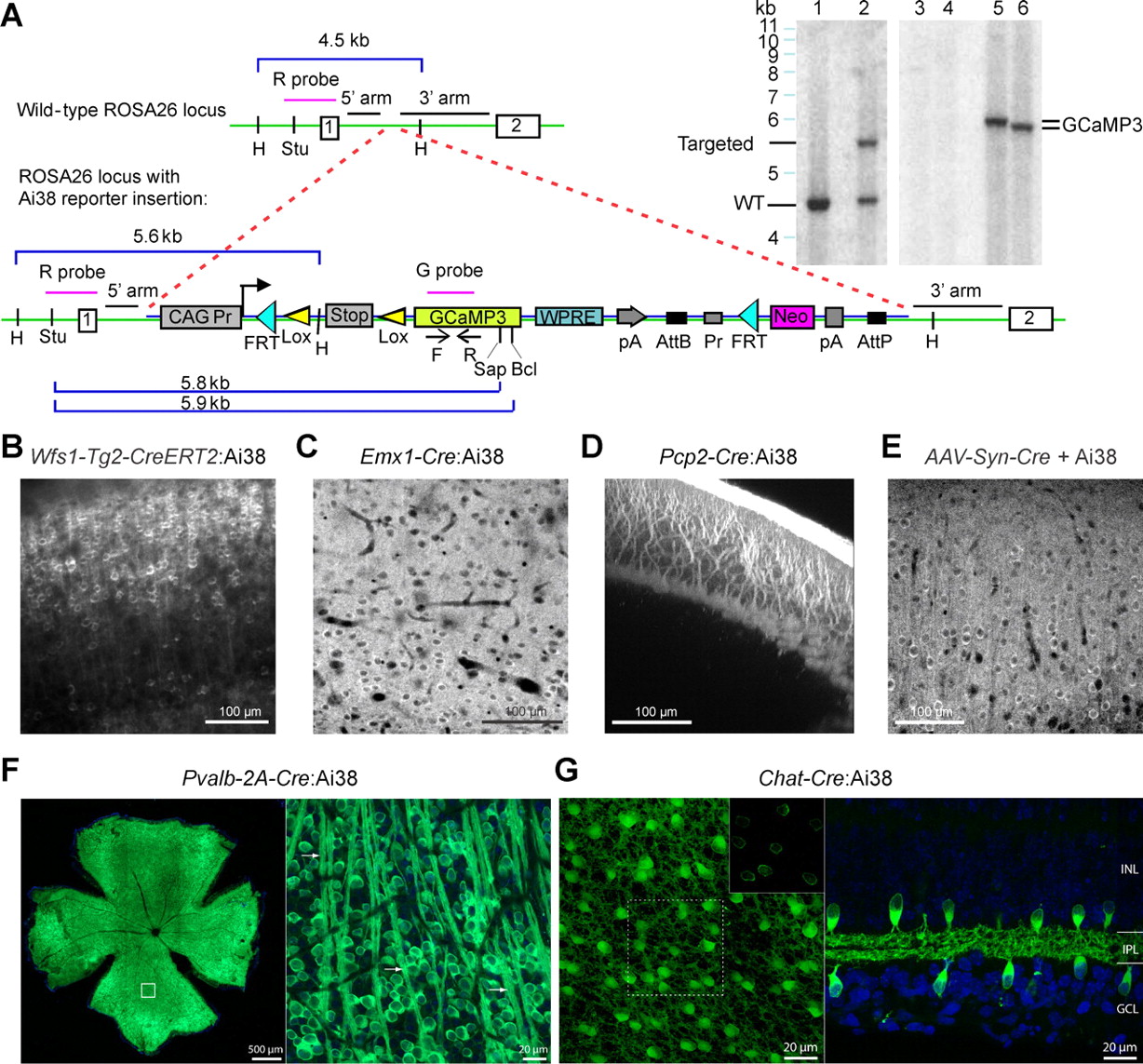
Expression of GCaMP3 in various cell populations in the Ai38 mouse. A, Schematic of the gene targeting strategy to generate the Ai38 reporter mice. GCaMP3-containing Cre reporter cassette (shown in between the dashed red lines) was targeted to the ROSA26 locus in the intron between endogenous exons 1 and 2. Locations of the 5′ and 3′ arms (1.1 and 4.3 kb, respectively) for homologous recombination are indicated by the black bars. Genotyping primers are located in the GCaMP3 sequence, as indicated by the open arrows (the F and R primers). Correct gene targeting was confirmed by Southern blot (top right, lanes 1–2) using the ROSA26 locus-specific probe (the R probe) immediately upstream of the 5′ arm on HindIII (H)-digested genomic DNA, which gives rise to a 4.5 kb band for the wild-type (WT) locus and a 5.6 kb band for the gene-targeted locus. Lane 1, HindIII on WT ES cell DNA. Lane 2, HindIII on Ai38 mouse tail DNA. The single-copy integration of the transgene was confirmed by the appearance of a single band in Southern blot (top right panel, lanes 3–6) using the GCaMP3-specific probe (the G probe) on StuI/BclI- or StuI/SapI-digested genomic DNA, which gives rise to a 5.9 kb or a 5.8 kb band, respectively. Lane 3, StuI/BclI on WT ES cell DNA. Lane 4, StuI/SapI on WT ES cell DNA. Lane 5, StuI/BclI on Ai38 mouse tail DNA. Lane 6, StuI/SapI on Ai38 mouse tail DNA. B, Native fluorescence of GCaMP3 in a coronal cortical section from a Wfs1-Tg2-CreERT2:Ai38 mouse. C, GCaMP3 fluorescence in a cortical section of a Emx1-Cre:Ai38 mouse. D, Pcp2-Cre:Ai38 mouse expressing GCaMP3 in cerebellar Purkinje cells, native GCaMP3 fluorescence. E, GCaMP3 fluorescence near the AAV-syn-Cre injection site in the cortex of an Ai38 mouse. F, Pvalb-2A-Cre:Ai38 mouse expressing GCaMP3 in retinal ganglion cells. The retina was radially incised to permit flattening on a microscope glass slide for confocal microscopy. Green, GCaMP3; blue, DAPI. Radial blood vessels (black) are visible, converging on the optic disk (center). Right, Z-projection of confocal stack through the fiber and ganglion cell layers (higher magnification of the boxed area). The image shows GCaMP3-expressing ganglion cells and bundles of GCaMP3-positive axons (arrows), oriented toward the optic disk. G, Ai38 crossed with Chat-Cre targets GCaMP3 expression to SACs. Left, Maximum intensity projection of a confocal stack through the ganglion cell layer (GCL), inner plexiform layer (IPL), and inner nuclear layer (INL) of a whole-mount retina (age P8). Inset, Single z-plane through somata of SACs in the INL. Right, Confocal image of a side-on view of the same retinal slice.
- Figure 2.
Stable expression levels in the Ai38 mouse over months. Native GCaMP3 fluorescence in layer 2/3 of visual cortex from Wfs1-Tg2-CreERT2:Ai38 mice (A, B) and adult wild-type mice infected with AAV-syn-GCaMP3 (C, D). E, Quantification of neuronal brightness. Error bars correspond to SEM.
- Figure 3.
GCaMP3 imaging in Ai38 mice in the visual cortex. A, Native in vivo GCaMP3 fluorescence of layer 2/3 cells in a Wfs1-Tg2-CreERT2:Ai38 mouse (160 μm below the pial surface). Regions of interest covering cytosolic regions of the cells were marked in red or green. B, Responses of three example cells (marked green in A) to eight oriented grating stimuli. C, Fluorescence change of a cell (cell 2 in A and B) before, during, and after visual stimulation at the preferred orientation. D, Visual responses (ΔF/F) of 77 responsive cells, rank ordered by signal level, to eight angles aligned in columns starting with preferred angle. E, Fourier spectra of ΔF/F(t) during the presentation of pref. stimuli. Gray, Individual cells; red, median across cells. F, Average of 320 visual responses from 65 cells normalized to stimulus offset. Inset, Distribution of half-decay times (T1/2) from all responses. G, Orientation tuning of a neuron fitted with sum of two Gaussians (see Materials and Methods). H, OSI, tuning width, and DSI of visually responsive cells.
- Figure 4.
Comparison of visual cortical responses for different GCaMP3 delivery methods and the synthetic calcium indicator OGB-1. A, Distribution of response amplitude (ΔF/F) at the preferred orientation. B, Average ΔF/F at preferred orientation for low responder (50th to 80th percentile), mid responder (80th to 97th percentile), and high responder (>97th percentile). C, Baseline brightness as a function of laser power. Each data point shows averaged baseline fluorescence of all cells in a given field (90–160 μm below the pial surface). D, Averaged visually evoked calcium transients normalized to the end of the stimulus period. E, Half fluorescence decay time. F, Fourier spectra of ΔF/F(t) during the presentation of pref. stimuli. G–I, Averaged OSI, tuning width, and DSI for all visually responsive neurons. J, Percentage of visually responsive cells as a function of neuropil compensation factor r (see Materials and Methods). Error bars correspond to SEM.
- Figure 5.
Visual cortical dynamics are not perturbed by long-term expression of GCaMP3 in Ai38 mice. A, OGB-1 responses (ΔF/F) of 799 visually responsive cells to eight angles aligned in columns starting with the preferred angle in Emx1-Cre:Ai38 mice. B, Comparison of the fraction of visually responsive cells recorded in wild-type (WT) mice with Emx1-Cre:Ai38 mice using OGB-1-AM dye.
- Figure 6.
Neuronal dynamics in genetically targeted neurons in the retina from Ai38 mice. A, Two-photon image of GCaMP3-expressing starburst amacrine cells in the ganglion cell layer of Chat-Cre:Ai38 mice at postnatal day 8. The arrowhead indicates the approximate direction of a wave of synchronized calcium activation. B, Spontaneous fluorescence changes recorded from the cells in A. Traces show transient calcium increases with SNR of ∼100. Rise time of the activation was temporally offset across the cell population (dotted line shown for reference), consistent with a traveling wave. C, Two-photon image of retinal ganglion cells in an adult Pvalb-2A-Cre:Ai38 mouse retina. D, Fluorescence responses recorded from the cells annotated in C. In 50% of trials (red traces), scan onset (arrowhead) was followed by a visible light flash (LED, 458 nm, 100 ms duration; indicated with blue bar); in the other 50% (black traces), no flash was presented. Most cells responded to the scan laser [due to activation of the photoreceptors underlying the scanned region (Borghuis et al., 2011)], or to the brief visible light flash, or both. Each trace represents the average response of 10 trials.
- Figure 7.
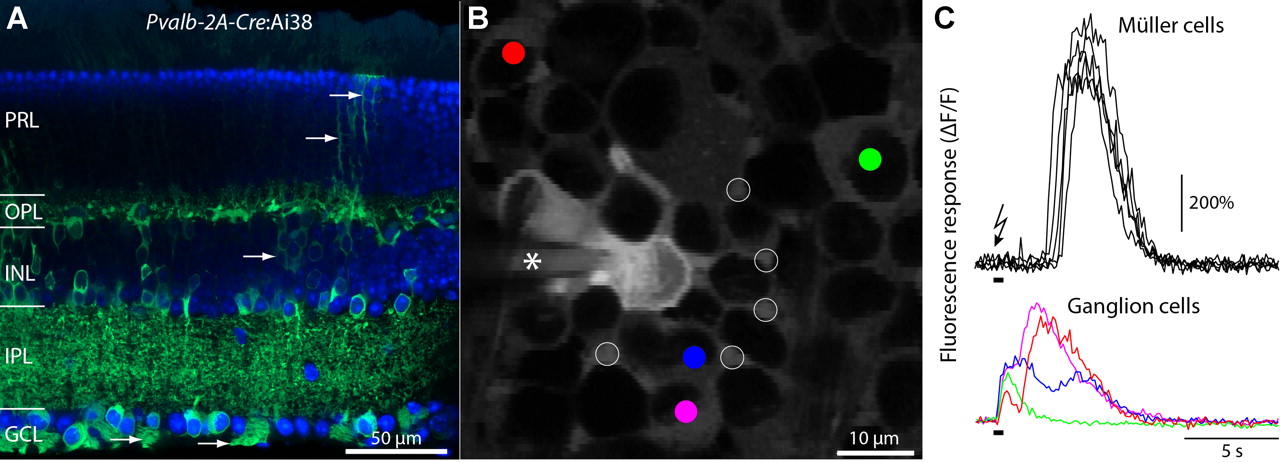
Electrical stimulation evokes robust calcium responses in Müller glia from Ai38 mice. A, Ai38 crossed with Pvalb-2A-Cre expressed GCaMP3 in several retinal neuron types and also in Müller glia (arrows). PRL, Photoreceptor layer; OPL, outer plexiform layer; INL, inner nuclear layer; IPL, inner plexiform layer; GCL, ganglion cell layer. B, Two-photon fluorescence image of the ganglion cell layer in an adult Pvalb-2A-Cre:Ai38 retina. The field of view includes several ganglion cells (colored circles), and also Müller glia processes (open circles). The electrical stimulus (see text) was delivered to the ganglion cell layer through a patch pipette (asterisk). C, Fluorescence responses of the Müller glia processes and the ganglion cells annotated in B. Each trace represents the change in fluorescence intensity referenced to the fluorescence intensity at scan onset. The electrical stimulus (timing indicated by black bar) was delivered >5 s after scan onset. Ganglion cell responses preceded the glial cell response by >1 s.
- Figure 8.
GCaMP3 expression patterns and functional signals in Pcp2-Cre:Ai38 mice. A, Single optical section (xy plane) in the cerebellum showing GCaMP3 expression pattern in the molecular layer of a Pcp2-Cre:Ai38 mouse. B, Maximum projection image (xy plane) of the Purkinje cell layer. The arrows denote Purkinje cell axons. C, Single optical section (xz plane) showing Purkinje cell somata and their dendrites. D, Responses from an isolated dendrite showing calcium transients at low rate (∼0.5 Hz). E, Subdendritic signals at two regions of interest defined by independent component analysis (ICA) (Hyvärinen, 1999). F, Various signals recorded from PC dendrites (ROIs selected with ICA are sorted left-to-right, and signals bottom-to-top).


