

Article Figures & Data
Figures
- Figure 1.
Lateral habenula (LHb) neurons show biphasic inhibitory-excitatory responses to cocaine. A, Intravenous cocaine reduces LHb firing 0–10 min postinjection (red traces) and increases firing in a subpopulation of LHb neurons 15–35 min postinjection (black traces). Inset shows average firing rates during these two time windows of LHb neurons showing only inhibition, and those showing biphasic inhibition/excitation. Intravenous saline injections are without effect (gray trace), and there is no average change in firing rate among nearby thalamic neurons (blue trace). B, Time course of responses of individual LHb neurons exhibiting biphasic responses to intravenous cocaine. C, Track along which neurons were recorded in one rat, shown against a Nissl counterstain. D, Map of individual recorded neurons in the LHb and adjacent thalamus. During the period 0–10 min postinjection, firing rates of neurons located within the LHb mostly show significant inhibition (red filled circles) or no change (asterisks) with only one neuron showing significant excitation (green filled circle). Neurons in the thalamus show equal proportions of inhibition versus excitation. E, At 15–35 min postinjection, LHb neurons are either significantly excited (green diamonds) or nonresponsive (asterisks), with only one inhibition (red diamond). Notably, none of the recorded thalamic neurons showed the shift from inhibition to excitation seen in LHb neurons. F, G, Many LHb units show firing rate changes to both cocaine and food-predictive auditory cues. Each of these units exhibited an inhibitory response to the food cue (relative to a nonfood-predictive cue) (F) and each neuron's response to cocaine was also inhibitory (G). Data in F are plotted in 100 μs bins, whereas data in G are shown in 15 s bins. *p < 0.05, two-tailed t test; **p < 0.02, two-tailed t test. Scale bar, 1 mm (C).
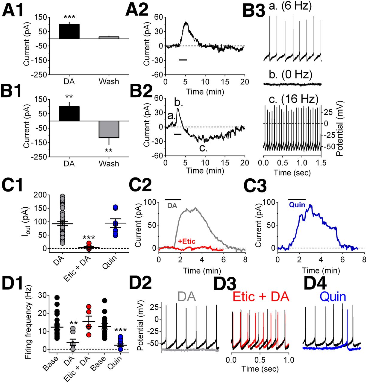
- Figure 2.
Effects of DA on LHb neurons in vitro. A1, Mean (n = 11 cells) dopamine-induced change in holding current (Current) in a subset of LHb neurons in brain slices. A significant outward (hyperpolarizing) current is observed when dopamine (4 μm) is applied to the LHb-containing brain slices, followed by a return to baseline during washout. A2, Time course of the effect of dopamine (black horizontal bar) in a single LHb neuron in which only an inhibitory outward current was observed. B1, Mean (n = 8 cells) dopamine-induced change in holding current in another subset of LHb neurons (biphasic cells) in which inhibitory outward currents were followed by depolarizing inward currents during dopamine washout. B2, A typical time course of the membrane current of a biphasic LHb neuron demonstrating a biphasic response to dopamine. Note the longer lasting delayed inward current following cessation of dopamine application (black horizontal bar). B3, Traces showing spontaneous action potential discharge for the cell shown in B2. Each trace (a–c) was recorded during the corresponding labeled period shown in B2. Dopamine hyperpolarized this cell and eliminated spontaneous firing, followed by an increase in firing rate above baseline during washout. C1, Mean DA agonist-induced inhibitory outward currents. Peak current amplitude for each cell tested under each of the indicated conditions is shown as well as the mean ± SEM for all cells (superimposed horizontal bars). C2, Example of the effect of DA (4 μm) on membrane current in a single LHb neuron, and its blockade by the D2 antagonist eticlopride (Etic, 200 nm). C3, Effect of the D2-like DA receptor agonist quinpirole (Quin, 1 μm) on membrane current in another LHb neuron. Note that the effects of DA were blocked by eticlopride and mimicked by quinpirole, indicating D2-like receptor involvement in the inhibition of LHb neurons. Etic alone had no effect on membrane current (data not shown). D1, Mean effects of DA (4 μm), Quin (1 μm), and Etic (200 nm) on spontaneous firing of LHb neurons. D2, Example of the effect of DA (gray trace) on firing in a single LHb neuron. D3, The effect of DA on cell firing is blocked by Etic (red trace) in the same cell shown in D2. D4, Effect of Quin (blue trace) on spontaneous firing in a single LHb neuron. In D2–D4, black traces correspond to baseline immediately before drug application. **p < 0.001, ***p < 0.0001, one-way ANOVA and Bonferroni post hoc test with multiple-comparison correction.
- Figure 3.
Cocaine effects on LHb neurons in vitro. A, Mean (± SEM) time course of cocaine (10 μm)-induced changes in neuron membrane current in a group of cells demonstrating only inhibitory outward currents, and blockade by the DA D2-like receptor antagonist eticlopride (200 nm). B, Mean time course of membrane current in five neurons demonstrating biphasic responses to cocaine. C, Representative current-clamp recordings of membrane responses from LHb neuron during cocaine (10 μm) application. Cocaine hyperpolarized this neuron and decreased the rate spontaneous action potential discharge. A similar action of cocaine was observed in another LHb neuron. D, Representative blockade of the inhibitory effect of cocaine on spontaneous firing by eticlopride (200 nm) in LHb neuron. These data and the ability of eticlopride to block the inhibitory effects of DA on LHb neuron firing (Fig. 2D) suggest that these effects are mediated by D2-like DA receptors. The term “Base” refers to recordings made during a stable baseline period.
- Figure 4.
Functional linkage of LHb and RMTg. We examined c-Fos in LHb neurons that were labeled by retrograde tracer injections into either the RMTg (A, site also spread to IPN) or VTA (B). Cocaine-induced c-Fos was found in 37 ± 9% of RMTg-projecting LHb neurons (C), but in a much lower percentage of VTA-projecting LHb neurons (D). Black label indicates c-Fos immunoreactivity; brown label indicates retrograde tracer immunoreactivity. Red filled arrows indicate neurons expressing both tracer and c-Fos, whereas open arrows indicate neurons expressing tracer only. Unilateral lesions of the posterior LHb and fasciculus retroflexus (FR, indicated by asterisk) (E), produce a twofold reduction in cocaine-induced c-Fos in the ipsilateral (lesioned) RMTg (G) relative to the contralateral (intact) RMTg (H), suggesting that cocaine-induced RMTg c-Fos is partly dependent on its LHb afferents. G, H Magnified from left and right boxed regions in F, where brown cytoplasmic label indicates immunoreactivity for the neuronal marker NeuN. Red filled arrows indicate neurons immunoreactive for both NeuN and c-Fos, whereas open arrows indicate cells expressing NeuN alone. Graphs show percentages of cocaine-induced double-labeled neurons in the LHb after retrograde tracer injections (I) or LHb/FR lesions (J). K, Animals show conditioned place aversion if placed into conditioning chambers 15 min after intravenous cocaine infusions, whereas placement 0 min postinfusion produces place preference, and placement 30 min postinfusion produces neither preference nor aversion. L, Activation of the RMTg by local AMPA injections produces conditioned place aversion. Scale bars, A, B, E, F, 1 mm; C, D, G, H, 50 μm. **p < 0.01, ***p < 0.001, ap < 0.05, one-tailed.
- Figure 5.
Histological confirmation of lesions of the RMTg and FR. A, B, The fiber-sparing excitotoxin quinolinic acid ablates the RMTg region while relatively sparing neurons in the adjacent MRN, as determined by NeuN immunostaining. C, Nissl-stained sections showing intact FR (asterisks). D, Nissl-stained brain section after bilateral electrolytic FR lesion (asterisks). Scale bars: A, B, 1 mm; C, D, 2 mm.
- Figure 6.
LHb and RMTg role in conditioned avoidance responses to cocaine. A, Unlesioned animals traversing a runway to obtain cocaine show progressive increases in run latencies (open triangles) reflecting conditioned avoidance of the cocaine-paired goal compartment. Animals with FR or RMTg lesions show no such increase (filled round and square symbols), suggesting a failure to develop conditioned avoidance. B, Unlesioned animals show progressive increases in alternations between approach and retreat, thought to reflect conflicting rewarding and aversive effects of cocaine, whereas FR or RMTg-lesioned animals lack this increase in reversals. Latencies and reversals are shown as medians for each trial. Scatterplots show latencies (C) and reversals (D) averaged over trials 4–7 for each individual rat; horizontal lines represent group medians. E, Representative path traversed by an individual unlesioned rat alternates between approach and retreat, whereas rats with LHb or RMTg lesions (F) show markedly fewer reversals and much faster latencies. Note large difference in time scales between E and F. **p < 0.01, ***p < 0.002.
- Figure 7.
Optogenetic characterization of RMTg role in conditioned avoidance response to cocaine. A, We tested behavioral effects of optical inhibition of RMTg area neurons after injections of AAV containing the light-sensitive proton pump ArchT. Virus also expresses GFP, indicated by dark immunoreactivity in RMTg region dorsal to the IPN and ventral to the decussation of the superior cerebellar peduncle (xscp). Optical fibers in this case are in the RMTg, but slightly rostral to the plane shown. B, Efficacy of ArchT expression is assessed by measuring locomotor distance traveled (in arbitrary units), while optogenetic inhibition of RMTg is applied during alternate 60 s epochs, producing repeatable and reversible increases in distance traveled. C, Graph showing ratio of distance traveled in even versus odd (light-on vs light-off) epochs; this ratio is significantly higher than 1.0 when virus and fiber are in the RMTg, but not when virus and fibers are located 0.6 mm dorsal. D, Animals receiving optogenetic inhibition of the RMTg 0–10 min after cocaine infusions showed progressive increases in run latencies, similarly to sham control rats and to animals receiving optical illumination without inhibitory opsins present. In contrast, animals expressing ArchT and receiving optical inhibition 15–25 min postinfusion did not show increased run latencies, suggesting that neural activation of the RMTg during the 15–25 min window after cocaine is critically required for conditioned avoidance behaviors. Notably, optical illumination is only delivered after animals reach the goal compartment and receive cocaine; hence, observed run patterns are presumably influenced by optical illumination that occurred during preceding trials. E, Optogenetic inhibition of the RMTg 15–25 min postdrug also reduced run direction reversals relative to the other two groups. F, G, Scatterplot of average latencies (F) and reversals (G) averaged over trials 4–7 for each individual rat. Horizontal lines are group medians. Scale bar, A, 1 mm. *p < 0.05, ***p < 0.00001.
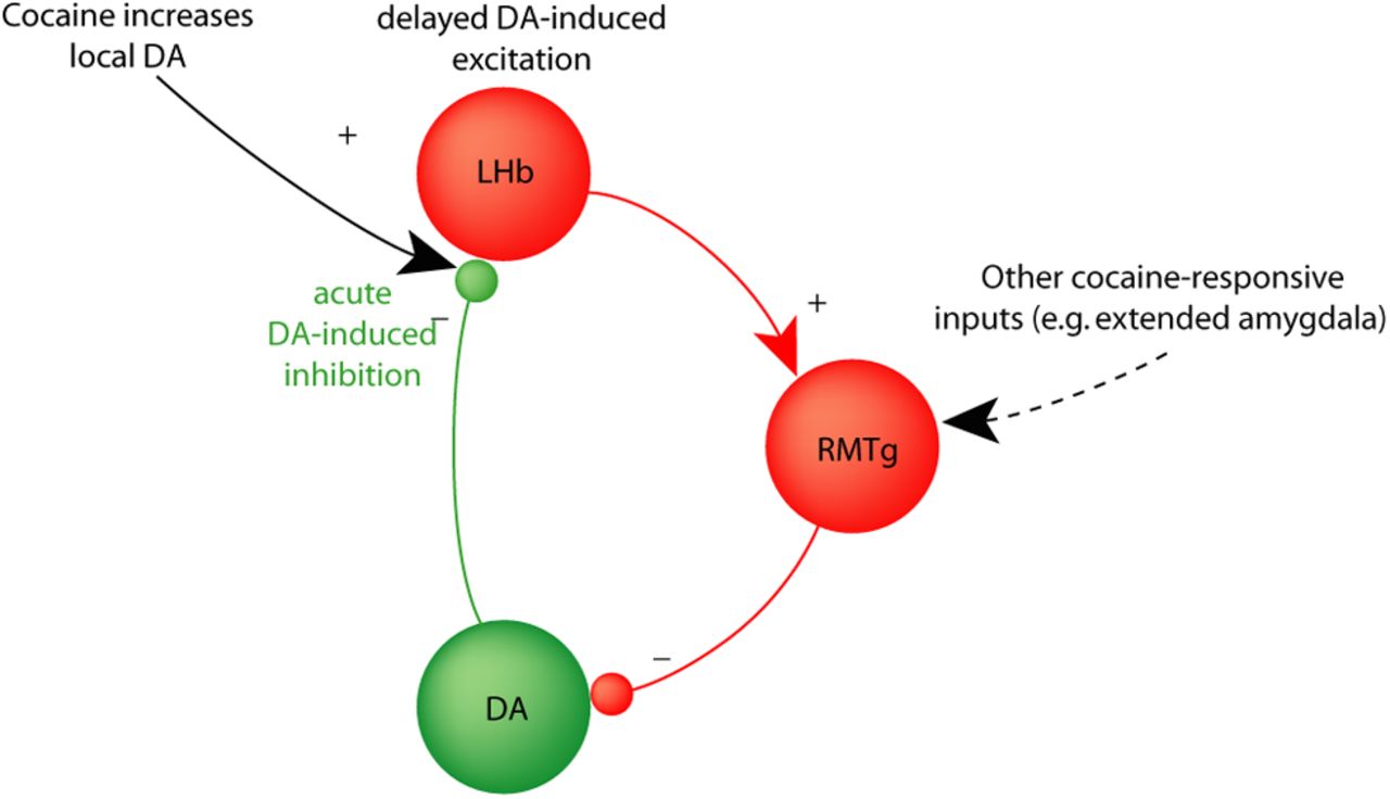
- Figure 8.
Schematic of feedback loops between LHb, RMTg, and VTA DA neurons. In this proposed circuit model, cocaine increases DA levels in the LHb, initially inhibiting LHb neurons (red) and contributing to reciprocally inhibitory interactions between DA and LHb neurons. DA-induced delayed excitation of the LHb (black text and arrow) converges with cocaine-induced responses in other RMTg afferents to drive a negative feedback signal opposing the initial actions of cocaine and other DA-mediated rewards.


