

Article Figures & Data
Figures
- Figure 1.
Endogenous buffer capacity is enhanced in aging CA1 pyramidal neurons. Widefield image of a CA1 pyramidal neuron loaded with 200 μm OGB-1 (bottom left). White box, Region of interest (ROI) where calcium transients were measured, ∼40 μm from the soma. Dotted white boxes, Background regions. Scale bar, 40 μm. A, Example calcium transients evoked by an action potential in neurons loaded with different concentrations of OGB-1 and measured using a CCD camera. B, Reciprocal amplitude of calcium transients (1/Δ[Ca2+]) plot against exogenous buffer capacity (κB). The R values and the slopes of the linear extrapolation were 0.771 and 0.000,124 (p values < 0.0001) for young, and 0.742 and 0.000,078 (p values < 0.0001) for aging. Each point represents one neuron loaded with 25, 50, 75, 100, 150, or 200 μm OGB-1 (29 neurons from 12 young adult rats, 24 neurons from 9 aged rats). C, No significant difference was observed in the peak amplitude of calcium transients evoked with single action potentials in CA1 neurons from young and aging rats that were loaded with 25 μm OGB-1 (5 neurons from 3 young adult rats, 6 neurons from 5 aged rats). The figure illustrates the data obtained for each individual neuron (small circles) and the mean ±SEM (larger circle). D, Resting calcium concentration in young and aging CA1 neurons (29 neurons from 12 young adult rats, 24 neurons from 9 aged rats).
- Figure 2.
Endogenous buffer capacity and unperturbed calcium transient measured in individual neurons. A, Maximum intensity projection of a neuron. Measurements were made using a line scan (dashed line) 40–80 μm from the soma. Example calcium transients taken during loading of 100 μm OGB-1, 7, 20, and 50 min after breaking whole-cell. B, Example of the increase in resting fluorescence (f0, arbitrary units) over time as OGB-1 diffused into and approached a steady-state concentration in the dendrite. Points are fit with an exponential. C, Example of endogenous buffer capacity calculated by back-extrapolation of the relationship between the reciprocal amplitude of calcium transients (1/Δ[Ca2+]) plot against exogenous buffer capacity (κB) for the results in (B). In this example neuron, there was an abrupt reduction in the amplitude of Ca2+ transients at 50 min, causing an upswing in the plot. The points after 50 min (open circles) were discarded before fitting a line through the results. D, The endogenous buffer capacity is significantly enhanced in neurons from aging rats (*p < 0.05, unpaired t test). E, Peak amplitude of the unperturbed calcium transient evoked with a single action potential is not significantly altered with aging (young, 8 neurons from 4 rats, 342 ± 65 nm; aging, 7 neurons from 5 rats, 206 ± 29 nm).
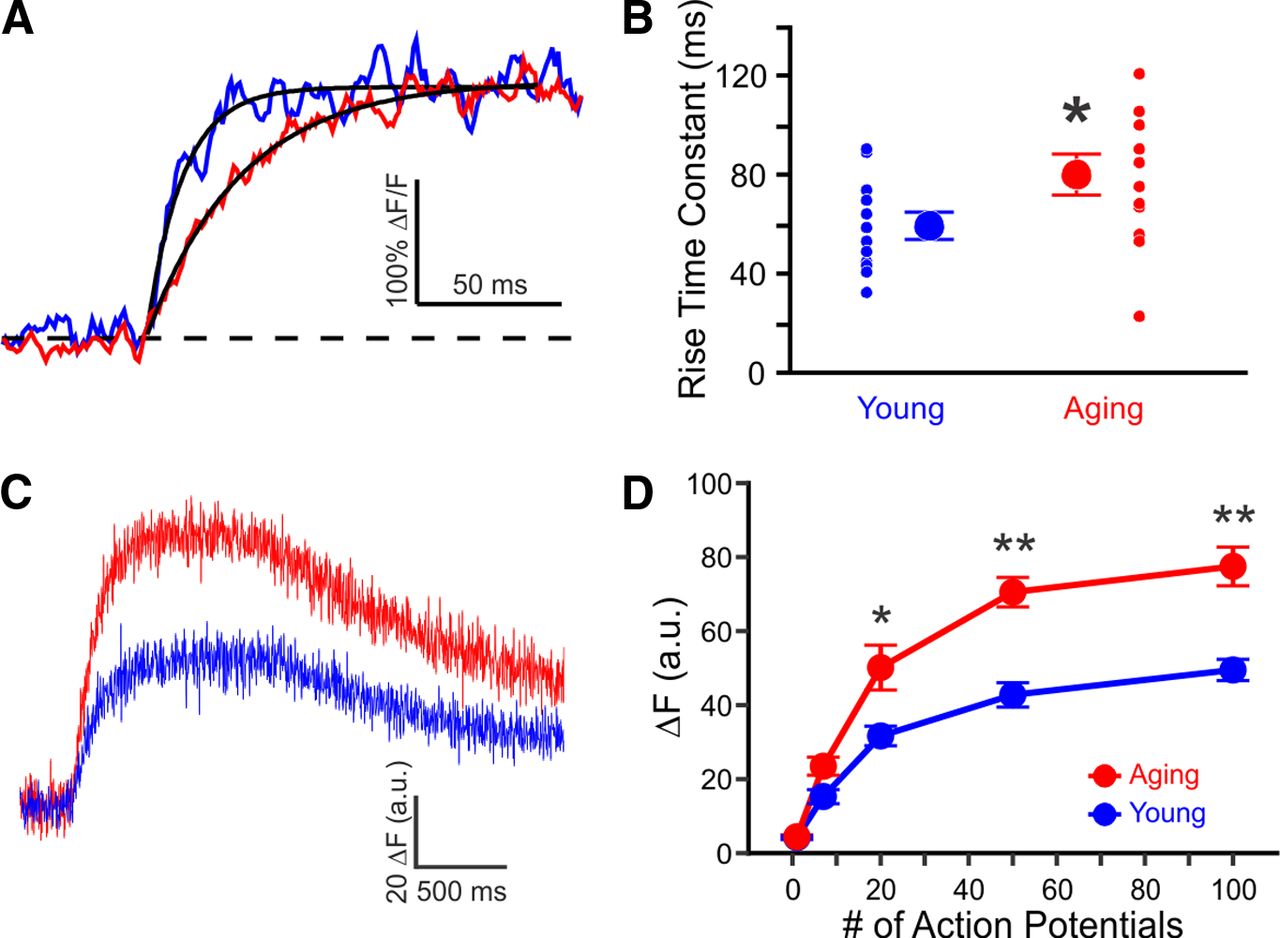
- Figure 3.
Rise time and amplitude of calcium influx are increased in aging CA1 neurons. A, Example measurements evoked with 50 Hz train of action potentials from young (blue) and aging (red) neurons loaded with 100 μm OGB-1. For illustrative purposes, only the initial rise to plateau phase is shown. B, Rise time constants in CA1 neurons from young (60 ± 5 ms, n = 12 from 9 rats) and aging (80 ± 8 ms, n = 12 from 10 rats) rats measured with 100 μm OGB-1 during a train of action potentials at 50 Hz. C, Example measurements from young (blue) and aging (red) neurons loaded with 500 μm OGB-1, during 100 action potentials at 100 Hz. D, Steady-state rise in fluorescence (ΔF) during 1 s trains of action potentials, measured with 500 μm OGB-1. Young, n = 4 from 3 rats. Aging, n = 5 from 4 rats. *p < 0.05, **p < 0.005, unpaired t test.
- Figure 4.
Calcium concentration during trains of action potentials. A, Mean calcium concentrations (shaded regions represent SEM) during 100 Hz trains in young (blue, n = 6 from 2 rats) and aging (red, n = 6 from 3 rats) neurons loaded with 150 μm OGB-6F. B, Calcium concentration during theta burst activity (5 action potentials at 100 Hz) at theta frequency (5 Hz). Significant differences were revealed with repeated ANOVAs for the 100 Hz and theta burst activity. Further analysis revealed significant differences between the age groups at various points during the train of activity. *p < 0.05.


Tables
Young 27 neurons Aging 22 neurons Resting membrane potential (mV) −66.6 ± 0.7 −66.3 ± 1.1 Input resistance (MΩ) 67.6 ± 3.0 56.2 ± 3.0 AP threshold (mV) −49.6 ± 1.3 −52.1 ± 1.1 AP amplitude (mV) 83.1 ± 2.2 86.7 ± 1.8 AP half width (ms) 0.85 ± 0.03 0.78 ± 0.03 A summary of membrane properties of CA1 pyramidal neurons in young and aging rats. All values are mean ± SEM


