

Article Figures & Data
Figures
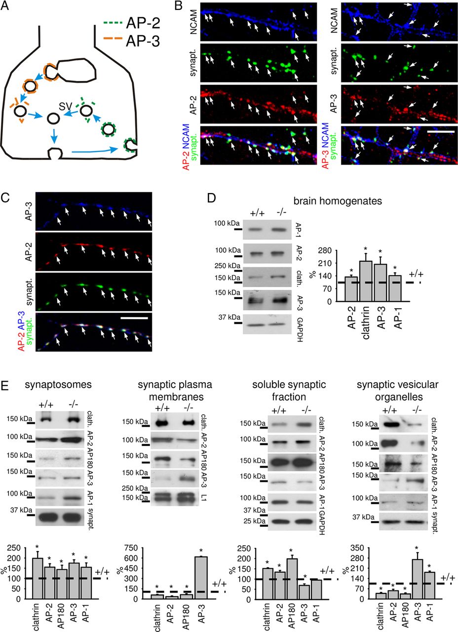
- Figure 1.
Targeting of AP-2, AP180, and clathrin to the synaptic plasma membrane is reduced in NCAM−/− synapses. A, A scheme showing the subsynaptic localization of the AP-2 and AP-3 adaptor proteins and their role in synaptic vesicle formation. Synaptic vesicles are formed via AP-2/clathrin mediated endocytosis from the presynaptic surface membrane or via AP-3 mediated budding from endosomes. B, Cell surface NCAM was labeled by indirect immunofluorescence of formaldehyde-fixed hippocampal neurons maintained in culture for 12 d. Neurons were then permeabilized and labeled with antibodies against synaptophysin and AP-2 or AP-3. Note that NCAM- and synaptophysin-positive clusters colocalize with AP-2 and AP-3 accumulations (arrows). C, Hippocampal neurons maintained in culture for 12 d were colabeled with antibodies against synaptophysin, AP-2, and AP-3. Note overlapping accumulations of AP-2 and AP-3, which colocalize with synaptophysin-positive clusters. D, E, NCAM+/+ and NCAM−/− brain homogenates (D) and synaptosomes, synaptic plasma membranes, soluble fraction from synaptosomes, and synaptic vesicular organelles (E) were probed by Western blot with the indicated antibodies. Note reduced levels of AP-2, AP180 and clathrin, and increased levels of AP-3 in NCAM−/− synaptic plasma membranes and vesicular organelles. Synaptophysin served as a loading control for synaptosomes and vesicular organelles, while labeling with antibodies against the plasma membrane associated cell adhesion molecule L1 and cytosolic marker protein GAPDH verified equal loading in synaptic plasma membranes and soluble fraction/brain homogenates, respectively. Graphs show quantitation of blots. Mean levels + SEM in NCAM−/− mice normalized to the levels in NCAM+/+ mice set to 100% (dashed lines) are shown. *p < 0.05, paired t test (compared to NCAM+/+; n = 3). Scale bars: 10 μm.
- Figure 2.
NCAM accumulates in the presynaptic membrane. A, Recycling synaptic vesicles were labeled by incubating live NCAM+/+ neurons with antibodies against the lumenal domain of synaptotagmin 1 applied in the culture medium containing 47 mm K+. Neurons were then fixed with formaldehyde and colabeled with antibodies against NCAM, PSD95 and synaptophysin. Note the clusters of NCAM overlapping with accumulations of synaptotagmin 1 and synaptophysin apposed to PSD95 clusters (arrows). B, Formaldehyde-fixed NCAM−/− neurons transfected with NCAM140 were labeled with antibodies against NCAM, the presynaptic marker protein synaptophysin and the postsynaptic marker protein PSD95. An axon of an NCAM140 transfected neuron is shown. Note that accumulations of synaptophysin colocalize with accumulations of NCAM140 in NCAM−/− neuron transfected with NCAM140 abutting onto NCAM−/− neurons (arrows). NCAM140/synaptophysin accumulations are apposed to PSD95 clusters. C, Brain homogenates, synaptic plasma membranes (membranes) and synaptic vesicular organelles (vesicular organelles) were probed by Western blot with the antibodies against the intracellular domain of NCAM140 and NCAM180, synaptophysin (synapt.), PSD95, and Na,K-ATPase. Arrows indicate NCAM isoforms detectable by the antibodies. Note that NCAM accumulates in the synaptic plasma membrane. Synaptic membranes, containing both presynaptic and postsynaptic portions, are also enriched in PSD95 and plasma membrane localized Na,K-ATPase. NCAM is also present in synaptic vesicular vesicles, which are highly enriched in synaptophysin but are free of PSD95 and Na,K-ATPase. NCAM180 detectable in the synaptic vesicular organelles is probably contained in postsynaptic transport organelles that are present in this fraction. NCAM immunoreactivity at ∼160 kDa most likely is a proteolytic fragment of NCAM180. The graph shows quantitation of blots (mean + SEM; n = 3) with the signal in homogenates set to 100%. *p < 0.05, paired t test. D, Synaptosomes and highly purified synaptic vesicles (synaptic vesicles) were probed by Western blot with polyclonal antibodies against a common epitope at the C terminus of NCAM140 and NCAM180, monoclonal antibody D3 against an NCAM180-specific epitope in the intracellular domain of NCAM 180, and CSP. Arrows indicate NCAM isoforms detected by the antibodies. Note that NCAM140 is detectable in immunopurified synaptic vesicles. A smear above the NCAM140 band was reproducibly detectable in our preparations and may represent glycosylated NCAM140 or NCAM140-containing protein complexes, which were not fully dissociated by SDS-PAGE. Note that NCAM180 is not detectable in synaptic vesicles with NCAM180-specific antibodies even after prolonged exposure of the blot, while NCAM180 is readily detectable in synaptosomes. Mock immunopurification (mock) of synaptic vesicles with nonimmune Igs served as control of immunopurification. In these experiments, 4–12% polyacrylamide gels were used to enable detection of both NCAM isoforms and CSP in one SDS-PAGE run, which was necessary to control for protein loading and specificity of synaptic vesicle immunopurification, resulting in NCAM140 and NCAM180 labeling patterns different from the data shown in C, in which 8% polyacrylamide gels were used and low molecular weight proteins were allowed to run out of the gel to achieve better separation of the NCAM bands. Scale bars: 10 μm.
- Figure 3.
AP-2 binds to the dileucine motif in the intracellular domain of NCAM140. A, NCAM immunoprecipitates (IP) from synaptosomes and 13% of the material used for NCAM immunoprecipitation (input, 13%) were probed by Western blot analysis with the antibodies against the extracellular domain of NCAM, AP-2, AP-3, and AP-1. Proteins were separated using 8% polyacrylamide gels to achieve better separation of high molecular weight proteins. Mock immunoprecipitation with nonimmune rabbit Igs was performed for control. Note that AP-2 but not AP-3 or AP-1 coimmunoprecipitate with NCAM. B, Amino acid sequence of a fragment of NCAM140 comprising transmembrane and membrane adjacent portions of the extracellular and intracellular domains of NCAM is shown. The LL motif in the intracellular domain of NCAM is shown in bold and underlined. C, NCAM immunoprecipitates from CHO cells transfected with NCAM140, nonmutated or mutated in the membrane-proximal LL dileucine motif, were analyzed by Western blot with antibodies against NCAM and AP-2. Mock immunoprecipitation with NCAM antibodies from CHO cells transfected with GFP only was performed for control. Note that mutation in the LL dileucine motif inhibits coimmunoprecipitation of AP-2 with NCAM140.
- Figure 4.
NCAM promotes binding of AP-2 versus AP-3 to synaptic plasma membranes. A, NCAM+/+ and NCAM−/− synaptic plasma membranes were treated with alkali to strip peripheral proteins. The membranes were then incubated with cytosol and recruitment of AP-2, clathrin, and AP-3 from the cytosol to the membranes was assessed by Western blot. Note that alkali removed AP-2, clathrin, and AP-3 from the membranes, but not L1, which served as a loading control. Note more efficient recruitment of AP-2 and clathrin to NCAM+/+ synaptic membranes and enhanced recruitment of AP-3 to NCAM−/− synaptic membranes. The graph shows the quantitation of blots. Mean levels + SEM of AP-2, clathrin, and AP-3, recruited to NCAM−/− synaptic membranes, are shown. The signals were normalized to NCAM+/+ levels (dashed line) set to 100%. *p < 0.05, paired t test (compared to NCAM+/+; n = 3). B, C, Recruitment of AP-2 and AP-3 to NCAM+/+ synaptic membranes as analyzed by Western blot. When indicated, the cytosol was preincubated with BFA or competitors, either the nonmutated intracellular domain of NCAM140 (140ID) or 140ID with the mutated membrane-proximal dileucine motif (LA140ID). Note that 140ID but not LA140ID reduces recruitment of AP-2 to the membranes (B). AP-3 recruitment is enhanced from the cytosol preincubated with 140ID, while AP-2 recruitment is enhanced from the cytosol preincubated with BFA (C). Note that these effects are blocked when the cytosol was preincubated with 140ID and BFA (C). No recruitment of AP-1 is observed. Brain homogenate (BH) is included to show AP-1 bands at the expected molecular weight (C). Graphs show quantitation of blots. Mean levels + SEM of AP-2 and AP-3, recruited to synaptic membranes from cytosol in the presence of BFA or competitors, are shown. The signals were normalized to the signal without BFA or competitors (dashed lines) set to 100%. *p < 0.05, paired t test (compared to the signal without BFA or competitors; n = 3). L1 (A, C) or syntaxin 1B (syn 1B; B) served as a loading control.
- Figure 5.
NCAM promotes substitution of AP-3 by AP-2 in synaptic plasma membranes during postnatal development. A, Levels of AP-2, AP-3, and AP180 in NCAM+/+ and NCAM−/− synaptic plasma membranes and synaptosomes from P7 or P60 mice were analyzed by Western blot. Note that levels of AP-2 and AP180 increase, while levels of AP-3 decrease with age in synaptic plasma membranes from NCAM+/+ mice. Developmental substitution of AP-3 by AP-2 and AP180 is reduced in NCAM−/− synaptic plasma membranes. Graphs show quantitation of blots. Mean ratios + SEM of protein levels in synaptic plasma membranes to their levels in synaptosomes are shown. Values were normalized to the values for P60 NCAM+/+ mice set to 100%. *p < 0.05, paired t test (n = 3). Labeling for L1 (for synaptic membranes) or GAPDH (for synaptosomes) served as a loading control. B, NCAM immunoprecipitates (IP) from brain lysates of P1, P7, P14, P21, and P60 NCAM +/+ mice were probed with antibodies against the extracellular domain of NCAM, AP-2, and AP-3 by Western blot. Brain homogenates (BH) from P60 mice were applied for comparison. Mock IP with nonimmune rabbit Igs was performed for control. A broad band of NCAM immunoreactivity observed at 200 kDa in brains of P1–P7 mice represents polysialylated NCAM (PSA-NCAM). Note that levels of nonpolysialylated NCAM140 and efficiency of the coimmunoprecipitation of AP-2 with NCAM increase with age. AP-3 does not coimmunoprecipitate with NCAM.
- Figure 6.
The amount of AP-2/AP180/clathrin-coated vesicles is reduced in NCAM−/− versus NCAM+/+ mice. A, Synaptic vesicles purified in a sucrose gradient were probed by Western blot with the antibodies against clathrin, AP-2, AP180, and synaptophysin (syn.). Note lower levels of clathrin, AP-2, and AP180 and unchanged levels of synaptophysin in NCAM−/− versus NCAM+/+ synaptic vesicles. The graph shows quantitation of the blots (mean + SEM; n = 3) with the signal in NCAM+/+ mice set to 100%. *p < 0.05, paired t test. B, Synaptic vesicles purified in a sucrose gradient were further purified by immunoprecipitation (IP) with antibodies against synaptophysin and probed by Western blot for synaptophysin, AP-2, and AP-3. Mock immunoprecipitation with nonimmune rabbit Igs was performed for control. Note unchanged levels of synaptophysin and reduced levels of AP-2 in NCAM−/− synaptic vesicles. Synaptic vesicles are negative for AP-3. C, Synaptophysin positive organelles were immunoprecipitated with antibodies against synaptophysin from the crude synaptic vesicular organelle (SVO) fraction and probed by Western blot for synaptophysin, AP-2, and AP-3. Note unchanged levels of synaptophysin, reduced levels of AP-2, and increased levels of AP-3 in NCAM−/− organelles. B, C, The crude synaptic vesicular organelle fraction was applied to the gel for comparison.
- Figure 7.
AP-3-mediated bulk membrane retrieval is enhanced in NCAM−/− synaptic boutons. A, NCAM+/+ and NCAM−/− cultured hippocampal neurons were allowed to take up HRP in the presence of 47 mm K+ applied for 90 s. Where indicated, neurons were pretreated with BFA for 1 h. Representative electron micrographs of synapses are shown. HRP-loaded vesicles are seen as dark core structures. Scale bar, 300 nm. HRP uptake is enhanced in NCAM−/− synaptic boutons when compared to NCAM+/+ synaptic boutons. The number of HRP positive organelles is increased in NCAM+/+ synaptic boutons and reduced in NCAM−/− boutons after BFA application. Higher magnification of the area outlined in NCAM−/− synapse (right) shows that HRP was taken up by synaptic vesicle-like structures (arrows) and larger endosome-like structures (arrowheads). B, Graphs show numbers of SV-like structures with the cross-section area <2000 nm2, endosome-like structures with the cross-section area >2000 nm2, and the total number of SVs including HRP-loaded and nonlabeled SVs counted per synapse section area. C, Graphs show numbers of HRP-loaded vesicles of the indicated size normalized to the total number of HRP-loaded vesicles per synaptic bouton section (top) and the total cross-section area of all SV-like structures or endosome-like structures normalized to the total cross-section area of all HRP loaded organelles per synaptic bouton section (bottom). In B and C, mean values + SEM are shown. *p < 0.05, t test (n > 100 synapses were analyzed from 4–6 coverslips). D, A scheme showing the hypothetical role of AP-2 and AP-3 in bulk membrane retrieval in NCAM+/+ and NCAM−/− synaptic terminals. AP-3-dependent steps blocked by BFA are indicated.
- Figure 8.
AP-3-dependent synaptic vesicle endocytosis and intrasynaptic processing are upregulated in NCAM−/− synaptic boutons. A, Presynaptic boutons of NCAM+/+ and NCAM−/− cultured hippocampal neurons that were either not treated (control) or treated as indicated with BFA and dynasore were loaded with the fixable analog of FM1–43 applied in the presence of 47 mm K+ for 90 s. After washing, neurons were fixed and colabeled with antibodies against the presynaptic marker protein SV2 to visualize presynaptic boutons. Graphs show quantification (mean + SEM) of FM1–43 fluorescence levels in SV2 accumulations (top) or the ratio of FM1–43 fluorescence levels to SV2 immunofluorescence levels (bottom). Values were normalized to the levels in control NCAM+/+ neurons set to 100%. Compared to NCAM+/+ neurons, FM1–43 uptake in NCAM−/− presynaptic boutons is reduced and increased in response to BFA. Note that BFA and dynasore have an additive effect on FM1–43 uptake in NCAM−/− but not NCAM+/+ presynaptic boutons. *p < 0.05 (t test compared to control neurons of the same genotype); §p < 0.05 (t test compared as indicated); n > 20 images of neurons with N > 200 synapses per image were analyzed. B, Graphs show quantification (mean + SEM) of FM1–43 release from presynaptic boutons of NCAM+/+ and NCAM−/− cultured hippocampal neurons in response to 47 mm K+. Values [I(t)] were normalized to FM1–43 levels before stimulation [I(0)]. Where indicated, neurons were preincubated with BFA. BFA inhibits FM1–43 release from the presynaptic boutons of NCAM−/− neurons. C, A diagram showing the roles of AP-2 and AP-3 adaptor proteins in synaptic vesicle endocytosis and intrasynaptic processing in NCAM+/+ and NCAM−/− synaptic terminals. AP-3- and AP-2-dependent steps blocked by BFA and dynasore, respectively, are indicated.
- Figure 9.
Endocytosis of VAMP2-pHluorin is reduced in NCAM−/− synaptic boutons. A, NCAM+/+ neurons were cotransfected with cherry and VAMP2-pHluorin. Neurons were stimulated with 1 ms bipolar current pulses at 10 Hz to yield fields of 10 V/cm. Gray scale images show pHluorin fluorescence in neurons before (0 s), during (45 s), and after (420 s) stimulation. Note an increase in pHluorin fluorescence intensity in synaptic boutons in response stimulation (arrows). B, VAMP2-pHluorin endocytosis monitored in synaptic boutons of NCAM+/+ and NCAM−/− cultured hippocampal neurons cotransfected (tr.) with an empty pcDNA3 vector, or NCAM+/+ neurons cotransfected with NCAM140LA in the pcDNA3 vector. Neurons were stimulated with 47 mm K+ and allowed to recover in 4 mm K+. C, Synaptic vesicles purified from NCAM+/+ and NCAM−/− brains were probed by Western blot with antibodies against the largest subunit (116 kDa) of proton ATPase. Note that the levels of proton ATPase are not changed in NCAM−/− versus NCAM +/+ synaptic vesicles. D, VAMP2-pHluorin endocytosis monitored in synaptic boutons of cultured NCAM+/+ and NCAM−/− hippocampal neurons cotransfected with control or AP-3 siRNA or NCAM140. Neurons were stimulated with 1 ms bipolar current pulses at 10 Hz to yield fields of 10 V/cm. In B and D, fluorescence intensity levels before stimulation were set to 0, and signals during the recovery time were normalized to the peak intensity reached during stimulation. Mean values ± SEM are shown (n = 14–25 synapses from 8–10 coverslips were recorded in each group). E, Mean ± SEM. values for pHluorin fluorescence recovery efficiency (Ir = (1 − Iend) * 100%) and for time constants (t1/2) are shown.*p < 0.05, one-way ANOVA with Dunnett's multiple comparison post-test.
- Figure 10.
Stoichiometry of AP-2 and AP-3 in CNS synapses differs from the stoichiometry of these adaptor proteins in neuromuscular junctions. A, NCAM+/+ synaptosomes (syn.; 5 μg total protein) and neuromuscular junctions (20 μg total protein) were probed by Western blot with the indicated antibodies. Note that both preparations are positive for synaptic markers VAChT, SNAP25, and VAMP, which are present at various levels in both preparations. Note also that the levels of AP-2 relative to AP-3 are higher in synaptosomes when compared to NMJs. B, NCAM+/+ synaptosomes (20 μg total protein) or NCAM+/+ and NCAM−/− neuromuscular junctions (20 μg total protein) were probed by Western blot with the antibodies against AP-2 and AP-3. GAPDH served as a loading control. Note that levels of AP-2 are decreased, while levels of AP-3 are increased in NCAM−/− NMJs compared to NCAM+/+ NMJs. The graph shows levels of AP-2 and AP-3 (mean + SEM) in NCAM−/− NMJs. Values were normalized to NCAM+/+ levels (dashed line). *p < 0.05, paired t test (compared to NCAM+/+; n = 3).


