

Article Figures & Data
Figures
- Figure 1.
Contrasting the effects of Kcnq2 and Kcnq3 conditional deletion on survival and ECoG activity. A, Targeting strategy for generation of Kcnq2fllfl and Kcnq3fllfl mice. Targeted axons (red, kcnq2; blue, Kcnq3) were flanked by two loxP sites. Inset, PCR validating Kcnq2fllfl and Kcnq3fllfl genotypes. Cerebral cortex deletion was achieved by crossing Kcnq2fllfl and Kcnq3fllfl with Emx1-ires-cre (Emx1) mice. B, Immunoblot analysis of hippocampal membrane fractions of KCNQ2 or KCNQ3 from cerebral cortex-specific Kcnq2 cKO and Kcnq3 cKO mice. Levels of KCNQ2 and KCNQ3 are reduced by ∼80% in the Kcnq2 cKO (n = 8) and Kcnq3 cKO (n = 7) mice, respectively. C, Survival curves of control (black, n = 10), Kcnq2 cKO (red, n = 17), and Kcnq3 cKO (blue, n = 8) mice. The survival graphs show that most Kcnq2 cKO mice die prematurely between P15 and P20. Control and Kcnq3 cKO mice did not die during the same time period. D, Left, Representative ECoG recordings from control, Kcnq2 cKO, and Kcnq3 cKO mice. Representative polyspike events were indicated by asterisk from Kcnq2 cKO and shown in extended time scale. Right, Polyspike events were counted every 30 s during the first 15 min of recordings from control (black, n = 5; P15–P19), Kcnq2 cKO (red, n = 4, P15–P19), and Kcnq3 cKO (blue, n = 5, P16–P19) mice.
- Figure 2.
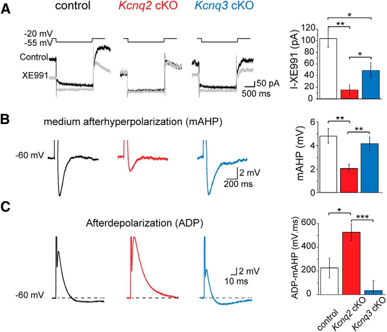
Contrasting the effects of Kcnq2 and Kcnq3 conditional deletion on the M current and mAHP. A, The M-current protocol and representative M-current traces. The M current was induced by a step hyperpolarization (1.5 s) to −55 mV from a holding potential of −20 mV in pyramidal neurons from either control, Kcnq2 cKO, or Kcnq3 cKO mice before (black) and after (gray) application of 20 μm XE991. Far right, Summary graph showing the effects of deleting either Kcnq2 or Kcnq3 on the M current (n = 8∼9). B, A representative mAHP followed by a 50 ms current injection (1 nA) in pyramidal neurons from either control, Kcnq2 cKO, or Kcnq3 cKO mice. Far right, Summary graph showing the effects of deleting either Kcnq2 or Kcnq3 on the mAHP (n = 12). Membrane potential was kept at −60 mV by injecting a small DC current through the recording pipette. C, A representative ADP induced by a 2 ms 1 nA current injection in pyramidal neurons from either control, Kcnq2 cKO, or Kcnq3 cKO mice. Far right, Summary graph showing the effects of deleting either Kcnq2 or Kcnq3 and ADP (n = 11–21). Membrane potential was kept at −60 mV by injecting a small DC current through the recording pipette. Data shown are means ± SEM. Statistical significance was determined by one-way ANOVA (*p < 0.05; **p < 0.001, ***p <0.0005), using Tukey's as post hoc test.
- Figure 3.
Elevated neuronal excitability of CA1 pyramidal neurons in Kcnq2-null but not Kcnq3-null neurons. A, Voltage responses to various current injection steps (1 s) in pyramidal neurons from either control (n = 18), Kcnq2 cKO (n = 18), or Kcnq3 cKO (n = 18) mice. Membrane potential was kept at −60 mV by injecting a small DC current through the recording pipette. Representative traces showing the effects of deleting either Kcnq2 (red) or Kcnq3 (blue) on pyramidal neuron excitability. B, Summary graphs showing the effect of Kcnq2 or Kcnq3 deletion on CA1 pyramidal neuron AP number, initial firing frequency, and final firing frequency. Data shown are means ± SEM. Statistical significance was determined by two-way repeated-measures ANOVA (*p < 0.05).
- Figure 4.
Contrasting the effects of Kcnq2 and Kcnq3 deletion on CA1 pyramidal neuron intrinsic properties. A, Left, Voltage responses to various current injection steps (1 s from 0 to −100 pA) in pyramidal neurons from either control, Kcnq2 cKO, or Kcnq3 cKO mice. Membrane potential was kept at −60 mV by injecting a small DC current through the recording pipette. Right, Summary graphs showing the effect of deleting Kcnq2 or Kcnq3 on the RN (n = 21–26). RN was determined by the slope of the line fitted to voltage versus current relationship. B, Left, Voltage responses to a current injection ramp (2.5 s ramp from −100 to +200 pA; 0.12 nA/s) recorded in pyramidal neurons from either control, Kcnq2 cKO, or Kcnq3 cKO mice. Dashed boxed area is shown in extended time scale to show a burst of APs from Kcnq2 cKO mice. Far right, Summary graph showing the effects of deleting Kcnq2 or Kcnq3 in the RN (n = 7–9). RN was measured from the slope between −63 mV and the voltage before the first AP. C, Representative single APs recorded for each genotype of CA1 pyramidal neurons elicited by 1 nA 2 ms current injection. Inset shows the first derivative of the AP from the different genotypes. Right, Summary graphs showing the effects of deleting Kcnq2 or Kcnq3 on the AP threshold (n = 12–15). For the box plots, the central line represents the median value, the boundaries of the box represent the SD, the square symbol represents the mean value, and whiskers represent the minimum and maximum data value. Statistical significance was determined by one-way ANOVA (*p < 0.05) using Tukey's as post hoc test.
- Figure 5.
Contrasting the effects of deleting Kcnq2 and Kcnq3 on KCNQ channel levels and activity. A, Left, Hippocampal membrane fractions from the indicated mouse lines were probed with antibodies against KCNQ2, KCNQ3, or KCNQ5 channels. Western blots were also probed with a β-actin antibody as a protein loading control (detected between the 37 and 50 kDa molecular weight standards; predicted molecular weight is 42 kDa). Right, Summary graph showing the change in KCNQ protein level in Kcnq2 cKO or Kcnq3 cKO mice in relation to control mice (n = 3–8). B, Sample traces of tonic firing activity in CA1 pyramidal neurons from the indicated mouse lines in the background presence of 8.5 mm [K+]o, and following application of either retigabine (Ret; 20 μm), ICA-27243 (ICA; 25 μm), or XE991 (20 μm). Left, Summary graph of firing rate changes induced by retigabine or ICA-27243 in either control, Kcnq2 cKO, or Kcnq3 cKO mice (n = 7–12). Cell-attached recordings took place in the voltage-clamp configuration. Data shown are means ± SEM. Statistical significance was determined by one-way ANOVA (*p < 0.05) using Dunnett's as post hoc test.
- Figure 6.
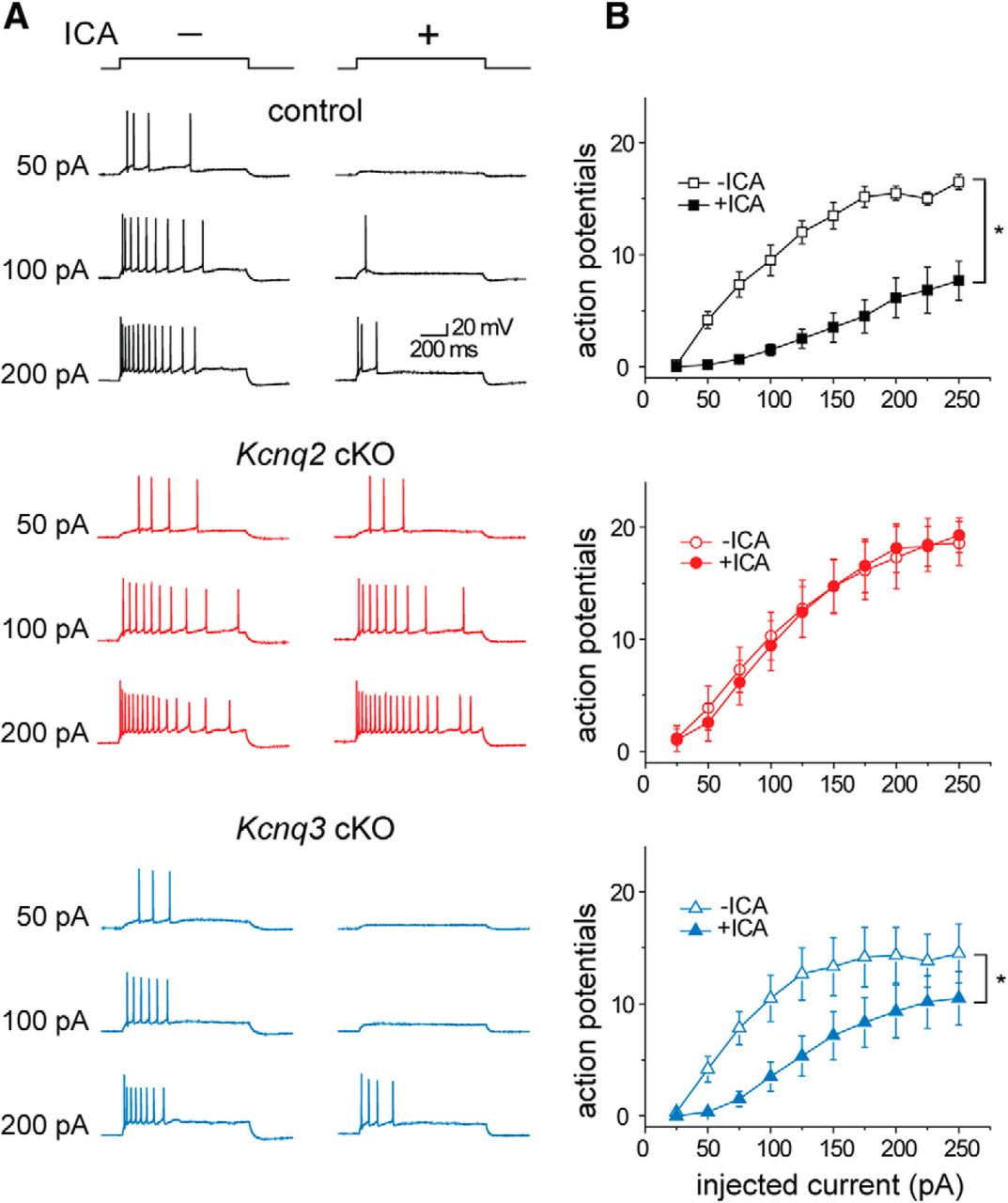
The KCNQ2 allosteric activator ICA-27243 inhibits AP firing in Kcnq3 cKO but not Kcnq2 cKO mice. A, Voltage responses to various current injection steps (1 s) in pyramidal neurons from either control, Kcnq2 cKO, or Kcnq3 cKO mice. Membrane potential was kept at −60 mV by injecting a small DC current through the recording pipette. Representative traces showing the effects of deleting either Kcnq2 (red) or Kcnq3 (blue) on pyramidal neuron excitability and in the presence or absence of ICA-27243 (25 μm). B, Summary graphs showing the effect of ICA-27243 on pyramidal neuron AP number in control (n = 6), Kcnq2 CKO (n = 7), or Kcnq3 cKO (n = 6) mice. Data shown are means ± SEM. Statistical significance was determined by two-way repeated-measures ANOVA (*p < 0.05).


