

Article Figures & Data
Figures
- Figure 1.
Caspr and Caspr2, respectively, dictate the location and membrane accumulation of Kv1 channels at the nodal environ. A, Caspr and Caspr2 are not detectable in dKO mice. Immunofluorescence of teased sciatic nerves isolated from WT, Caspr-null (Caspr−/−), Caspr2-null (Caspr2−/−), and dKO mice using antibodies to Caspr, Caspr2, and Na+ channels (NaCh). The location of the nodes and the juxtaparanodal region are labeled with asterisks and arrowheads, respectively. Note that Caspr2 is mislocalized at the paranodes in the absence of Caspr, whereas the distribution of Caspr is unchanged in the absence of Caspr2. B, C, Absence of both Caspr and Caspr2 does not lead to myelin abnormalities. Immunofluorescence analysis using antibodies to P0, neurofilament (NF) and NaCh (B), as well as electron microscopy analysis of cross-sections (C) of sciatic nerves isolated from the indicated genotypes. D, E, Absence of Caspr or Caspr2 results in altered distribution of juxtaparanodal components. Shown is immunofluorescence labeling of teased sciatic nerves isolated from the indicated genotypes using antibodies to NaCh and Kv1.2 potassium channels (D) or CNTN2 (E). The location of the nodes of Ranvier is marked with an asterisk in all panels. In the absence of Caspr2, Kv1.2 channels are not accumulated at the juxtaparanodal region, but are occasionally detected at the double mesaxonal lines that passed through this region (arrow). Arrowhead marks the presence of abnormal patches of Kv1 channels at the edge of the juxtaparanodal region in the dKO nerves. Scale bars: A, B, D, E, 10 μm; C, 5 μm.
- Figure 2.
Absence of Caspr and Caspr2 results in the disruption of Kv1.2 localization along the mesaxonal line. A, Immunofluorescence of teased sciatic nerves isolated from WT, Caspr-null (Caspr−/−), Caspr2-null (Caspr2−/−), and dKO mice using antibodies to Kv1.2 channels. In WT mice and the single mutants, these channels are present at the juxtamesaxonal line, as well as in radial rings (asterisks) located beneath the Schmidt Lanterman incisures. In contrast, Kv1.2 channels are found in large aggregates (arrowheads) that are distributed along the internodes. B, Cross-sections of sciatic nerves isolated from the indicated genotypes labeled using antibodies to Kv1.2, dystroglycan (DG), and neurofilament (NF). Kv1.2 aggregates (arrowheads) are present in proximity to the axolemma. C, Western blot analysis of sciatic nerves reveals similar levels of Kv1.2 in WT, Caspr−/−, Caspr2−/−, and dKO mice. D, Absence of Caspr and Caspr2 does not affect the distribution of protein 4.1B. E, F, Distribution of Kv1.2 aggregates in relation to Schmidt-Lanterman incisures. Teased sciatic nerves isolated from the four different genotypes were labeled using antibodies to Kv1.2 and MAG (E) or E-cadherin (Ecad; F). Arrowheads mark the location of Kv1.2 aggregates. Scale bars, 10 μm.
- Figure 3.
Deletion of both Caspr and Caspr2 results in widening of the nodes. A, Double immunofluorescence labeling of sciatic nerves isolated from the indicated genotypes using antibodies to neurofilament (blue) and axonodal components: Na+ channels (NaCh), NF186, ankyrin G, or βIV spectrin. Bars highlight the wider nodal region in the dKO nerves. B, Quantification of axonodal width-to-height ratio showed a widening of the dKO node compared with WT mice and the single mutants. Error bars indicate SEM of n = 4 mice for each genotype (**p < 0.05). C, Double immunofluorescence labeling of sciatic nerves isolated from the indicated genotypes using antibodies to neurofilament (blue) and glial microvilli proteins: gliomedin, phospho-ERM (pERM), or Syndecan-3. D–F, Electron microscopy images showing a longitudinal view of the nodes of Ranvier in sciatic nerves of WT (D) and dKO (E,F) mice. In WT nerves, the node is bordered by the PNJ and Schwann cell microvilli (asterisk) are confined to the nodal gap (arrows). In dKO nerves, Schwann cell microvilli processes are detected between the axolemma and the detached paranodal loops (E,F, arrowheads). Asterisks in E and F mark microvilli and the accumulation of mitochondria at the nodes, respectively. Scale bars: A, C, 10 μm; D–F, 1 μm.
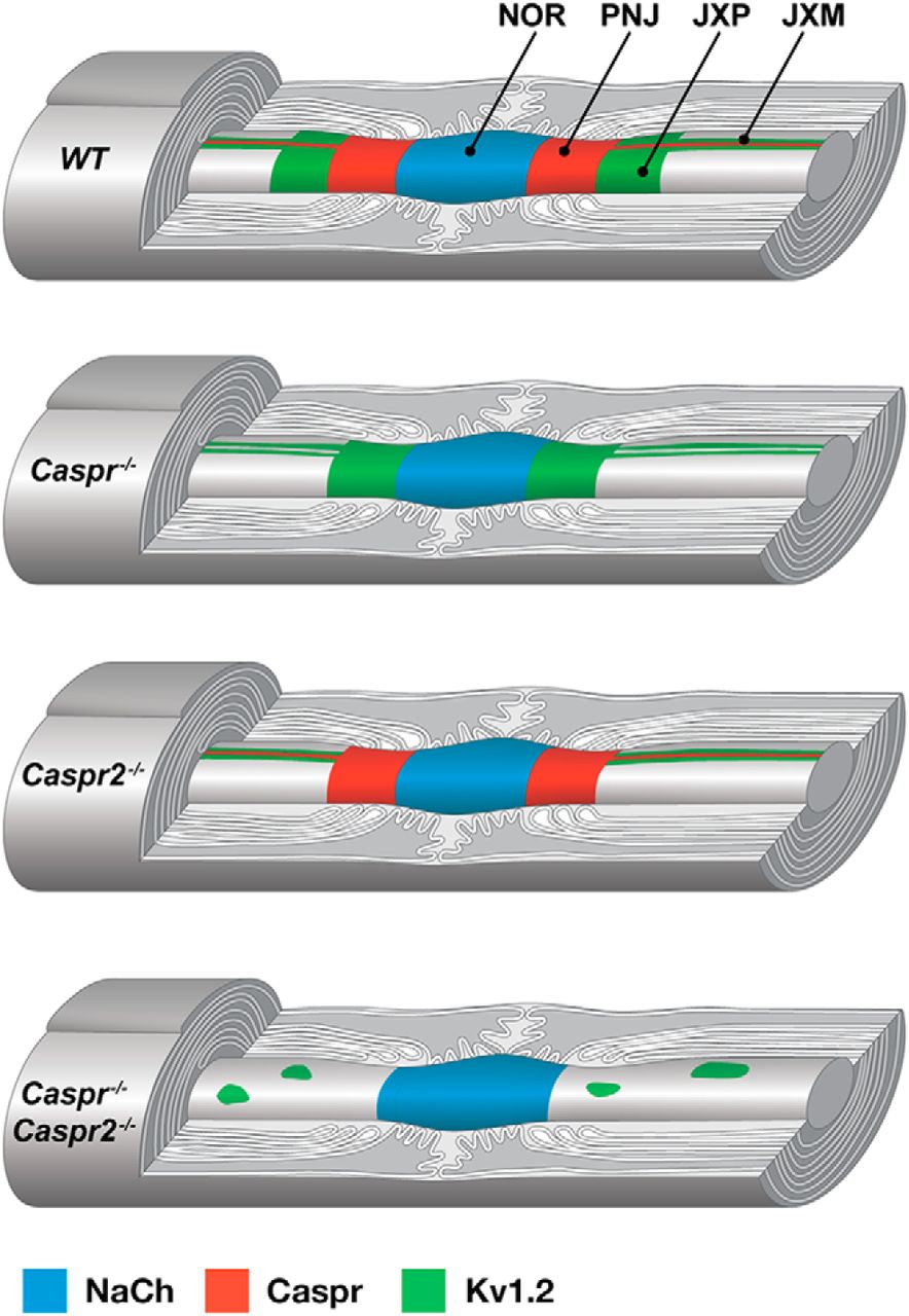
- Figure 4.
Caspr proteins organize myelinated axons. A schematic drawing illustrating the distribution of Na+ channels (NaCh), Caspr, and Kv1.2 channels along myelinated PNS axons in WT, Caspr−/−, Caspr2−/−, and double Caspr−/−/Caspr2−/− mutant mice. Whereas in Caspr−/− or Caspr2−/− nerves, Kv1.2 channels accumulate at the JXM, in the absence of both Caspr proteins, these channels form large aggregates along the internodal axolemma. NOR indicates node of Ranvier; PNJ, paranodal junction; and JXP, juxtaparanodal region.


