

Article Figures & Data
Figures
- Figure 1.
Microbead-induced elevations in mouse intraocular pressure. A, Daily mean IOP in mmHg (n ≥ 20 measurements each day, mean ± SEM) for a 37 d period in cohorts of C57 vs Trpv1−/− mice (n = 10 each) before (day 0) and after (days ≥1) a single unilateral injection of polystyrene microbeads (1.0 μl) into the anterior chamber. The fellow eye in each animal received an equivalent volume saline injection as an internal control. B, Microbead-injected eyes exhibited a significant increase in IOP following injection (mean ± SEM for days ≥1) compared with saline-injected eyes in both C57 mice (19.77 ± 0.76 vs 14.75 ± 0.65 mmHg) and Trpv1−/− mice (19.79 ± 0.80 vs 14.96 ± 0.72 mmHg). *p < 0.001, two-sided t test for each cohort. Between the two cohorts, IOP was similar for both saline-injected (p = 0.842) and microbead-injected (p = 0.980) eyes (two-sided t test for each).
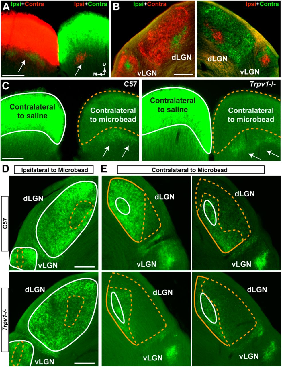
- Figure 2.
Microbead-induced IOP elevation causes deficits in anterograde transport to central brain structures. A, Coronal section shows both superior colliculi (SC) near rostral pole following bilateral intravitreal injection of CTB into left (green CTB) and right (red CTB) eyes of C57 mouse. Fluorescent signal for dominant contralateral projection is overexposed purposely to reveal sparser ipsilateral projection (arrows) for each eye. Medial (M) and dorsal (D) orientations indicated. B, Dorsal LGN (dLGN) and ventral LGN (vLGN) nuclei from same C57 brain show dominant contralateral with smaller ipsilateral projections. C, Coronal sections through the rostral pole of C57 (left) and Trpv1−/− (right) SCs show intact CTB signal (green) in the SC contralateral to saline injection (white outline). CTB signal in the SC contralateral to microbead injection (dashed yellow outline) is reduced after 5 weeks of elevated IOP. In SC, contralateral to microbead injection, the CTB signal in ipsilateral projections (arrows) remain intact, as expected. D, E, Coronal sections through LGN from C57 mice (top row) and Trpv1−/− mice (bottom row) following bilateral intravitreal injections of CTB (green). D, In the LGN ipsilateral to microbead-injected eye, CTB signal from saline-injected eyes remains intact (solid white outline), while the signal from microbead-injected eyes shows deficits embedded in both dLGN and vLGN (dashed yellow outline). Deficits appear worse in the Trpv1−/− LGN. E, In LGNs contralateral to microbead-injected eye, CTB signal from saline-injected eyes is again intact (solid white outline). In the dLGN, CTB signal from microbead-injected eyes (yellow outlines) shows a range of deficits (dashed yellow). Once again, deficits appear worse in Trpv1−/− dLGN. The Trpv1−/− vLGN also appears to have a lower CTB signal. Scale bars, 200 μm.
- Figure 3.
Trpv1−/− exacerbates deficits in anterograde axonal transport. A, Top row, Coronal sections through SC following intravitreal injection of CTB (green) into saline- and microbead-injected eyes of C57 mice. Microbead-induced IOP elevation (Fig. 1) induced deficits in anterogradely transported CTB (dotted lines). Bottom row, Retinotopic maps reconstructed from serial sections of SC with optic disc gap indicated (circles). Density of the signal from transported CTB ranges from 0% (blue) to 50% (green) to 100% (red). Medial (M) and rostral (R) orientations are indicated. B, Top row, Sagittal SC sections from Trpv1−/− mice with microbead-induced IOP elevations show worse deficits in CTB transport compared with C57. Corresponding retinotopic maps (bottom row) demonstrate nearly complete loss of CTB transport. OD, Optic disc representation (no RGCs). Scale bars: A, B, 500 μm. C, Fraction of the retinotopic map with intact RGC axonal transport (defined by ≥70% CTB signal) to individual C57 and Trpv1−/− saline-injected (n = 10 and 8, respectively) and microbead-injected (n = 10 and 10, respectively) superior colliculi. D, Transport of CTB from saline-injected eyes was near 100% and similar for C57 (93.8 ± 2.5%) and Trpv1−/− (90 ± 3.5%) cohorts (mean ± SEM; p = 0.46, two-sided t test). Intact transport in SC following IOP elevation was reduced in both cohorts compared with the saline-injected eyes (*p < 0.001, two-sided t test), but the decrease in Trpv1−/− (26.6 ± 4.0%) was twice as severe as in C57 mice (51.7 ± 4.1%; †p < 0.001, two-sided t test). Legend is as in C.
- Figure 4.
Trpv1−/− mice exhibit more severe optic nerve pathology following elevated IOP. A, Cross sections of C57 optic nerve from saline- and microbead-injected eyes. Microbead-induced elevated IOP yielded a modest reduction in the packing density of intact axons and increased the incidence of degenerating axonal profiles (arrowheads). B, Cross section through Trpv1−/− optic nerve from microbead-injected eye shows severely diminished axon packing, overt gliosis (arrows), and far more degenerating profiles compared with C57 nerves (arrowheads). Saline-injected eye nerves from the two cohorts appear similar. Scale bars: A, B, 10 μm. C, Density of intact axons in cross sections through individual C57 and Trpv1−/− saline-injected (n = 10 and 9, respectively) and microbead-injected (n = 9 and 7, respectively) optic nerves quantified from cross sections, as shown in A and B. D, Mean (±SEM) axon density in C57 and Trpv1−/− optic nerves. Diminished density from microbead-induced IOP elevation in Trpv1−/− nerves is more than twice that of C57 nerves (†p < 0.001, two-sided t test). *Significance for microbead compared with corresponding saline nerve (*p = 0.001 for C57; p < 0.001 for Trpv1−/−, two-sided t test). E, Cross-sectional area (mean ± SEM) of Trpv1−/− optic nerves shrinks with elevated IOP compared with saline-injected eyes. *p = 0.005, two-sided t test. F, Number of axons (mean ± SEM) calculated as a product of nerve area and axon density. Trpv1−/− nerves have nearly twice the loss compared with C57 nerves with elevated IOP (†p < 0.001, two-sided t test), though both groups have fewer axons compared with saline-injected nerves (*p = 0.03 for C57 and p = 0.04 for Trpv1−/−, two-sided t test). Number of axons in saline-injected nerves was similar in two cohorts (p = 0.70, two-sided t test). Legend applies to C–F.
- Figure 5.
Progression to RGC body loss accelerated in Trpv1−/− mice. A, B, Confocal micrographs of C57 (A), and Trpv1−/− (B) retinas show RGCs labeled both by CTB uptake (red) and with antibodies against phosphorylated neurofilaments (green). In both cohorts, there is little apparent change in cell number with microbead-induced IOP elevations. Importantly, nearly every RGC is labeled by both markers, indicating intact RGC uptake of CTB. C, C57 (left) and Trpv1−/− (right) retinal peripheries show accumulation of phosphorylated neurofilaments in RGC dendritic arbors (arrowheads) with IOP elevation. Tendency is more robust in Trpv1−/− retinas, with axonal accumulation as well (dashed lines). Scale bars: A–C, 20 μm. D, Density of RGC bodies (RGCs/mm2) labeled for phosphorylated neurofilaments (cells/mm2) expressed as the ratio of microbead-injected to saline-injected retinas (mean ± SEM) for increasing eccentricities from the optic disc. At each location, cells were scored as described above (see Materials and Methods) in images that were 0.101 mm2 in area (n = 5 retina each). For superior, inferior, and temporal quadrants, neither cohort had significant RGC body loss at any eccentricity (p ≥ 0.08, two-sided t test). In the nasal quadrant, Trpv1−/− microbead-injected retinas showed moderate (20–40%) accelerated loss compared with saline-injected retinas at 0.75 and 2.25 mm eccentric (*p = 0.03 and p = 0.05, respectively, two-sided t test).
- Figure 6.
Microbead-induced anterograde transport deficits in SCs are retinotopically sectorial. A, B, Retinotopic maps of C57 (A) and Trpv1−/− (B) SCs from microbead-injected eyes show progression of transport deficits (shaded regions) by retinal quadrant and eccentricity. Deficits in C57 mice (43% and 68%, left and right, respectively) progress from the nasal (N) fields toward other sectors and the representation of optic disk (*). Deficits in Trpv1−/− SCs are more severe (69% and 81%, left and right, respectively), covering nearly all of the nasal quadrant from inferior (I) to superior (S) fields. The temporal (T) field is most often the last affected in both mouse strains. SC maps transformed into retinal quadrant and eccentricity coordinates are as specified by Siminoff et al. (1966) and Dräger and Hubel (1976).
- Figure 7.
TRPV1 antagonism accelerates RGC axonal transport deficits in rats. A, IOP in two cohorts of rats was elevated for <5 weeks (30–33 d) following microbead injection (5.0 μl); the opposing eye received an equivalent volume of saline. Elevation (mean ± SEM) compared with saline-injected eyes in both vehicle (27.04 ± 0.14 vs 20.20 ± 0.14 mmHg) and drug (27.99 ± 0.26 vs 21.01 ± 0.10 mmHg) cohorts was significant (*p < 0.001, two-sided t test for each cohort). Between the two cohorts, IOP was similar for microbead-injected eyes (p = 0.10), but was slightly higher in the drug saline-injected eye (†p = 0.001, two-sided t test for each). B, Coronal SC sections from rats treated with TRPV1 antagonist show worse deficits in CTB transport with microbead-induced IOP elevations compared with vehicle rats. C, Corresponding retinotopic maps demonstrate accelerated progression in transport deficits for the drug group. The color scheme is as in Figure 3. D, Fraction of the retinotopic SC map with intact transport (defined by ≥70% CTB signal) from saline-injected eyes was similar for vehicle (92.4 ± 1.2%) and drug (92.3 ± 2.4%) cohorts (mean ± SEM; p = 0.93, two-sided t test). Intact transport in SC following IOP elevation was reduced in both cohorts compared with the saline-injected eye (*p ≤ 0.04, two-sided t test), but the decrease with drug treatment (39.2 ± 18.9% intact) was more severe than with vehicle (76.3 ± 6.4% intact; †p = 0.04, two-sided t test). Scale bars: B, C, 500 μm.
- Figure 8.
Trpv1−/− modifies RGC physiology with elevated IOP. A, IOP in C57 and Trpv1−/− mouse eyes used for RGC physiology remained elevated for ∼4 weeks (25–28 d) after microbead injection compared with saline injection (*p < 0.001, two-sided t test for each cohort). IOP (mean ± SEM) was similar in C57 and Trpv1−/− mice for both saline-injected (14.63 ± 0.78 vs 14.73 ± 0.79 mmHg, respectively; p = 0.523) and microbead-injected (19.47 ± 0.97 vs 19.77 ± 0.94 mmHg; p = 0.559) eyes (two-sided t test for each). B, A C57 RGC filled with 1% Lucifer yellow during whole-cell patch-clamp recording after IOP elevation shows ramified dendrites and axon (arrow). Scale bar, 50 μm. C, Microbead-induced IOP elevation increases spontaneous firing of action potentials under current-clamp in C57 but not Trpv1−/− RGCs. D, Spontaneous firing rate (mean ± SEM) increased significantly for RGCs in C57 microbead-injected retinas (12.43 ± 1.88 Hz, n = 27) compared with saline-injected retinas (5.65 ± 1.20 Hz, n = 14; *p = 0.025, two-sided t test). RGCs in Trpv1−/− microbead-injected retinas demonstrated significantly less spontaneous activity compared with their C57 counterparts (4.44 ± 0.77 Hz, n = 14; †p = 0.006, two-sided t test), but were similar to their own saline-injected RGCs (n = 20; p = 0.33, two-sided t test). E, Traces show RGC action potentials induced by 25 or 50 pA depolarizing current steps for cells with low spontaneous rates (<0.5 Hz); RGCs in Trpv1−/− retinas typically required more depolarization to reach a threshold rate of firing >3 Hz. F, Depolarizing current (mean ± SEM) required to induce action potentials in RGCs with low spontaneous rates (< 0.5 Hz) was greater in Trpv1−/− microbead-injected retinas (62.50 ± 9.32 pA, n = 13) compared with Trpv1−/− saline-injected retinas (34.62 ± 6.03 pA, n = 10; *p = 0.004, two-sided t test) and compared with C57 microbead-injected retinas (42.50 ± 7.72 pA, n = 20; †p = 0.011, two-sided t test). The threshold current was the same for C57 microbead- and saline-injected retinas (41.30 ± 8.11 pA, n = 23) vs Trpv1−/− saline-injected retinas (p = 0.86, one-way ANOVA on ranks).


