

Article Figures & Data
Figures
- Figure 1.
Generation of a KI mouse with deletion of the Reelin CTR. A, Schematic of the wild-type allele, targeting vector, Neo-ΔC allele, and ΔC allele. Neo-ΔC mice were bred with CAG-Cre transgenic mice to eliminate the neomycin-resistance cassette. the FLAG-epitope coding exon is highlighted in yellow. E, S, and X indicate recognition sites for EcoR I, SpeI, and XbaI, respectively. B, The genotypes were confirmed by Southern blotting. Genomic DNA from the tail of the wild-type (+/+), heterozygote (+/ΔC), or ΔC-KI (ΔC/ΔC) mice was digested with XbaI or EcoR I and hybridized with 5′- or 3′-probes (shown in A), respectively. C, Genotype analysis using PCR. Using the P1 and P2 primers (shown in A), 380 and 320 bp fragments were amplified from wild-type and ΔC alleles, respectively. D, Schematic of wild-type Reelin (ReelinWT) and ReelinΔC-FLAG proteins. Solid black box indicates a FLAG epitope. E, The expression pattern of Reelin and density of Cajal–Retzius cells in MZ were not affected in ΔC-KI mice. Brain sections from the indicated mice at P7 were immunostained with anti-Reelin antibody. Nuclei were stained with Hoechst 33342. Arrows indicate Cajal–Retzius cells in which accumulation of Reelin is observed. Scale bar, 200 μm. Quantification of the density of Cajal–Retzius cells in the MZ is shown at the right side of the panels. Error bars indicate mean ± SEM (n = 3). F, The amount of Reelin was increased in ΔC-KI mice. The homogenates of P3 mouse cerebral cortices from Reelin-deficient reeler mouse (rl/rl), wild-type (+/+), or ΔC-KI (ΔC/ΔC) mice were analyzed by WB using the antibody indicated on the left of the panel. Asterisks indicate full-length Reelin. Arrowhead indicates the N-terminal fragment generated by N-t cleavage (NR2). Molecular mass markers (kDa) are shown on the right of the panels. Arrows indicate nonspecific bands. Quantification of the amount of full-length Reelin and NR2 is shown below the panels. Error bars indicate mean ± SEM. *p < 0.05 (one-sample t test). N.S., Not significant. n = 4. G, The amount of Reelin secretion was approximately equivalent in wild-type and ΔC-KI mice. The supernatants and cell lysates from primary cultured cortical neurons of wild-type (+/+), or ΔC-KI (ΔC/ΔC) mice were analyzed by WB. Asterisk indicates full-length Reelin. Arrowhead indicates NR2. Quantification of the amount of full-length Reelin and NR2 is shown below the panels. Error bars indicate mean ± SEM. *p < 0.05 (one-sample t test). N.S., not significant. n = 3. H, The amount of Dab1 was increased in ΔC-KI mice. Homogenates of P3 mouse cerebral cortices were analyzed by WB. Anti-α-tubulin was used as the loading control. Quantification of the amount of Dab1 is shown below the panels. Error bars indicate mean ± SEM. F(2,9) = 10.66. **p < 0.01 (Tukey's test). *p < 0.05 (Tukey's test). N.S., Not significant. n = 4.
- Figure 2.
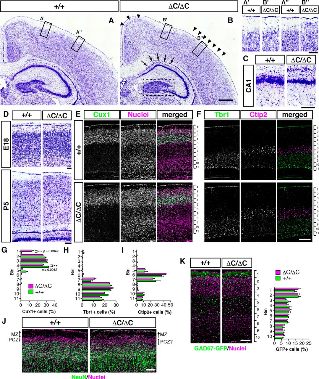
Formation of the MZ and upper layers is abnormal in mice lacking the Reelin CTR. A–C, Nissl staining of sections of the wild-type (+/+, A) and ΔC-KI (ΔC/ΔC, B) mice brain at P7. In ΔC-KI mice, the MZ was narrow (B, arrowheads), the CA1 pyramidal cell layer was split into two layers (B, arrows; C), and the dentate granule cell layer (B, surrounded by broken line) was less packed compared with wild-type mice. Scale bars: B, 400 μm; B″, C, 100 μm. D, The MZ is narrower in ΔC-KI mice than in wild-type mice at P5, but not at E18. Scale bars: top, 50 μm; bottom, 100 μm. E, F, Brain sections from the indicated mice at P3 were immunostained with the indicated antibodies. Nuclei were stained with Hoechst 33342 (E, middle column). Scale bar, 200 μm. G–I, Quantification of the indicated layer markers demonstrates that Cux1+ cells are affected in ΔC-KI mice. Graphs represent the percentage of cells in each of the 11 equal-size vertical bins. Error bars indicate mean ± SEM. **p < 0.01 (unpaired t test). n = 3. J, Formation of PCZ is abnormal in ΔC-KI mice. Brain sections from indicated mice at P2 were immunostained with anti-NeuN (green), and nuclei were stained with Hoechst 33342 (magenta). The MZ/PCZ border and PCZ/cortical plate border are clear in wild-type mice, and both are blurred in ΔC-KI mice. Scale bar, 100 μm. K, The distribution of GAD67-GFP+ interneurons was normal in ΔC-KI mice. Brain sections from indicated mice bearing GAD67-GFP allele at P3 were immunostained with anti-GFP (green), and nuclei were stained with Hoechst 33342 (magenta). Scale bar, 100 μm. Graph represents the percentage of GFP+ cells in each of the 10 equal-size vertical bins. Error bars indicate mean ± SEM (n = 3).
- Figure 3.
Development of apical dendrites is abnormal in mice lacking the Reelin CTR. A, In utero electroporation of GFP expression vectors was performed at E14.5, and mice were fixed at P7. The upper region of cerebral cortical slices is shown. II/III, Layer II/III. In ΔC-KI mice, the border between MZ and layer II/III is blurred. Scale bar, 100 μm. B, Schematic drawing represents the two plasmids used for sparse labeling of neurons (pDCX-Cre and pCAGGS-RG, left), and the plasmid resulting from Cre-mediated recombination (right). In the absence of pDCX-Cre, pCAGGS-RG expresses DsRed because of the presence of an SV40 polyadenylation sequence (Stop) after the DsRed, whereas in the presence of pDCX-Cre, Cre-mediated recombination excises the DsRed expression cassette and the Stop sequence through two loxP sites, and the resulting plasmid expresses GFP. To reduce the probability of recombination, the concentration of pDCX-Cre was reduced to one-five-hundredth compared with that of pCAGGS-RG, resulting in a small number of GFP-positive cells being observed. C, Sparse labeling by pCAGGS-RG/pDCX-Cre coelectroporation clearly shows the misorientation and hypertrophy of apical dendrites in ΔC-KI mice. Three representative images are shown. Scale bar, 50 μm. D, Quantification of the total apical dendrite length (p = 3.729 × 10⁻⁸, >30 neurons from each of three different brains per group were analyzed). E, Line graph of the percentage of GFP+ cells versus total apical dendrite length. F, Quantification of branching points of apical dendrites (p = 5.949 × 10⁻⁷, >30 neurons from each of three different brains per group were analyzed). G, Line graph of the percentage of GFP+ cells versus branching points of apical dendrites. H, Quantification of the percentage of GFP+ cells with apical dendrites that cross the border between the cortical plate and MZ (p = 5.307 × 10⁻¹³, >50 neurons from each of three different brains per group were analyzed). I, Relative bin position of labeled neurons. Bin positions were calculated by measuring the distance from the ventricular surface to labeled cells and from the ventricular surface to the top of the cortical plate (>100 neurons from each of three different brains per group were analyzed). D, F, H, Error bars indicate mean ± SEM. ***p < 0.001 (Mann–Whitney U test).
- Figure 4.
Reelin is cleaved within its CTR by PC family proteases. A, Schematic of Reelin protein and amino acid sequence of the CTR. Boxed region highlights the consensus sequence for PC family proteases. Arrows indicate two known cleavage sites. Arrowheads indicate epitopes of anti-Reelin antibodies. B, Schematic of Reelin mutants with a FLAG-tag. Solid black boxes represent FLAG-tags. C, Secreted ReelinC-FLAG did not retain its FLAG-tag. The culture supernatants of HEK293T cells expressing the above Reelin mutants were analyzed by WB with the indicated antibodies. The reactivity of ReelinFLAG+C to anti-FLAG antibody is weaker than ReelinΔC-FLAG for unknown reason (Kohno et al., 2009b). D, Intracellular ReelinC-FLAG retained the FLAG-tag. The culture supernatants and whole-cell lysates of ReelinC-FLAG-expressing HEK293T cells were analyzed by WB. E, Reelin is cleaved by a PC family protease(s). The culture supernatants from ReelinC-FLAG-expressing HEK293T cells (lanes 1–3) or COS-7 cells (lanes 4–6) cultured without inhibitors (lanes 1 and 4), with FI-1 (lanes 2 and 5), or FI-2 (lanes 3 and 6) were analyzed by WB with G10 (top) or anti-FLAG (bottom). Asterisks indicate full-length Reelin. Arrowheads indicate Reelin fragments generated by cleavage within RR3 and cleavage between RR6 and RR7, respectively. FI-1 inhibits cleavage at both sites (Kohno et al., 2009a). Secreted ReelinC-FLAG protein retained the FLAG-tag in the presence of the PC inhibitors (lanes 2, 3, 5, and 6). F, LoVo cells, which bear no endogenous PC activity, do not cleave Reelin at the WC site. The expression vector for ReelinC-FLAG or ReelinC-Venus was transfected into LoVo cells with or without a furin vector. The culture supernatant was collected and analyzed by WB with the indicated antibodies. G, Schematic of AP-RR78CF and its mutants. The consensus sequence of PC family proteases is underlined. Mutated amino acids are shown in bold. H, R3455 is necessary for cleavage by PC family proteases. The culture supernatants of HEK293T cells expressing the indicated proteins with or without FI-1 were analyzed by WB. I, Monoclonal antibodies 12C10 and 1D4 specifically recognize ReelinFL. Wild-type Reelin (WT), ReelinC-FLAG, or a mutant Reelin lacking the last 6 residues (Δ6) were expressed in HEK293T cells cultured with or without FI-1. The culture supernatants were analyzed by WB with the indicated antibodies. J, FI-1 inhibits WC cleavage by CGNs. CGNs were cultured with the indicated inhibitors, and the culture supernatants were analyzed by WB. ReelinWT and ReelinΔ6 from HEK293T cells were run as references. K, The ratio of endogenous Reelin protein cleaved at the WC site is dependent on developmental stage. Reelin proteins were immunoprecipitated using anti-Reelin CR-50 antibody from the brain homogenate at the indicated stages, analyzed by WB, and quantified. Error bars indicate mean ± SEM. F(2,6) = 19.42. **p < 0.01 (Tukey's test). N.S., Not significant. n = 3. L, WC cleavage had little effect on the ability of Reelin to induce Dab1 phosphorylation in cultured cerebral cortical neurons in the standard assay. Cerebral cortical neurons were incubated for 20 min at 37°C with conditioned medium containing ReelinWT collected from HEK293T cells in the presence of FI-1 (ReelinFL) or ReelinΔ6 collected in the presence of FI-1. Whole-cell lysates were separated by SDS-PAGE followed by WB using anti-phosphotyrosine 4G10 (top) and anti-Dab1 (bottom) antibodies. Molecular mass markers (kDa) are shown on the left of the panel. Error bars indicate mean ± SEM. F(2,6) = 50.21. **p < 0.01 (Tukey's test). N.S., Not significant. n = 3. M, ReelinFL localizes to the MZ. Sections of the cerebral cortices of wild-type (+/+, top) or reeler (rl/rl, bottom) mice at P1 were immunostained with AF3820 (antibody against the N-terminal region of Reelin, red) and 12C10 (green). Nuclei were stained with Hoechst 33342 (blue). In sections from wild-type mice, the 12C10 antibody stained Cajal–Retzius cells in the MZ. Arrowheads indicate signals positive for AF3820 only. Scale bar, 50 μm. F, I, K, Only the bands of full-length Reelin protein (430 kDa) are shown.
- Figure 5.
RR78 with intact CTR binds to neuronal cell membrane. A, Schematic of the secreted AP (SEAP), AP-RR78C protein collected in the presence of DMSO (as a vehicle, AP-RR78C-Δ6), and AP-RR78C collected in the presence of FI-1 (AP-RR78C-FL). B, SEAP or AP-RR78C was expressed in HEK293T cells in the presence of DMSO or 50 μm FI-1. Culture supernatants were analyzed by WB using anti-AP and 1D4 antibodies. Molecular mass markers (kDa) are shown on the left of the panel. C–K, AP staining of primary cultured cortical neurons. Primary cultured cerebral cortical neurons were incubated with SEAP (C, F, I), AP-RR78C-Δ6 (D, G, J), or AP-RR78C-FL (E, H, K) in the presence of anti-myc antibody as the control IgG (C–E), 12C10 (F–H), or 1D4 (I–K). The interaction between AP-RR78C-FL and the neuronal cell membrane was inhibited by 12C10 or 1D4 antibodies (H and K, respectively). Scale bar, 100 μm.
- Figure 6.
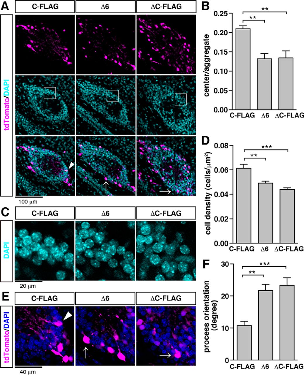
Reelin with an intact CTR is required for formation of the MZ structure. A, Neuronal aggregates induced by ectopic expression of Reelin. Mouse embryos were electroporated with the indicated vector and tdTomato vector (magenta) at E14.5 and fixed at P3. Nuclei were stained with DAPI (cyan). Scale bar, 100 μm. B, Quantification of relative areas of cell-body-sparse regions evaluated by the ratio of center to aggregate area (n = 7). C, Magnified images of the boxed areas in A. Scale bar, 20 μm. D, Quantification of cell density of neuronal aggregates (n = 7). E, Magnified images of a neuronal aggregate in A. Arrowhead and arrows indicate neurons that have processes oriented to the center of the neuronal aggregates and misoriented processes, respectively. Scale bar, 40 μm. F, Quantification of neuronal process angles in neuronal aggregates (n = 7). B, D, F, Error bars indicate mean ± SEM. Tukey's test was used to test for statistical significance: B, F(2,18) = 10.73; D, F(2,18) = 15.71; F, F(2,201) = 10.30. ***p < 0.001. **p < 0.01.


