

Article Figures & Data
Figures
- Figure 1.
Brain ischemic tolerance model using mouse MCAO. A, The MCA was occluded for various periods (from 15 min to 1.5 h), and 3 d later brain damage was assessed by TTC staining of coronal brain sections. A 15 min MCAO period caused no damage, whereas a 1 h MCAO period induced severe injury, mainly in the striatum and cortex. The 15 min MCAO was used for PC, while the 1 h MCAO represented severe MCAO. Scale bar, 2 mm. Sham, sham-operated mice. B, As shown in the experimental protocols, mice received PC 1 d (P1), 3 d (P3), or 6 d (P6) before severe MCAO. The images on the right depict a typical infarct evoked by severe MCAO, showing the effects of PC, which are summarized in C. The severe MCAO-evoked damage in the striatum was significantly reduced when mice received PC 3 d or 6 d earlier (P3 and P6), whereas damage in the cortex was significantly reduced only 6 d after PC (P6). PC 1 d prior (P1) had no effect on brain damage induced by severe MCAO in either brain region. Values are expressed as means ± SEM; *p < 0.05, **p < 0.01 versus severe MCAO alone; n = 4–9. C, Control. D, There was no difference in number of NeuN (green)-positive neurons between Sham and PC groups. Similar to TTC staining, the reduction in number of NeuN-positive neurons after severe MCAO was significantly inhibited by PC (P3). Scale bar, 50 μm. Values are shown as means ± SEM; **p < 0.01; n = 3–4.
- Figure 2.
PC-induced activation of glial cells. A, Open squares show the area in the striatum and cortex used for microscopic examination. Immunostaining with anti-GFAP (green) and anti-Iba1 (red) antibodies in the contralateral (B) and ipsilateral (C) hemispheres. Scale bars: main images, 100 μm; insets, 10 μm. D, The immunofluorescence intensity was quantified. Values are shown as means ± SEM; **p < 0.01 versus contralateral side (Contra); n = 3–4. GFAP-positive and Iba1-positive signals were not affected by PC in the contralateral cortex or striatum (B). Ipsilaterally, PC induced the activation of astrocytes (i.e., upregulation of GFAP immunoreactivity) 3 and 6 d after PC in the striatum, and 6 d after PC in the cortex. Activation lasted for at least 8 weeks (data not shown). PC induced the activation of microglia (i.e., upregulated Iba1 immunoreactivity and promoted the appearance of amoeboid morphology) 1 d after PC, which lasted at least 6 d after PC in the striatum. PC caused no activation in the cortex.
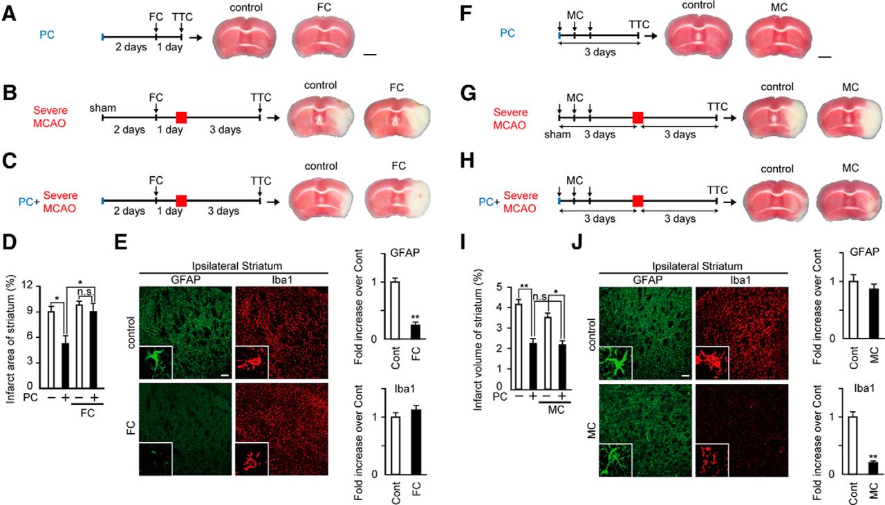
- Figure 3.
PC-induced activation of astrocytes, but not microglia, is essential for ischemic tolerance. A, As shown in the experimental protocol, after PC, mice received an intrastriatal injection of FC (1 pmol/site). Brain damage was assessed by TTC staining. FC injection after PC caused no damage. Scale bar, 2 mm. B, FC had no effect on severe MCAO (1 h MCAO)-induced ischemic injury, but FC abolished PC-induced ischemic tolerance (C; please compare with results in Fig. 1). Results are summarized in D. Values are shown as means ± SEM; *p < 0.05; n = 4–6. E, The morphology of astrocytes (GFAP: green) and microglia (Iba1: red) in the ipsilateral striatum were analyzed by immunohistochemical staining 3 d after PC. The immunofluorescence (IF) intensity was quantified. PC-induced activation of astrocytes was suppressed by FC injection, whereas that of microglia was not affected. Scale bars: main images, 100 μm; insets, 10 μm. Values are shown as means ± SEM; **p < 0.01 versus control (Cont); n = 3. F, As shown in the experimental protocols, after PC, mice received three intraperitoneal injections of MC (45 mg/kg). MC injections after PC caused no damage. Scale bar, 2 mm. MC affected neither severe MCAO (1 h MCAO)-induced ischemic injury (G) nor PC-induced ischemic tolerance (H). Results are summarized in I. Values are shown as means ± SEM; *p < 0.05, **p < 0.01; n = 5–11. J, The morphology of microglia (Iba1: red) and astrocytes (GFAP: green) in the ipsilateral striatum was assessed by immunohistochemical staining 3 d after PC and the IF intensity was quantified. PC-induced activation of microglia was suppressed by MC injection, whereas that of astrocytes was not affected. Scale bars: main images, 100 μm; insets, 10 μm. Values are shown as means ± SEM; **p < 0.01 versus Cont; n = 3.
- Figure 4.
P2X7 receptors are upregulated in activated astrocytes by PC. A, The localization of P2X7 receptors was analyzed by double immunohistochemical staining for P2rx7-EGFP (green), GFAP (red), and Iba1 (red) in the ipsilateral striatum after PC or sham operation. Scale bars: main images, 60 μm; insets, 27 μm. Results are summarized in B. Values are shown as means ± SEM; **p < 0.01 versus sham operation, n = 3–4. C, The time dependency of upregulation of P2rx7 mRNA in the ipsilateral striatum, assessed by quantitative RT-PCR (n = 3–4). P2X7 receptors were significantly upregulated 1, 3, and 6 d after PC. Values are shown as means ± SEM, **p < 0.01 versus naive mice (nv). sh, Sham-operated mice. D, The upregulation of P2rx7 mRNA by PC was significantly suppressed by FC, a metabolic inhibitor of astrocytes. Values are shown as means ± SEM; *p < 0.05, **p < 0.01, n = 3.
- Figure 5.
Absence of PC-induced ischemic tolerance in P2X7−/− mice. A, PC itself had no effect on TTC staining in either WT or P2X7−/− mice. There was no significant difference in severe MCAO-evoked brain damage (the size of the infarct) between WT and P2X7−/− mice. PC-induced ischemic tolerance was abolished in P2X7−/− mice. Results are summarized in B. Scale bar, 2 mm. Values are shown as means ± SEM; *p < 0.05, **p < 0.01; n = 7–8. C, D, There was no difference in PC-evoked glial activation between WT and P2X7−/− mice. Both WT and P2X7−/− mice showed similar morphologies for astrocytes (GFAP: green) and microglia (Iba1: red) in the ipsilateral striatum 3 d after PC. Scale bars: main images, 100 μm; insets, 10 μm. The PC-evoked upregulation of mRNAs for GFAP and Iba1 (Aif1) in P2X7−/− mice was almost identical to that in WT mice. Values are shown as means ± SEM; *p < 0.05, **p < 0.01; n = 3–5.
- Figure 6.
PC-evoked upregulation of astrocytic HIF-1α and EPO require the P2X7 receptor. A–C, Immunohistochemical analysis of HIF-1α in the ipsilateral striatum. Typical pictures and quantitative data are shown in A–C, respectively. Representative immunohistochemical pictures stained with anti-HIF-1α (red), anti-GFAP (green), and anti-NeuN (green) antibodies in the ipsilateral striatum. At 1 d after PC, HIF-1α expression was increased mainly in neurons, and this upregulation was independent of the activation of P2X7 receptors; i.e., upregulation of HIF-1α was observed in both WT and P2X7−/− mice. In contrast, 3 and 6 d after PC, HIF-1α expression was increased in astrocytes. This upregulation was dependent on the P2X7 receptor; i.e., astrocytic upregulation of HIF-1α on day 3 and 6 after PC was significantly higher in WT mice than in P2X7−/− mice. For quantitative analysis, we used the index of percentage of HIF-1α-positive cells (see the Materials and Methods section for details). Scale bars: A, B, main images, 30 μm; insets, 12 μm. Values are shown as means ± SEM; **p < 0.01 versus WT-d3; ##p < 0.01 versus WT-d6; n = 3–6. sh, Sham-operated mice; D, PC significantly upregulated mRNA for EPO, a well known target molecule of HIF-1α, at 3 and 6 d after PC in WT mice, but not in P2X7−/− mice. Values are shown as means ± SEM; *p < 0.05, **p < 0.01; n = 3–6. nv, Naive mice.
- Figure 7.
BzATP-evoked upregulation of HIF-1α in primary cultures of astrocytes. A, Cortical astrocytes derived from WT or P2X7−/− mice were treated with BzATP, a P2X7 receptor agonist, for 24 h at the indicated concentrations, and then Western blotting was performed. HIF-1α protein was increased by BzATP in WT astrocytes, but not in P2X7−/− astrocytes. Data are representative of four independent experiments. C, control. B, The BzATP-evoked increase in HIF-1α protein. Increase in HIF-1α was normalized to β-actin. Data show the fold increase over control (no treatment). Values are shown as means ± SEM; **p < 0.01 versus WT-control; ## p < 0.01 versus WT-50 μM; n = 4–6. C, The expression of P2X7 receptors in cortical astrocytes was assessed using Western blotting. The P2X7 receptor was expressed in cultured astrocytes obtained from WT mice, but not from P2X7−/− mice.
- Figure 8.
Schematic diagram of the mechanisms underlying astrocyte-mediated brain ischemic tolerance. Severe ischemia induces neuronal death (without PC), whereas PC does not cause ischemic injury. Although neuron-derived HIF-1α is upregulated 1 d after PC, this event has no effect on brain injury by severe ischemia. In contrast, 3 d after PC, the upregulation of P2X7 receptors, followed by the upregulation of HIF-1α and neuroprotective molecules (EPO, etc.), is observed in activated astrocytes. Upregulation of P2X7 receptors provides neuroprotection against severe ischemia and induces ischemic tolerance.


Tables
- Table 1.
Time course of the changes in number of GFAP- and Iba1-P2X7 receptor-positive cells in the ipsilateral striatum after PC
Mean cell number (cells/field) Sham Day 1 Day 3 GFAP (+) 1.1 ± 0.4 4.8 ± 2.3 125.3 ± 8.9** P2rx7-EGFP/GFAP (+) 0.3 ± 0.2 0.9 ± 0.5 37.8 ± 10.3** Iba1 (+) 55.8 ± 4.6 100 ± 11.1** 107.9 ± 13.7** P2rx7-EGFP/Iba1 (+) 4.8 ± 1.9 54.4 ± 6.3** 43.8 ± 11.2** We randomly chose three microscopic fields (600 × 600 μm) of the striatum, and then counted the number of GFAP (+), P2rx7-EGFP/GFAP (+), Iba1 (+), and P2rx7-EGFP/Iba1 (+) cells at each time point. Values are shown as means ± SEM;
↵**p < 0.01 versus sham-operated mice; n = 3–4 (three fields per mouse). GFAP (+), GFAP-positive cells; Iba1 (+), Iba1-positive cells; P2rx7-EGFP/GFAP (+), GFAP/P2X7 receptor-double positive cells; P2rx7-EGFP/Iba1 (+), Iba1/P2X7 receptor-double positive cells.


