

Article Figures & Data
Figures
- Figure 1.
Task design, implantation, and behavior. a, Illustration of a monkey in the experimental setup. The cues were presented on a masked monitor and reflected by a mirror such that cues appeared superimposed on the grasping handle. b, Delayed grasping task with two grip types (top, power grip; bottom, precision grip). Trials were presented in pseudorandom order in darkness and with the handle in the upright position. c, d, Plots of RT and MT against delay length for all successful trials of both monkeys. Boxes represent the median and 25th/75th percentiles for each delay bin, while whiskers mark ∼±2.7 SDs. Outliers are shown as individual points. e, f, Array locations for Monkeys S (e) and B (f). Two arrays were placed in F5 on the bank of the arcuate sulcus (AS) and two were placed in the AIP toward the lateral end of the intraparietal sulcus (IPS). In Monkey B two more arrays were placed on the bank of the central sulcus (CS). These were not used in this study. The cross shows medial (M), lateral (L), anterior (A), and posterior (P) directions. Note that Monkey S was implanted in the left hemisphere and Monkey B was implanted in the right hemisphere.
- Figure 2.
Example average firing rate curves of single units for delayed (1300 ms) versus nondelayed (0 ms) grasps. a–c, Example single units from area F5 of both monkeys showing (a) a suppressed cue response during nondelayed grasps, (b) an identical cue response for either delay, and (c) a large condition-independent response restricted to the delay period. d, e, Similar single-unit examples from the AIP of both monkeys. f, A typical movement period response selective for grip type. Delayed data were aligned to two events, grip cue onset and movement onset, and are separated by a gap, which marks the go cue. Nondelayed data were only aligned to movement onset. The later cue period was only present for nondelayed grasps. The cue was always presented for 300 ms regardless of delay. Curves and shaded bands represent mean and SEM, respectively.
- Figure 3.
Targeted dimensionality reduction of neural trajectories in F5 and the AIP. Population data of all conditions, for each area and dataset separately, were projected into a seven-dimensional task space as determined by targeted dimensionality reduction (see Materials and Methods). A single-session trial-averaged example is shown for Monkey S (Session S4, top) and Monkey B (Session B2, bottom). Trajectories begin 100 ms before the grip cue and end at movement onset. For the no-movement condition, data is plotted from 100 ms before the grip cue until reward onset.
- Figure 4.
Point-to-curve distance between delayed (1000 ms) and nondelayed (0 ms) trajectories. a, Minimum Euclidian distance in the full neural space between each time point on the delayed trajectory (in steps of 10 ms) and the entire nondelayed trajectory over time for two example datasets (B2-Power, S6-Precision) from both areas and monkeys. The black line represents the minimum point-to-curve distance between the delayed and nondelayed trajectory, while the gray line represents the chance level (see Materials and Methods). Black bars along the top of plots denote times when the distance is significantly greater than chance level (bootstrapping procedure with 1000 resamples, p = 0.01; see Materials and Methods). Error bars represent the fifth and 95th percentiles of the distances generated by the bootstrapping procedure. b, Fraction of significant distances over all datasets and grip types (6 datasets × 2 grip types). Error bars represent the SEM over datasets and grip types. c, Difference in onset of grip and delay separation over all datasets and grip types (6 datasets × 2 grip types).
- Figure 5.
Neural trajectory stability over the course of no-movement trials. a, Mean Euclidean distance in the full neural space for the no-movement trials between all pairs of time points over both grip types for example datasets in each monkey (Sessions B5, S3). Cue, cue epoch; Mem, mem epoch; Rew, reward epoch. All plots are clipped at 8 spikes/s for visualization. b, For each pair of time points, distance results were tested for a significant difference using a bootstrapping procedure (1000 resamples in steps of 50 ms, p = 0.01). Panels show time pairs where a significant difference was found in the dataset of a for both grip types (white), one grip type (gray), or in no condition (black). c, Percentage of time points showing a significant difference over all datasets and grip types (6 datasets × 2 grip types) separately for each monkey and area. d, Mean distance between all time points during the stable portion of the memory period (650–1800 ms after cue onset; a, dashed box) for all individual datasets and grip types (6 datasets × 2 grip types) across areas and paired according to recording session. Stars indicate a significant difference (Wilcoxon sign-rank test, p < 0.001).
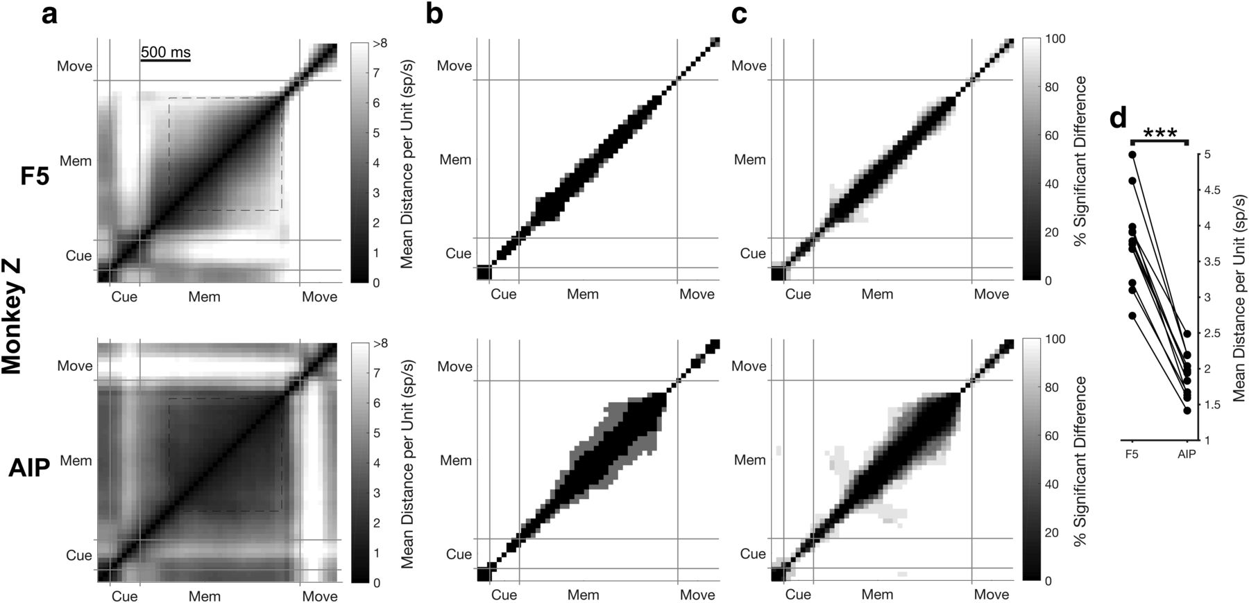
- Figure 6.
Neural trajectory stability over the course of instructed trials for an additional experiment. Same layout as Figure 5. a, Mean Euclidean distance in the full neural space for the Instructed trials between all pairs of time points over both grip types for an example dataset in Monkey Z (see Materials and Methods). Cue, cue epoch; Mem, memory epoch; Move, movement epoch. All plots are clipped at 8 spikes/s for visualization. b, For each pair of time points, distance results were tested for a significant difference using a bootstrapping procedure (1000 resamples in steps of 50 ms, p = 0.01). Panels show time pairs where a significant difference was found in the dataset of a for both grip types (white), one grip type (gray), or in no condition (black). c, Percentage of time points showing a significant difference over all datasets and grip types (6 datasets × 2 grip types). d, Mean distance over the stable portion of the memory period (650 ms after cue onset; go cue; a, dashed box) for all individual datasets and grip types (6 datasets × 2 grip types) across areas and paired according to recording session. Stars indicate a significant difference (Wilcoxon sign-rank test, p < 0.001).
- Figure 7.
Representation of subjective anticipation across F5 and the AIP. a, Illustration of the probability of a go cue at all times during the delay (binned into 25 ms bins for visualization purposes), the hazard rate (Eq. 4), and the subjective anticipation function (Eq. 5 substituted into Eq. 4). b, Subjective anticipation axis as determined by targeted dimensionality reduction for all single trials of the no-movement condition across both brain regions (Session S5). c, Projection of F5 and the AIP onto the subjective anticipation axis 100 ms before the go cue correlated with single-trial RT for two delay bins in the same example dataset. d, Summary of RT prediction over all datasets. Stars denote a significant difference between areas (Wilcoxon sign-rank test, p < 0.05). e–g, Same as b–d for Monkey B (Session B4).
- Figure 8.
Clustering of movement-initiation activity in F5. a, Example individual unit activity in F5 over all linearly spaced delays (0–1000 ms) for an example dataset from each monkey (Sessions S5-Precision, B2-Precision), aligned to movement onset. b, Euclidean distance between all pairs of delays in the full neural space for two example time points of the example dataset, including identified clusters, using a clustering analysis that finds community structure (see Materials and Methods). c, Clusters identified in the distance matrices over time (in steps of 10 ms) for the example dataset. Black significance bar shows time points where the modularity statistic exceeded chance level (permutation test, p < 0.01). d, Same analysis as c averaged over all datasets and grip types (6 datasets × 2 grip types).
- Figure 9.
Clustering of movement initiation activity in the AIP. Same layout as Figure 8. a, Example individual unit activity in the AIP over all linearly spaced delays (0–1000 ms) for an example dataset from each monkey (S3-Power, B2-Precision), aligned to movement onset. b, Euclidean distance between all pairs of delays in the full neural space for two example time points of the example dataset, including identified clusters, using a clustering analysis that finds community structure (see Materials and Methods). c, Clusters identified in the distance matrices over time (in steps of 10 ms) for the example dataset. Black significance bar shows time points where the modularity statistic exceeded chance level (permutation test, p < 0.01). d, Same analysis as c averaged over all datasets and grip types (6 datasets × 2 grip types).


Tables
Session Trial count Correct performance Units recorded in F5 Units in F5 meeting retention criteria Units Recorded in the AIP Units in the AIP meeting retention criteria B1–B6 B1 485 91% 78 48 36 26 B2 685 96% 86 53 38 28 B3 586 96% 57 35 32 18 B4 814 96% 60 38 26 19 B5 775 96% 66 34 27 18 B6 745 97% 65 44 33 21 Mean 682 95.3% 68.7 42.0 32.0 21.7 S1–S6 S1 502 98% 108 74 139 102 S2 514 97% 116 75 134 100 S3 571 97% 132 99 127 102 S4 658 99% 133 92 131 109 S5 590 99% 139 103 136 100 S6 546 98% 129 98 130 98 Mean 564 98.0% 126.2 90.2 132.8 101.8


