

Article Figures & Data
Figures
- Figure 1.
Rescue of ERK activity with an ERK pathway inhibitor in 16p11.2del mice at E14.5 and P10. a, IHC of E14.5 coronal sections from mice treated with vehicle or ERK inhibitor. Veh-treated 16p11.2del mice shows upregulation of ERK activity in the dorsomedial cortex * (anti-pERK; green). This is abrogated after 5 d of inhibitor treatment starting at E10.5. b, Western analysis of E14.5 veh- or inh-treated cortical lysates. c, Quantification of Western analysis showing a significant increase in ERK1 and ERK2 activity in 16p11.2del animals (pERK1: ****p < 0.0001, pERK2: *p < 0.05), which is restored to normal level after inhibitor treatment (##p = 0.0087; #p < 0.05; veh-treated: nWT = 19, nDel = 13; inh-treated: nWT = 19, nDel = 12). ERK1 total levels are decreased in vehicle deletion animals (**p < 0.01) and inhibitor animals (****p < 0.0001; veh-treated: nWT = 8, nDel = 7; inh-treated: nWT = 10, nDel = 9). D, Western analysis of P10 veh- or inh-treated cortical lysates, and (e) Western analysis of P10 veh or inh prenatally treated cortical lysates, quantified in (e) ERK1 and ERK2 activity are elevated in deletion animals at P10 (pERK1: **p < 0.01, pERK2: p**<0.01), which is normalized in embryonic inhibitor treatment (#p < 0.05, ###p < 0.001). All values normalized to loading control GAPDH or CoxIV and reported as a fold-change. p Values are from Bonferroni post hoc analysis (*compares WT to deletion; #compares vehicle deletion to inhibitor deletion).
- Figure 2.
Reversal of deficits in cortical neurogenesis in the 16p11.2del mice after treatment with ERK pathway inhibitor at E14.5. IHC of coronal sections and Western analyses of cortical lysates at E14.5 a, IHC with proliferation marker, BrdU injected 30 min. before kill. a′, The number of BrdU+ progenitors was analyzed (veh-treated: nWT = 8, nDel = 6; inh-treated: nWT = 9, nDel = 9; *p < 0.05, #p < 0.05). b, IHC with intermediate progenitor marker, Tbr2 (green). b′, Quantification of Tbr2+ progenitors (veh-treated: nWT = 11, nDel = 4; inh-treated: nWT = 9, nDel = 16; **p < 0.0012, ##p < 0.0052). b″, Quantification of Western analysis (veh-treated: nWT = 29, nDel = 37; inh-treated: nWT = 23, nDel = 21; ****p < 0.00001, #p < 0.0388). c, IHC for layer V marker, Ctip2 (red). c′, Quantification of Ctip2+ neurons (veh-treated: nWT = 5, nDel = 4; inh-treated: nWT = 4, nDel = 6; ***p = 0.0005, ####p < 0.0001, *p = 0.026). c″, Quantification of Western analysis (veh-treated: nWT = 15, nDel = 9; inh-treated nWT = 8, nDel = 11; *p = 0.0186, ##p = 0.0073). d, IHC with layer VI marker, Tbr1 (green). d′, Quantification of Tbr1+ neurons (veh-treated: nWT = 9, nDel = 9; inh-treated: nWT = 5, nDel = 5; *p = 0.025, ###p = 0.0004). d″, Quantification of Western analysis (veh-treated: nWT = 15, nDel = 7; inh-treated: nWT = 11, nDel = 12; *p = 0.038, ##p = 0.0017). All Western analyses data represented as a fold-change, normalized to a loading control. p Values are from Bonferroni post hoc analysis (*compares WT to deletion; #compares vehicle deletion to inhibitor deletion).
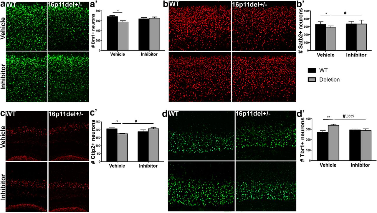
- Figure 3.
Prenatal treatment with an ERK pathway inhibitor stably restores normal cortical cytoarchitecture 16p11.2del mice. a, Mice were evaluated at P2 by IHC with layer II–IV marker Brn1 (green). a′, Quantification of Brn1+ neurons shows a rescue in somatosensory cortex of 16p11.2del mice (veh-treated: nWT = 11, nDel = 13; inh-treated: nWT = 17, nDel = 17; *p < 0.05). b, IHC with layer 2–4 marker, Satb2 (red). b′, Quantification of Satb2+ neurons (veh-treated: nWT = 14, nDel = 13; inh-treated: nWT = 10, nDel = 15; *p = 0.033, #p = 0.0105). c, IHC with layer V marker, Ctip2 (red). c′, Quantification of Ctip2+ neurons (veh-treated: nWT = 21, nDel = 14; inh-treated: nWT = 9, nDel = 13; *p = 0.014, #p = 0.033). d, IHC with layer VI marker, Tbr1+ (green). d′, Quantification of Tbr1+ neurons (veh-treated: nWT = 8, nDel = 9; inh-treated: nWT = 12, nDel = 10; **p = 0.0076, #p = 0.0535). p Values are from Bonferroni post hoc analysis (*compares WT to deletion; #compares vehicle deletion to inhibitor deletion).
- Figure 4.
Treatment with ERK pathway inhibitor normalizes the levels of the downstream ERK effectors: p27 Kip1 and cyclin D1. IHC of coronal sections and Western analyses from WT and 16p11.2del mice at E14.5 a, Immunostaining against CyclingD1 antibody (green); a′, Western blot analysis, quantified in a″ (veh-treated: nWT = 16, nDel = 7; inh-treated: nWT = 16, nDel = 13; ***p < 0.001). b, Immunostaining against p27Kip1(green); b′, Western blot analysis, quantified in b″ (veh-treated: nWT = 8, nDel = 10; inh-treated: nWT = 8, nDel = 10; **p < 0.01, *p < 0.05). p Values are from Bonferroni post hoc analysis.
- Figure 5.
Prenatal ERK inhibitor treatment partially rescues ventral hippocampal and lateral septal volume in 16p11.2del mice. Mice were treated for 5 consecutive days starting at E10.5 and evaluated at P90. A, TBM analyses revealed significant increased volume of SC, PAG and Hypo in 16p11.2del mice compared with WT littermates (t test, p < 0.01 FWE cluster-corrected, with cluster defining threshold of |t| > 2.3). In 16p11.2 deletion mice, we also observed an increased volume of the RS ctx, as well as reduced volume of vHPC, LS, Ent ctx, and Amy (t test, p < 0.01 FWE cluster-corrected, with cluster defining threshold of |t| > 2.3). b, Comparison between treated and nontreated 16p11.2del mice shows that ERK-inhibitor produces bilateral foci of increased volume in the vHPC and LS (t test, p < 0.01 FWE cluster-corrected, with cluster defining threshold of |t| > 2.3). c, A composite illustration of a and b revealed that foci of increased gray matter volume (red/yellow; from b) are spatially located in the same hippocampal and septal regions exhibiting reduced gray matter volume in 16p11.2 del mice (blue/light blue; from a), suggesting a partial anatomical rescue of volumetric deficits upon treatment with ERK inhibitor. d, e, Consistent with TBM results, anatomical labeling revealed reduced relative volume in vHPC (t test: t(17) = 3.78, p = 0.001) and LS (t test: t(17) = 2.21, p = 0.041) in 16p11.2del mice compared with WT littermates (one-way ANOVA of vHPC: F(3,34) = 8.083, p < 0.001; one-way ANOVA of LS: F(3,34) = 1.692, p = 0.1872). Treatment with ERK inhibitor in 16p11.2del mice partially restored morphoanatomical volume in these brain regions (vHPC, t test: t(16) = 2.79, p = 0.013), although the effects in LS did not reach full statistical significance (t test, t(16) = 1.78, p = 0.078). Amy, Amygdala; Ent ctx, entorhinal cortex; Hypo, hypothalamus; LS, lateral septum; PAG, periaqueductal gray; RS, retrosplenial cortex; SC, superior colliculus; vHPC, ventral hippocampus. *p < 0.05,**p < 0.01.
- Figure 6.
Reversal of Behavioral Impairment of 16p11.2del mice after prenatal ERK pathway inhibitor treatment. WT or 16p11.2del 3-month-old male or female mice treated with Veh or Inh at E10.5 for 5 d. a, Elevated plus maze shows a no change in percentage of time in open arm, but decreased closed arm time in 16p11.2del mice that is rescued by inhibitor treatment (*p < 0.05, #p < 0.05; veh-treated: nWT = 25, nDel = 22; inh-treated: nWT = 27, nDel = 28). b, Open Field shows increased time spent in center in 16p11.2del mice that is rescued with inhibitor treatment (**p < 0.01, #p < 0.05; veh-treated: nWT = 23, nDel = 20; inh-treated: nWT = 19, nDel = 18). c, Fear conditioning shows increased freezing in 16p11.2del animals that improves with inhibitor treatment (**p < 0.01; veh-treated: nWT = 5, nDel = 6; inh-treated: nWT = 11, nDel = 11), conditioned stimulus: 85 dB sound at 2800 Hz for 30 s; US: 0.56 mA. Retention test performed 18 h later for 5 min in the absence of tone. d, NOR was evaluated in WT and 16p11.2del animals (#p < 0.05; veh-treated: nWT = 16, nDel = 12; inh-treated: nWT = 8, nDel = 16). e, Naive females were exposed to 3 WT pups placed in three corners of the cage, time to retrieve pups was recorded (first pup: **p < 0.01; second pup: **p < 0.01; third pup: ***p = 0.001; veh-treated: nWT = 21, nDel = 17; inh-treated: nWT = 6, nDel = 6). f, Mice were food deprived for 24 h, then placed in a cage containing one food pellet (Teddy Graham) buried under normal cage bedding; time to retrieve was recorded (**p < 0.01, ####p < 0.0001; veh-treated: nWT = 22, nDel = 22; inh-treated: nWT = 22, nDel = 7). p Values are from Bonferroni post hoc analysis (*compares WT to deletion; #compares vehicle deletion to inhibitor deletion).
- Figure 7.
Postnatal treatment with ERK pathway inhibitor partially restores behavioral deficits in adult 16p11.2del mice. WT or 16p11.2del 3-month-old male or female mice treated with veh or inh at P90 for 5 d. a, Elevated plus maze analysis of percentage of time in open arm (*p < 0.05), entries in open arm, immobility time (##p < 0.01), and total distance traveled (veh-treated: nWT = 15, nDel = 11; inh-treated: nWT = 12, nDel = 7). b, Open-field analysis of entries into center (##p < 0.01), time spent in center and total distance traveled (veh-treated: nWT = 22, nDel = 17; inh-treated: nWT = 23, nDel = 9). c, ELISA performed on whole brain lystaed of P90 mice (**p < 0.05; #p < 0.05; veh-treated: nWT = 10, nDel = 8; inh-treated: nWT = 7, nDel = 5). D, Mice were food deprived for 24 h, and then placed in a cage containing one food pellet (Teddy Graham) buried under normal cage bedding; time to retrieve (latency) was analyzed. p Values are from Bonferroni post hoc analysis (*compares WT to deletion; #compares vehicle deletion to inhibitor deletion).


