
In the article “The Spatial Patterning of Mouse Cone Opsin Expression Is Regulated by Bone Morphogenetic Protein Signaling through Downstream Effector COUP-TF Nuclear Receptors” by Shinya Satoh, Ke Tang, Atsumi Iida, Mariko Inoue, Tatsuhiko Kodama, Sophia Y. Tsai, Ming-Jer Tsai, Yasuhide Furuta, and Sumiko Watanabe, which appeared on pages 12401–12411 of the October 7, 2009 issue, there were erroneous duplications of images in Figures 1 and 5. In Figure 1, S-opsin day 7 (left column #3) and M-opsin day 14 (right column #4) are the same picture, flipped vertically. Additionally, the image in M-opsin day 14 is not correct. In Figure 5B, the same images were used for the control for COUPI KO and the control for COUPII CKO. Additionally, the images in COUPII CKO are not correct. The corrected Figures 1 and 5 are shown here. The original figure legends are correct.

Opsin expression patterns in retinal explant cultures. Retinal explants prepared from E17 or E12 mice were cultured for the indicated number of days. Whole-mount immunostaining was performed for S-opsin and M-opsin expression, and the signals were visualized using secondary antibodies conjugated to Alexa-488 (green). Photos were taken from the bottom of the explants (ONL). The pictures show the top of the dorsal side. D, Dorsal; V, ventral. Scale bars, 500 μm.
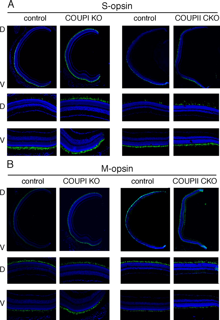
Expression of S-opsin and M-opsin in COUP-TFI-null and retinal-specific COUP-TFII-null mouse retina. A,B, S-opsin (A) and M-opsin (B) expression was examined by immunostaining using frozen-sectioned eyes from COUP-TFI knock-out mice (COUPI KO) at P23 and COUP-TFII retina-specific knock-out mice (COUP-TFIIfx/fx;Rx—Cre; COUPII CKO) at 3 months of age. The signals were visualized using a secondary antibody conjugated to Alexa-488 (green). The nuclei were visualized by DAPI staining (blue). The middle and bottom are enlarged views of the dorsal and ventral periphery. The control samples were taken from each littermate of the COUP-TFI KO and COUP-TFII CKO mice. D, Dorsal; V, ventral.