

Article Figures & Data
Figures
- Fig. 1.
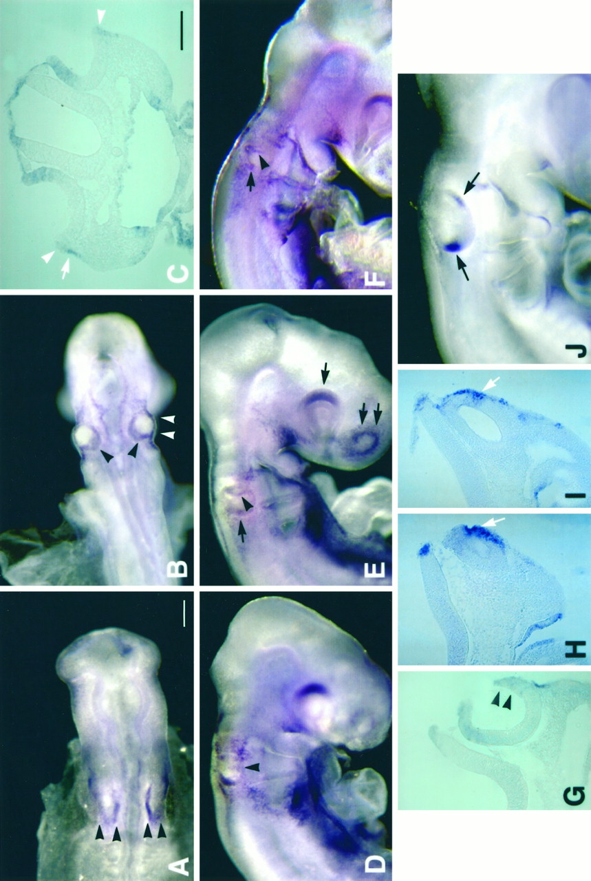
Gene expression of BMP4 in developing chick inner ear, stages 11–19, by whole-mount in situhybridization. The stages of embryos illustrated are as follows: (A) 12 somites, stage 11; (B,C) 21 somites, stage 13; (D,G) 26 somites, stage 16; (E) 28 somites, stage 16; (F, H, I) 30 somites, stage 17; and (J) 40 somites, stage 19. At stage 11 (A), BMP4 expression was detected in the medial and posterior margin of the otic placode (arrowheads). By stage 13 (B), BMP4 expression was present in the dorsal and posterior margin of the otic cup (black arrowheads). Some transcripts were detected in the ventral margin of the otic cup (white arrowheadsin B), but the majority of this hybridization signal was found to be contributed by ectoderm adjacent to the otic cup shown in a transverse section (C) of an embryo the same age asB (stage 13). Arrowheads inC point to hybridization signal within the otic epithelium, and the arrow points to signal in the adjacent ectoderm. At 26 somites (stage 16; D), the expression in the dorsal and posterior margin remained whereas the expression in the ventral portion of the otic epithelium had expanded (arrowheads in D, G). By 28 somites (stage 16; E), a positive streak appeared in the anterior otocyst (arrowhead) and the posterior hybridization signal became more restricted (black arrow). In addition, the dorsal portion of retina (single arrow) and the olfactory placode (double arrows) were also positive. A slightly older embryo (30 somites, stage 17; F) showed a similar pattern, with the arrowhead pointing to the anterior streak and the arrow pointing to the beginning of the posterior focus. In this embryo (F), the otocyst was closed as indicated by the notch shown in a transverse section (H). The hybridization signal from the anterior streak was present within the otic epithelium (arrow inH). Hybridization signal in the posterior focus was also within the otic epithelium, as shown in a more posterior section (arrow in I). By stage 19, two BMP4-positive foci were evident in the otocysts (arrowsin J). Scale bars: A,B, D–F, J, 200 μm; C, G–I, 100 μm.
- Fig. 2.
Gene expression of BMP4 in developing inner ear, stages 19 (E3) to stage 30 (E6.5), by in situhybridization of frozen sections. A–Dare horizontal sections of chick otocysts. At stage 19 (E3;A) only two BMP4-positive areas were present [anterior (sc) and posterior (p)]. The BMP4 expression in macula sacculi (s) is shown at stage 21 (B); lateral crista (lc) at stage 22 (C) and macula utriculi (u) at stage 24 (D). E and F are transverse sections of chick otocysts of stage 27 (E5) and stage 30 (E6.5), respectively. In addition to the BMP4-positive sensory organs as indicated in E and F, BMP4 was also expressed in the mesenchyme surrounding the dorsal portion of the inner ear (E), the tip of the first pharyngeal pouch (arrow in E), and the roof of the ampulla (arrow in F). Orientation:A, anterior; D, dorsal; L, lateral. ed, Endolymphatic apparatus; bp, basilar papilla; la, lagena. Scale Bar, 50 μm.
- Fig. 3.
Three-dimensional reconstruction of BMP4 gene expression of a stage 27 (E5) chick inner ear. The right inner ear is shown from an anterior (A) and a posterior (B) view. BMP4-positive areas are displayed in different colors, which include superior crista (sc) inblue, lateral crista (lc) inyellow, macula utriculi (u) inred, macula sacculi (s) inorange, basilar papilla (bp) and lagena (la) traced as one object in fuchsia, and posterior crista (pc) inbluish-green. Image in Awas tilted dorsally to reveal all of the positive areas. This specimen was reconstructed from 20 alternate sections of 12 μm thickness. Only the inner borders of the otic epithelium from each section were traced to give the contour of the inner ear. However, the entire outlines of each positive area were traced, which included the inner and outer borders of the positive epithelium. As a result, positive areas appear as bulged objects situated on the reconstructed fluid ducts of the inner ear. ed, Endolymphatic apparatus;psc, primordia for the superior and posterior semicircular canals. Scale bar, 100 μm.
- Fig. 4.
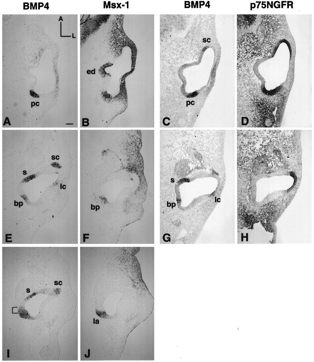
Gene expression pattern of BMP4, Msx-1, and p75NGFR in a stage 23 (E4) chick otocyst. A andB, E and F, andI and J are pairs of adjacent 12 μm sections from one embryo, with A and Bbeing the most dorsal pair and I and Jthe most ventral. C and D andG and H are pairs from another embryo. The C/D pair was chosen from a level similar to the A/B pair, and theG/H to E/F.A, C, E, G, and I were probed for BMP4 mRNA; B, F, and J were probed for Msx-1; and D and H were probed for p75NGFR. In the most dorsal sections, the posterior BMP4-positive area was Msx-1-positive (B) as well as p75NGFR-positive (D), which marked the presumptive posterior crista (pc) area. More ventral sections were still positive for Msx-1 (F) but negative for p75NGFR (H), indicating the presumptive basilar papilla (bp) region. The positive p75NGFR area inH included the presumptive lateral crista area and was continuous with the superior crista area in D. In the ventral portion of this broad BMP4-positive area (I, J), part of the area was strongly positive for Msx-1 marking the presumptive lagena. Area marked with bracketin I, which was positive only for BMP4, was part of the presumptive basilar papilla. Scale bar, 100 μm.
- Fig. 5.
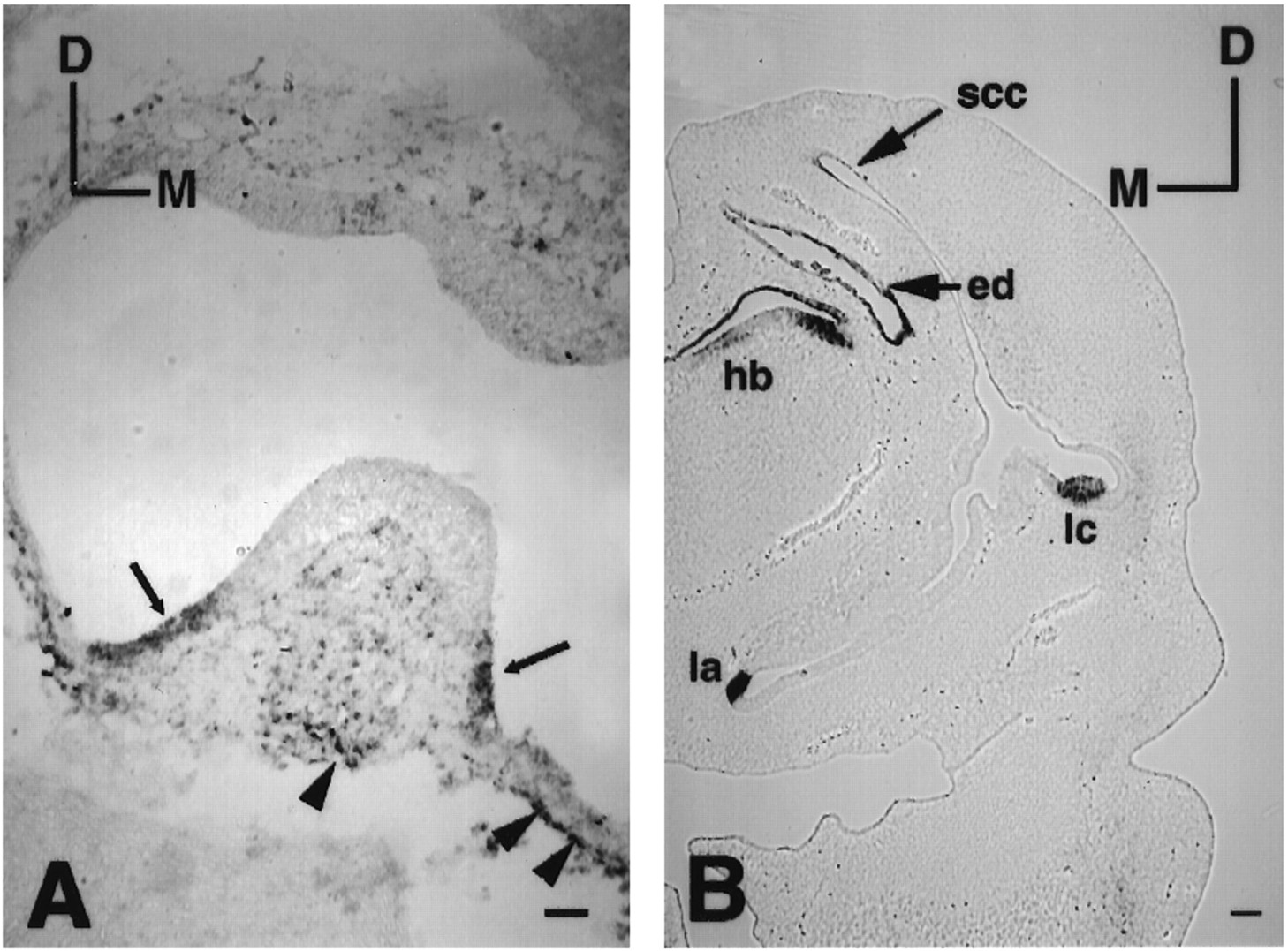
Gene expression of p75NGFR and Msx-1 in the developing inner ear. Transverse sections of an E12 (stage 38;A) and an E6 (stage 29; B) inner ear were probed for p75NGFR and Msx-1, respectively. At E12, p75NGFR expression was expressed in the three cristae of the inner ear. A part of the lateral crista ampullaris is shown in A. P75NGFR expression was located in the peripheral portion of the crista (arrows in A) as well as many areas of mesenchyme next to the otic epithelium (arrowheads). At E6 (stage 29), as illustrated in B, Msx-1 was expressed in the lagena (la), three cristae ampullaris [lateral crista is shown (lc)], the endolymphatic apparatus (ed), and some portions of the semicircular canals (ssc). Orientation: D, dorsal;M, medial; hb, hind brain. Scale bars:A, 50 μm; B, 100 μm.
- Fig. 6.
BMP4 and Msx-1 expression in the presumptive basilar papilla and macula neglecta. A–Cand F were probed for Msx-1. D andE were probed for BMP4. A andB are transverse sections of a stage 26 embryo (E4.5–E5) showing that the Msx-1 expression was absent in the anterior portion of basilar papilla (arrowhead inA) but present in the posterior portion of the papilla (arrowhead in B). The arrow inA points to part of the positive area in the lagena, which is more obvious in a posterior section shown in B(la). The Msx-1 expression in the papilla decreased over time (stage 27, E5; C) and eventually disappeared with transcripts remaining in the lagena (la) only (see Fig.5B). D and E are transverse sections indicating BMP4 expression in macula neglecta (mn) at stage 29 (E6) and stage 31 (E7), respectively.F is an adjacent section of E probed for Msx-1. Arrowheads in D andE point to positive hybridization signals from the posterior ampulla. The proximal tip of the basilar papilla (bp), which was BMP4-positive, is also shown inD–F. Orientations: D, dorsal; M, medial; hb, hind brain. Scale bars: A–C, 100 μm;D–F = 50 μm.


Tables
- Table 1.
Summary of sensory organ generation based on BMP4, Msx-1, and p75NGFR gene expression patterns
Sensory organ BMP4 Msx-1 p75NGFR Stage of generation1_a Superior crista + + + 19 Macula sacculi + − − 20 Lateral crista + + + 22 Macula utriculi + − − 24 Posterior cluster + 18 Posterior crista + + + 19 Basilar papilla Anterior arm + − − 23 Posterior arm + +1_b − 23 Lagena + + − 23 Macula neglecta + + − 29 ↵F1_a The generation age for each sensory organ is defined when discrete expression of each molecular marker listed was activated for that particular sensory organ. The only exception is the macula neglecta: at stage 29 (E6), only BMP4 expression was restricted, and Msx-1 expression did not become restricted until stage 31 (E7).
↵F1_b Msx-1 expression in the posterior arm of the basilar papilla disappeared by stage 29.


