

Article Figures & Data
Figures
- Fig. 1.
Western blot characterization of anti-mouse PDGF α-R, antibody in protein extracts from P1, P7, and adult mouse brain. The anti-mouse PDGF-αR recognizes 170 and 140 kDa protein bands, equivalent to the estimated size of the mature form and the precursor form, respectively, of the PDGF α-R in adult (lane 1), P7 (lane 2), and P1 (lane 3) mouse brain, and mouse 3T3 cells (lane 4), used as a positive antigen control. The relative migration positions of the molecular weight standards (myosin, 200,000; phosphorylase b, 97,000; bovine serum albumin, 66,000), run in parallel, are indicated.
- Fig. 2.
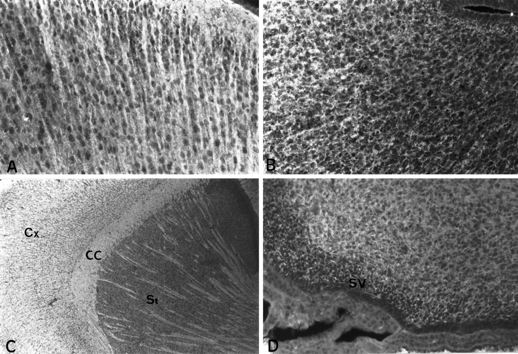
Expression of PDGF-αR in the P1 mouse brain.A, Immunodetection of PDGF-αR protein in the prefrontal cortex. PDGF-αR is localized on neurites and neuronal cell bodies. B, Immunoreactivity for PDGF-αR in the entorhinal cortex. PDGF-αR is widely expressed in the cerebral cortex (Cx) as well as in presumptive white matter structures, as illustrated for the corpus callosum (CC) and fibers of the striatum (St) in C. Striatal neurons are not stained with the polyclonal anti-PDGF-αR.D, Immunostaining of cells of the periventricular zone of the lateral ventricle. Magnification: A, B, D, 200×;C, 50×.
- Fig. 6.
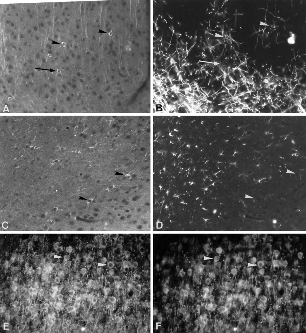
Immunocharacterization of the cell types expressing PDGF-αR. Double immunostaining with anti-PDGF-αR (A) and Rip (B) on P21 sagittal brain section. PDGF-αR-positive cells (arrowheads inA) are weakly stained with the Rip antibody (B). Often, there is not strict colocalization between PDGF-αR and Rip expression (arrow), suggesting that these single-labeled cells could be immature cells of the oligodendrocyte lineage. P21 sagittal brain section immunolabeled for PDGF-αR (C) and GFAP (D).Arrowheads in C indicate PDGF-αR-positive cells that do not express GFAP in D. P7 sagittal brain section through the cerebral cortex stained for PDGF-αR (E) and MAP2 (F). Neurons (arrowheads) stained with the anti-MAP2 antibody (E) express PDGF-αR (F). Magnification, 224×.
- Fig. 3.
Evolution of PDGF-αR immunoreactivity in the developing cerebellum. Immunodetection of PDGF-αR in the cerebellum at P1 (A, B), P7 (C, D), and P15 (E, F). PDGF-αR immunoreactivity is detected in the Purkinje cell layer, the granule cell layer, and the presumptive white matter (A). High-magnification view showing PDGF-αR immunoreactivity on Purkinje and granule cells. Note the lack of expression in the external germinal layer (B). The expression of PDGF-αR is also widely distributed in the P7 cerebellum (C). High-magnification view of immunostained Purkinje and granule cells (D). At this period of development, the protein is well evidenced in the Purkinje cell soma and dendrites. At P15, PDGF-αR immunoreactivity is considerably decreased in the granule cell layer and is not detected on white matter fibers (E, F). The expression is always observed in the soma and dendritic tree of Purkinje cells (arrows inF) and in oligodendrocyte progenitors (arrowheads in F). Magnification:A, C, E, 100×; B, D, F, 200×.EGL, External germinal layer; ML, molecular layer; PL, Purkinje cell layer;GL, granule cell layer; WM, white matter.
- Fig. 9.
Expression of PDGF-αR in the adult cerebellum and brainstem nuclei. In the adult cerebellum, the expression of PDGF-αR is detected only in the Purkinje cell layer (PL), whereas the molecular (ML) and granule cell layers (GL) remained unstained with the anti-PDGF-αR (A). B, High-magnification view of the immunoreactivity in the soma and dendritic processes of Purkinje cells. Note that PDGF-αR immunoreactivity is not found in Purkinje cell axons. C, Immunodetection of PDGF-αR in neurons of the interpositus cerebelli nucleus. D, High-power view of PDGF-αR-positive neurons of the interpositus cerebelli nucleus. E, PDGF-αR immunoreactivity in the facial nucleus. F, High-magnification view of the staining of facial nucleus neurons. Magnification: A, C,110×; B, D, 220×; E, 130×;F, 260×.
- Fig. 4.
In situ hybridization and IHC of PDGF-αR on P15 brain and spinal cord tissue sections. Expression of PDGF-αR transcripts in the cerebral cortex (A). High-magnification view of the hybridization signal showing the localization of PDGF-αR mRNA in the soma of cortical neurons (arrows in B). Detection of PDGF-αR transcripts in the hippocampus (C) and in motoneurons (arrowheads) of the spinal cord (D). Longitudinal spinal cord section, hybridized with the digoxigenin-labeled antisense probe complementary to murine PDGF-αR, showing the localization of the transcripts in motoneurons (arrowheads) and oligodendrocyte precursors (arrows; E). Adjacent section immunolabeled with the anti-PDGF-αR antibody (F). Note that motoneurons and immature cells of the oligodendrocyte lineage express PDGF-αR transcripts and protein.CA1, CA3, Hippocampal fields;DG, dentate gyrus. A–D, E, Bright-field and phase-contrast; F, indirect immunofluorescence. Magnification: A, C, E, F, 124×; B, D, 248×.
- Fig. 5.
Expression of PDGF-αR by immature oligodendrocytes in the P21 mouse brain. Spreading of PDGF-αR-positive cells (arrowheads), belonging to progenitor stages of the oligodendrocyte lineage, from the corpus callosum (CC) to the cerebral cortex (A). High-power view showing the morphology of these PDGF-αR-positive cells in the corpus callosum (B). View of the cerebral cortex, showing the concomitant expression of PDGF-αR by neurons (arrows) and immature cells of the oligodendrocyte lineage (arrowheads) extending several processes in contact with PDGF-αR-positive neurites (C). Expression of PDGF-αR by neurons (arrow) and oligodendroglial cells (arrowheads) in the subiculum (D). Oligodendrocyte progenitors, stained with the anti-PDGF-αR antibody, in the thalamus (E) and in cerebellar white matter (arrowhead in F). Magnification:A, D, 124×; B, C, E, F, 248× .
- Fig. 7.
Immunodetection of PDGF-αR in stem cells and oligodendrocyte progenitors of the adult mouse CNS. Neural stem cells of the subependymal/ependymal layer of the olfactory bulb are stained for PDGF-αR (A). These cells also express PSA-NCAM (B, adjacent section). In the adult CNS, very few oligodendrocyte progenitors (arrows) stained with the anti-PDGF-αR antibody were detected in the cerebral cortex (C) and spinal cord white matter (D). Magnification, 200×.
- Fig. 8.
Localization of PDGF-αR immunoreactivity in the adult mouse brain. Immunodetection of the PDGF-αR protein in neurons (arrows) of the cerebral cortex (A), hippocampus (B), subiculum (C), globus pallidus (D), and substantia nigra (E). High-power view of PDGF-αR-positive neurons of the substantia nigra pars reticulata showing the localization of the protein in the neuronal cell bodies, dendrites, and initial segment of the axons (F). A, B, Immunoperoxidase;C, D–F, immunofluorescence. DG, Dentate gyrus; CA3, hippocampal field. Magnification: A, B, C, E, 124×; D, F, 248×.
- Fig. 10.
Detection of PDGF-αR transcripts in the adult mouse CNS by nonradioactive in situ hybridization. PDGF-αR mRNA are widely expressed in neuronal populations, as illustrated for the hippocampus (A), subiculum and entorhinal cortex (B), substantia nigra pars compacta (C), Purkinje cell layer of the cerebellum (D), and neurons of the vestibular nucleus (E). Hybridization signal is not observed in white matter structures, such as the corpus callosum (A, B).F, Control in situ hybridization with the PDGF-αR digoxigenin-labeled oligonucleotide sense probe performed on sagittal brain tissue section through the cerebellum.A–F, Bright-field and phase-contrast.DG, Dentate gyrus; CA1,CA3, hippocampal fields; CC, corpus callosum; ML, molecular layer; PL, Purkinje cell layer; GL, granule cell layer. Magnification: A, 110×; B, D, F, 220×;C, E, 300×.


Tables
- Table 1.
Neuronal distribution of PDGF-αR immunoreactivity in developing and adult mouse CNS
Location Age P1 P7 P15 P120 Olfactory system Periglomerular layer +++ +++ +++ +++ Internal granular layer +++ +++ ++ + Mitral cell layer + ++ +++ +++ Olfactory ventricle +++ +++ ++ ++ Anterior olfactory nucleus +++ +++ +++ +++ Olfactory tubercle +++ +++ +++ +++ Cerebral cortex +++ +++ ++ ++ Striatum − − − − Globus pallidus nd +++ ++ ++ Hippocampus CA1–CA4 +++ +++ ++ ++ Dentate gyrus +++ +++ ++ ++ Subiculum nd nd ++ ++ Thalamus +++ +++ ++ ++ Substantia nigra Reticular nd nd ++ ++ Compact nd nd ++ ++ Colliculus nd nd ++ ++ Cerebellum Molecular layer − − − − Purkinje cell layer ++ ++ ++ +++ Granule cell layer +++ +++ + − Cerebellar nucleus nd nd ++ +++ Pons–medulla Facial nucleus nd nd ++ ++ Vestibular nucleus nd nd ++ ++ Trigeminal nucleus nd nd ++ ++ Other nucleus nd nd ++ ++ Spinal cord Dorsal horn (layers I and II) nd nd ++ ++ Motoneurons (ventral horn) nd nd ++ +++ Relative immunoreactivity was evaluated on brain and spinal cord tissue sections, processed for immunohistochemistry as described in Materials and Methods. The relative immunoreactivity for PDGF-αR was subjectively rated as follows: −, absent; +, low; ++, moderate; +++, high; nd, not determined.


