

Article Figures & Data
Figures
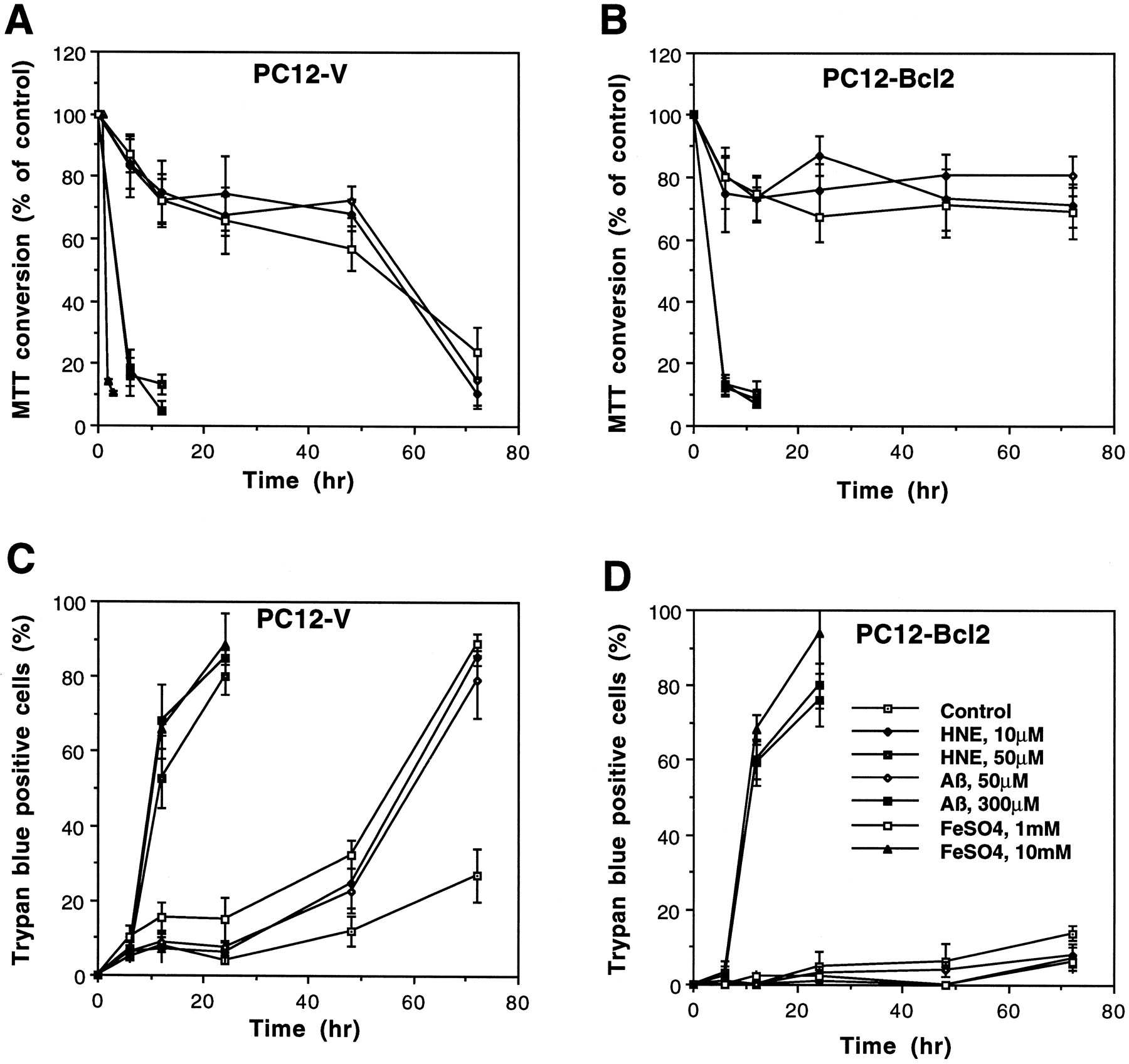
- Fig. 1.
Oxidative insults and HNE induce rapid and delayed cell death in PC12 cells: Bcl-2 prevents delayed cell death.A, B, PC12-V (A) and PC12-Bcl2 (B) cells were exposed to the indicated concentrations of HNE, Aβ, or FeSO4 for the indicated time periods, and levels of MTT reduction were quantified (mean and SEM of determinations made in four culture wells/condition).C, D, PC12-V cells (C) and PC12-Bcl2 cells (D) were exposed to vehicle (Control) or the indicated concentrations of HNE, Aβ, orFeSO4, and trypan blue-positive and nonstaining cells were counted. Values are the mean percentage of trypan blue-positive cells in four separate cultures.
- Fig. 2.
Oxidative insults and HNE induce apoptotic nuclear and plasma membrane alterations in PC12 cells: prevention by Bcl-2.A, Cultures of PC12-V cells and PC12-Bcl2 cells were exposed to vehicle (Control) or the indicated concentrations of HNE, Aβ, orFeSO4. At the indicated time points cells were stained with Hoescht dye, and the percentage of cells with condensed and fragmented (apoptotic) nuclei was determined (mean and SEM of determinations made in four separate cultures). No cells expressing Bcl-2 exhibited condensed and fragmented nuclei (PC12-Bcl2line represents combined data from cells exposed to each treatment condition). B, Annexin-V-positive cells were counted in PC12-V and PC12-Bcl2 cultures exposed for 4 hr to 10 μm HNE or for 16 hr to vehicle (Control) or 10 μm HNE. Values are the mean and SEM of determinations made in four separate cultures. *p < 0.001 compared with values for control cultures and PC12-Bcl2 cultures (ANOVA with Scheffé’spost hoc tests). C, PC12-V cell cultures were exposed for 48 hr to vehicle (Control) or the indicated aldehydes (10 μm). Cells were stained with Hoescht dye, and the percentages of cells with condensed and fragmented nuclei were determined. Values are the mean and SEM of determinations made in four separate cultures. *p < 0.001 compared with each of the other values (ANOVA with Scheffé’spost hoc tests). D, Confocal laser scanning microscope images of propidium iodide fluorescence in untreated control PC12-V cells (left), PC12-V cells exposed for 48 hr to 10 μm HNE (middle), and PC12-Bcl-2 cells exposed for 48 hr to 10 μm HNE (right). Note that many PC12-V cells treated with HNE exhibit DNA condensation and fragmentation (arrowheads), whereas cells expressing Bcl-2 did not.
- Fig. 3.
Oxidative insults and HNE induce apoptosis in primary hippocampal neurons: prevention by GSH. A, Cultures were exposed to 2 μm of each aldehyde (see Fig. 2C for aldehyde structures) or 0.2% ethanol (Control) for 16 hr, and percentages of neurons with condensed and fragmented nuclei were quantified. Values are the mean and SEM of determinations made in four separate cultures. *p < 0.05, **p < 0.001 compared with control value (ANOVA with Scheffé’s post hoc tests). B, Cultures were exposed for 16 hr to 0.2% ethanol (Control) or the indicated treatments, at which time cells were stained with Hoescht dye and the percentages of neurons with condensed and fragmented nuclei were quantified. Treatment concentrations were HNE, 10 μm; Aβ, 10 μm;FeSO4, 2 μm;GSH, 1 mm. Values are the mean and SEM of determinations made in four separate cultures. *p< 0.01 compared with control value; **p < 0.01, ***p < 0.05 compared with corresponding values for cultures not cotreated with GSH (ANOVA with Scheffé’spost hoc tests). C, Images of Hoescht dye fluorescence in hippocampal neurons from cultures exposed for 16 hr to 0.2% ethanol (Control), 2 μmHNE, 1 mm GSH-ethyl-ester plus 2 μm HNE (GSH+HNE), or 10 μmAβ. HNE and Aβ induced nuclear condensation and fragmentation in most neurons (arrowheads), whereas fluorescence remained uniformly distributed throughout the nucleus in neurons in control cultures and neurons in cultures cotreated with GSH and HNE (arrowheads).
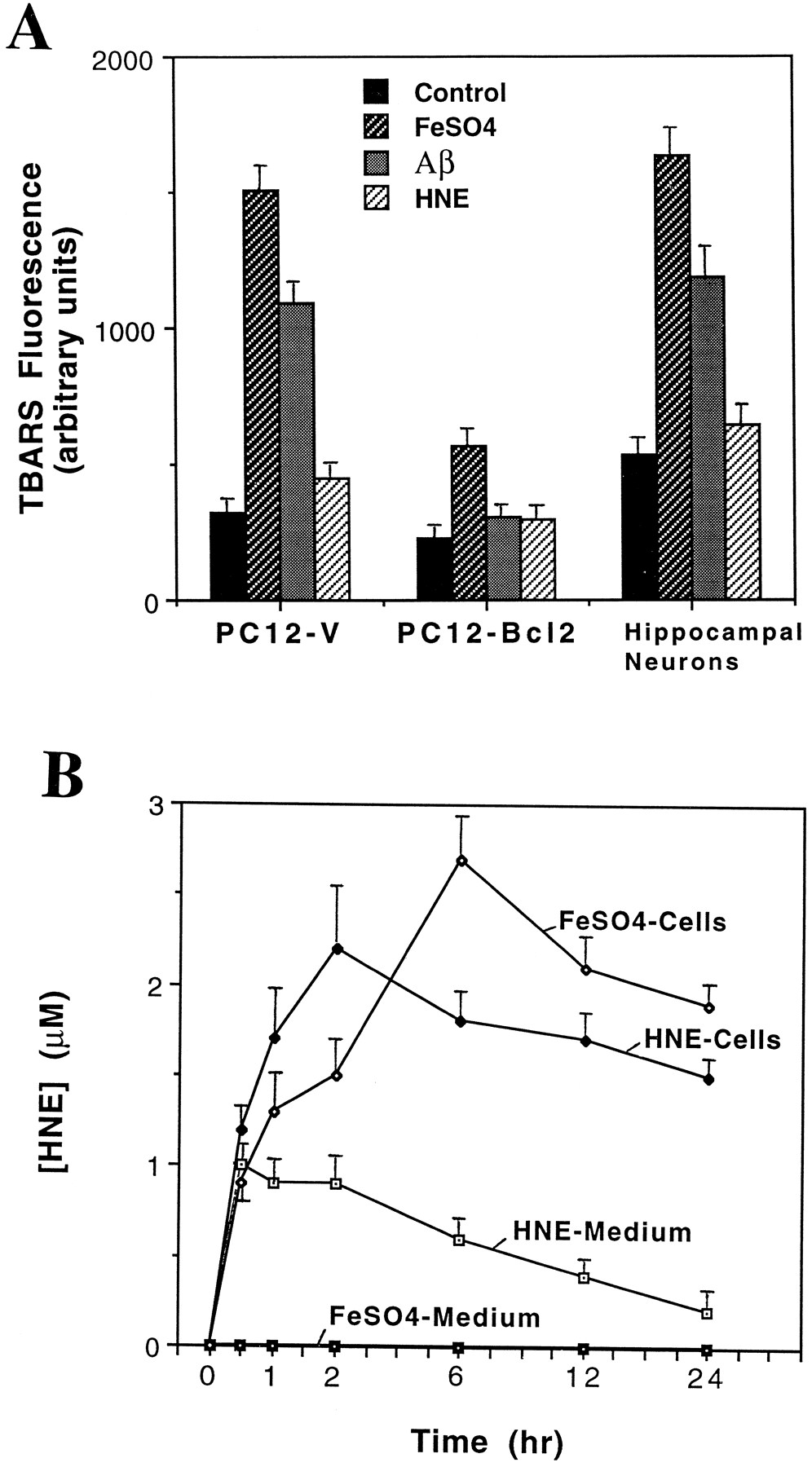
- Fig. 4.
Oxidative insults induce membrane lipid peroxidation in neural cells: suppression by Bcl-2. A, Levels of TBARS fluorescence were quantified in PC12-V cells, PC12-Bcl2 cells, and hippocampal neurons after 4 hr of exposure to vehicle (Control), FeSO4 (1 mm PC12 cells, 2 μm hippocampal neurons),Aβ (50 μm PC12 cells, 10 μm hippocampal neurons), or HNE (10 μm PC12 cells, 2 μm hippocampal neurons). Values represent the mean and SEM of determinations made in four to six separate cultures. B, Hippocampal cell cultures were exposed to 1 μm HNE or 2 μmFeSO4, and HNE levels in the culture medium and cells were quantified at the indicated time points. Values represent the mean and SEM of determinations made in four separate cultures.
- Fig. 5.
Oxidative insults induce formation of HNE–protein conjugates in neural cells: attenuation by GSH and Bcl-2.A, Hippocampal cultures were exposed for 2 hr to 0.2% ethanol (Control), 2 μmFeSO4, 2 μmHNE, or 1 mm GSH plus 2 μm HNE (GSH+HNE). Then cells were fixed and immunostained with HNE antibody. Arrowheads point to neuronal cell bodies.B, PC12-V cells and PC12-Bcl-2 cells were exposed for 2 hr to vehicle (0.2% ethanol) or 10 μm HNE. Then cells were fixed and immunostained with anti-HNE antibody. Note the much greater level of HNE immunoreactivity in PC12-V cells exposed to HNE, as compared with PC12-Bcl-2 cells exposed to HNE. C, Cell homogenates from untreated control PC12-V cultures (c), PC12-V cultures exposed to 10 μm HNE or 1 mmFeSO4 (v), and PC12-Bcl2 cultures exposed to 10 μm HNE or 1 mm FeSO4(b) were separated by SDS-PAGE (100 μg protein/lane). Then proteins were transferred to a nitrocellulose sheet and immunoreacted with an antibody against HNE–protein conjugates. Note that HNE and FeSO4 induced the appearance of many HNE–protein conjugates in PC12-V cells, but not in the PC12-Bcl2 cells. D, PC12-V cells, PC12-Bcl2 cells, and primary hippocampal neurons were exposed for 2 hr to 0.2% ethanol (Control), FeSO4 (1 mm for PC12 cells and 2 μm for hippocampal neurons), Aβ (50 μm for PC12 cells and 10 μm for hippocampal neurons), or HNE (10 μm for PC12 cells and 2 μm for hippocampal neurons). Then cells were fixed and immunostained with HNE antibody, and relative levels of HNE immunoreactivity were quantified (see Materials and Methods). Values represent the mean and SEM of determinations made in four separate cultures per condition (100 cells scored/culture).
- Fig. 6.
GSH protects PC12 cells against apoptosis induced by oxidative insults and HNE. A, Cultures of PC12-V cells were exposed for 70 hr to Vehicle, 10 μmHNE, 50 μmAβ, or 1 mmFeSO4 in the absence (Control) or presence of 1 mmGSH. Then cultures were fixed and stained with Hoescht dye, and the numbers of cells with condensed and fragmented nuclei were counted. Values are the mean and SEM of determinations made in four separate cultures. *p < 0.01 compared with vehicle control value; **p < 0.01 compared with corresponding control value (ANOVA with Scheffé’s post hoc tests). B, Cultures were exposed for 6 hr to vehicle (Cont), 50 μmAβ, 1 mmFeSO4, 10 μmHNE, or 300 μm buthionine sulfoximine (BSO). Then relative levels of GSH were determined by using the monochlorobimane fluorescence method. Values are the mean and SEM of determinations made in four separate cultures. *p < 0.01 compared with corresponding value for PC12-V cells (ANOVA with Scheffé’s post hoctests).


Tables
- Table 1.
Effects of macromolecular synthesis inhibitors, an endonuclease inhibitor, and antioxidants on apoptotic cell death induced by oxidative insults and HNE in PC12 cells
Insult Vehicle Cells with apoptotic nuclei (%) Cyclohex Act-D ATA VitE PG Cont (2 hr) 3 ± 1.2 1 ± 0.3 2 ± 0.6 1 ± 0.3 0 ± 0.3 1 ± 0.3 Cont (72 hr) 24 ± 4.7 11 ± 2.5 10 ± 3.3 9 ± 2.4 10 ± 2.1 13 ± 3.1 FeSO4 75 ± 3.2* 16 ± 2.71-160 22 ± 3.31-160 13 ± 3.31-160 13 ± 2.81-160 21 ± 2.11-160 Aβ 72 ± 3.3* 21 ± 3.41-160 23 ± 4.31-160 12 ± 2.21-160 11 ± 3.51-160 14 ± 3.21-160 HNE 71 ± 6.7* 29 ± 4.31-160 29 ± 4.01-160 26 ± 3.21-160 59 ± 3.3 65 ± 3.8 Cultures were pretreated for 2 hr with the indicated agents: 0.2% ethanol (Vehicle), 10 μm cycloheximide (Cyclohex), 5 μm actinomycin-D (Act-D), 100 μmaurintricarboxylic acid (ATA), 50 μg/ml vitamin E (VitE), 10 μm propyl gallate (PG), or 1 mmglutathione-ethyl ester (GSH). Some cultures were fixed at that point (Cont, 2 hr), while parallel cultures were exposed for 72 hr to 0.2% ethanol (Cont 72 hr), 1 mm FeSO4, 50 μm Aβ, or 10 μm HNE. Cells then were fixed and stained with Hoescht dye, and percentages of cells exhibiting nuclear condensation and fragmentation were determined. Values are the mean and SEM of determinations made in three or four separate cultures.
↵* p < 0.001 compared with value for control vehicle-treated (72 hr) cultures;
↵F1-160 p < 0.01 compared with value for vehicle-treated cultures exposed to the same insult. ANOVA with Scheffé’s post hoc tests. Preliminary studies showed that 10 μm cycloheximide reduced levels of protein synthesis by >90% during a 24 hr exposure period (data not shown).


