

Article Figures & Data
Figures
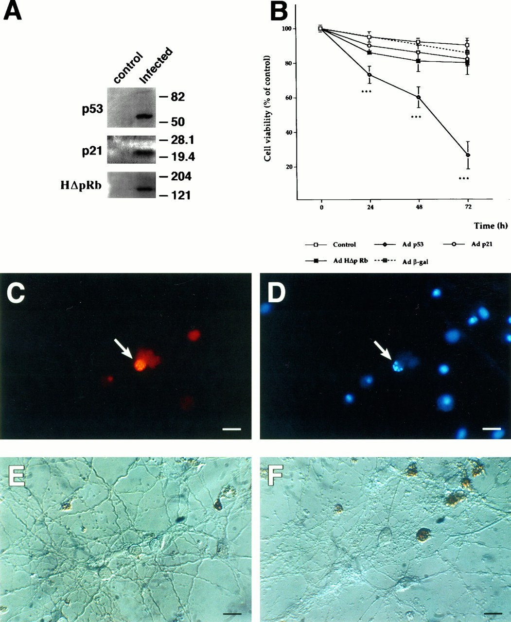
- Fig. 1.
Overexpression of p53 induces neurotoxicity in rat hippocampal pyramidal neurons. Hippocampal cultures were infected at 7 DIV, using 100 MOI of each virus. A, Western blots showing overexpression of p53, p21, and HΔpRb after 48 hr of infection. Hippocampal cultures were infected on 7 DIV, using 100 MOI of each virus. Similar results were found in three separate experiments. B, Time course plot of cell viability after Adp53, Adp21, Adβgal, or AdHΔpRb infection of hippocampal pyramidal neuron cultures. Results represent the mean ± SEM of 12 coverslips. ***p < 0.001 versus control conditions (no virus); ANOVA and Tukey’s test. C,D, Pictures showing colocalization of human p53 expression (C) and chromatin fragmentation measured using Hoechst 33342 (D, n> 200 cells). E, F, Cultures stained using the TUNEL technique illustrating the degree of double-stranded DNA breaks in control cultures (E) and 48 hr after Adp53 infection (F). Scale bar, 20 μm.
- Fig. 2.
p53-Induced gene expression. Hippocampal pyramidal neurons (7 DIV) (A) or PC-3 cells (B) were infected with Adp53 at 100 MOI. Later (8, 12, or 24 hr), the cells were harvested. Result shown illustrate effects at 12 hr, but similar patterns were seen at other time points. Similar results were found in three separate experiments. In addition, no increase in bax was observed at a 48 hr time point in hippocampal cultures.C, No changes in levels of the proteins were found in extracts from x-irradiated hippocampal neurons (500 cGy; immunoblots show the 24 hr time point).
- Fig. 3.
Time course plot of cell viability after exposure of the hippocampal pyramidal neuron cultures to 200, 500, and 1000 cGy total doses of x-irradiation. Cultures were irradiated at 11 DIV. Each point represents the mean ± SEM of 9 coverslips. **p < 0.01; ***p < 0.001 versus non-x-irradiated control conditions; ANOVA and Tukey’s test.
- Fig. 4.
Apoptosis after x-irradiation of hippocampal pyramidal neuron cultures. The cultures were exposed to a 500 cGy dose of x-irradiation at 11 DIV and analyzed 24 hr later. A, Nomarski picture showing morphological changes (n = 8). B, Propidium iodide and fluorescein diacetate staining (n = 8). C, TUNEL staining illustrating the degree of double-stranded DNA breaks (n = 3). D, Chromatin of hippocampal nuclei from x-irradiated cultures stained with Hoechst 33342.Arrows in A–D illustrate examples of nuclei with condensed chromatin. E,F, Control (E) and 500 cGy irradiated hippocampal cultures (F) stained for p53 5 hr after x-irradiation (n = 2). Scale bar, 20 μm.
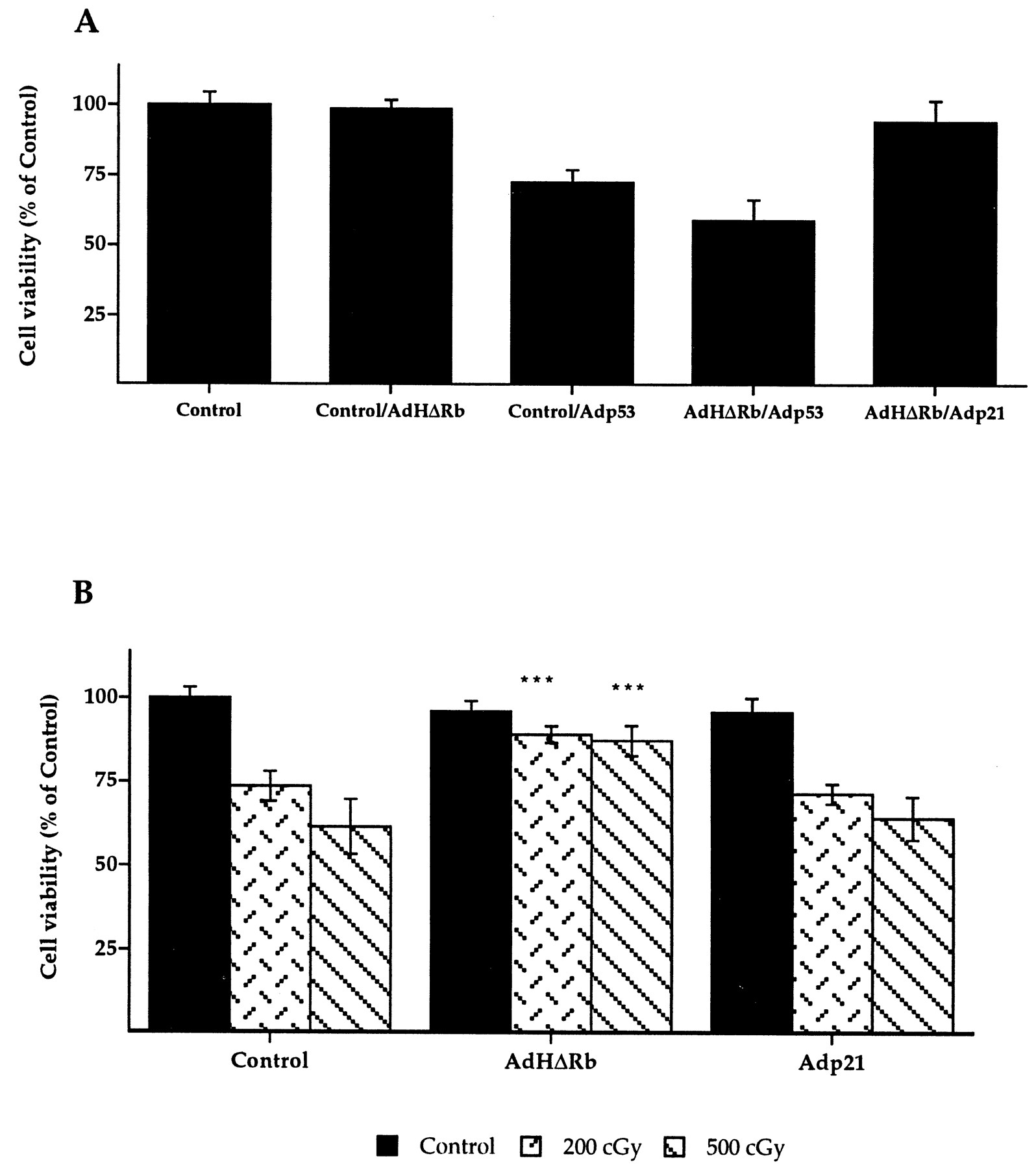
- Fig. 5.
Effects of the overexpression of HΔpRb on p53- and x-irradiation-induced cell death. Cultures were infected with 100 MOI AdHΔpRb at DIV 7, and 3 d later were again infected with Adp53 or exposed to x-irradiation. Cell viability was analyzed 48 hr (Adp53) or 24 hr (X-irradiation) later. A, Overexpression of HΔpRb failed to protect the cultures against cell death induced by p53 expression (AdHΔpRb/Adp53). B, Overexpression of HΔpRb was able to block x-irradiation-induced death of hippocampal neurons (AdHΔpRb), whereas p21 overexpression did not (Adp21). Data represent mean ± SEM of 9 coverslips. ***p < 0.01, versus Adp53 or irradiated control conditions; ANOVA and Tukey’s test.
- Fig. 6.
Effect of TGF-β1 (1 ng/ml) on neuronal death induced by either p53 overexpression or different doses of x-irradiation. A, 24 Hr pretreatment with TGF-β1 (Adp53/TGFβ(1X)) failed to protect the cultures 48 hr after 100 MOI Adp53 infection. Even the daily addition of the TGF-β1 for 3 consecutive days (Adp53/TGFβ(3X)) did not protect neurons 48 hr after Adp53 infection. Cultures were infected at 100 MOI, at 7 DIV.B, The 4 hr previous addition to the culture media of TGF-β1 (1 ng/ml) protected hippocampal pyramidal neuronal cultures against x-irradiation-induced neurotoxicity. Data represent mean ± SEM of 9 coverslips. **p < 0.01 versus Adp53 or irradiated control conditions; ANOVA and Tukey’s test.


