

Article Figures & Data
Figures
- Fig. 1.
Characterization of the anti-phospho-c-Jun antiserum. a, GST-c-Jun(1–223) (lanes 1, 2) or GST-c-Jun(1–223, A63/73) (lanes 3, 4) were (+) or were not (−) phosphorylated by recombinant JNK2 in the presence of [32P]ATP.(Top to bottom) first panel, Autoradiogram of32P-labeled proteins exposed either overnight (o/n) or (second panel) exposed only for 2 min. Third panel, The same blot was probed with affinity-purified phospho-c-Jun (α-P-cJun) antibody. Fourth panel, The blot was stripped and reprobed with the c-Jun antiserum (α-cJun).b, Samples (0.1 μg, lanes 2, 3; 1.0 μg, lanes 1, 4) of recombinant full-length c-Jun (Deng and Karin, 1992) were not (lanes 1, 2) or were phosphorylated (lanes 3, 4) with recombinant JNK2 in the presence of [32P]ATP (lanes 3, 4). (Top to bottom) first panel, Autoradiogram of the 32P-labeled proteins. Second panel, Immunoblotting with the phospho-c-Jun antiserum (α-P-cJun). Third panel, The blot was stripped and treated with buffer containing heat-inactivated calf intestinal phosphatase (
 ) or (fourth panel) native CIP (40 U/ml).Fifth panel, After final stripping, the blot was reprobed with the c-Jun antiserum (α-cJun).c, Immunodetection of phosphorylated c-Jun in immortalized 3T3 fibroblasts derived from wild-type (lanes 1, 2) or c-jun−/− mouse embryos (lanes 3, 4) (Hilberg et al., 1993) or c-jun−/−cells stably transfected with a human c-jun expression vector (lanes 5, 6), which were (+) or were not (−) UV-irradiated. The membrane was probed with the phospho-c-Jun antiserum (α-P-cJun) or c-Jun antibody (α-cJun). d, Detection of phosphorylated c-Jun in nuclear cortical extracts from untreated rats (lane 1) or after ischemia with 24 hr reperfusion (lane 2) by immunoblotting with the anti-phospho-c-Jun or anti-c-Jun antiserum.
 - Fig. 2.
Immunodetection of c-Jun N-terminal phosphorylation in the adult rat brain. c-Jun-IR (a–d) and phospho-c-Jun-IR (e–h) in the mamillary nucleus (mm) are shown: a, e, untreated rats;b, f, 5 d after transection of the mamillothalamic tract; c, g, competition by preincubation of the antibodies with 100 pmol of the phosphorylated c-Jun peptide; d, h, preincubation of the section with 1.2 μU alkaline phosphatase before incubation with the antiserum; longitudinal (i) and (j) coronal aspect of the location site of the medial forebrain bundle (MFB) and mamillothalamic tract (MT) transection at bregma −2.3 and 1.5 mm lateral from midline. Scale bar, 200 μm.
- Fig. 3.
Phospho-c-Jun-IR in the SNC. a–c, c-Jun-IR and (d–f) phospho-c-Jun-IR in the SNC of (a, d) untreated animals, (b, e) 5 d or (c, f) 20 d after transection of the medial forebrain bundle. The dotted line separates the pars compacta (p.c.) and the pars reticularis (p.r.). Arrows mark labeled nuclei. Scale bar, 100 μm.
- Fig. 4.
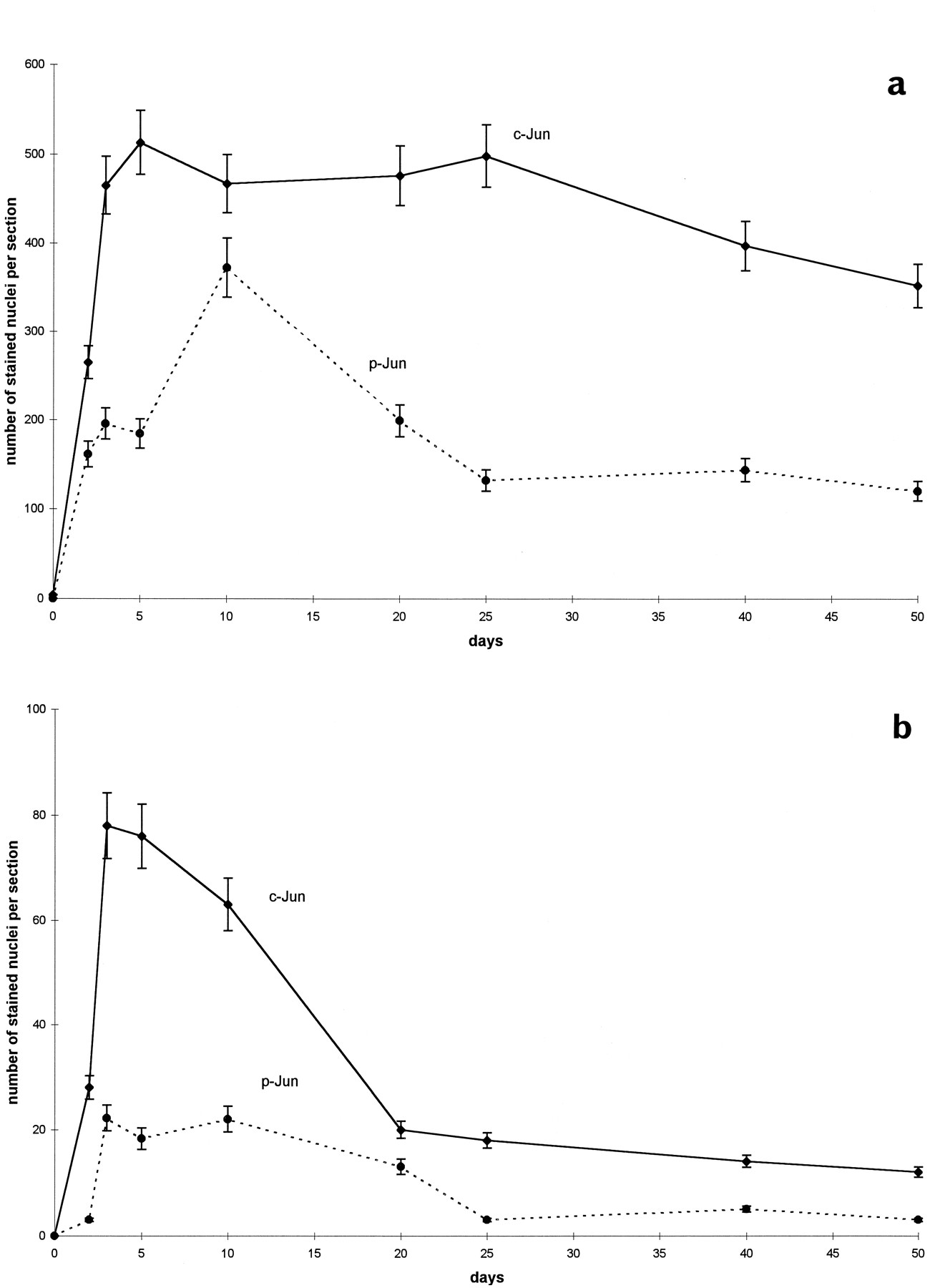
Time course of c-Jun and phosphorylated c-Jun in SNC and MnM after axotomy. Mean (±SD) of nuclei (per 50 μm section) labeled by (a) c-Jun and (b) phospho-c-Jun in the SNC (dotted line) and MnM (solid line) after transection of the medial forebrain bundle and mamillothalamic tract, respectively.
- Fig. 5.
Expression and phosphorylation of c-Jun, and TUNEL staining after MCA occlusion. Shown are (a, b) c-Jun-IR, (c, d) phospho-c-Jun-IR, and (e, f) TUNEL reaction in the ipsilateral (a, c, e) and contralateral (b, d, f) piriform cortex of consecutive sections after MCA occlusion with reperfusion for 3 d. Scale bar, 75 μm.
- Fig. 6.
Co-labeling of c-Jun phosphorylation and TUNEL. Shown is double-immunofluorescence of (a, b) phospho-c-Jun and (c, d) TUNEL in the superficial layer of the ipsilateral piriform cortex 12 hr (a, c) and 3 d (b, d) after MCA occlusion.Arrows indicate some of the double-labeled nuclei.e, Numbers of neurons labeled by TUNEL (white bars) and phospho-c-Jun (black bars) in the piriform cortex ipsilateral to the site of ischemia (between bregma −1.30 and −2.30). The time course gives the reperfusion period after MCA occlusion, which lasted 90 min. The numbersrepresent mean (±SD) calculated from nine 35-μm-thick sections (three sections each of three rat brains per time point). Thegray bars give the proportion of TUNEL or phospho-c-Jun-labeled neurons that are co-labeled with phospho-c-Jun or TUNEL, respectively.
- Fig. 7.
c-Jun expression and phosphorylation after pentylenetetrazole-induced seizures. a, b, Expression of c-Jun and (c, d) phosphorylation of c-Jun in the dentate gyrus (dg) of (a, c) untreated rats and (b, d) 15 min after injection of PTZ. py, Pyramidal layer. Scale bar, 200 μm.
- Fig. 8.
Expression of Fas-ligand in the penumbra after ischemia. Fas-ligand immunoreactivity in the piriform cortex (a) adjacent to the necrotic area that is marked by the dotted line and (b) in the contralateral intact cortex. Scale bar, 200 μm.
- Fig. 9.
Phospho-c-Jun, Fas-ligand, and TUNEL in the SNC. Shown are (a, b) phospho-c-Jun immunoreactivity, (c, d) Fas-ligand immunoreactivity, and (e, f) TUNEL staining in the ipsilateral SNC 3 d after (a, c, e) MCA occlusion or (b, d, f) 10 d after transection of the MFB. Scale bar, 100 μm.
- Fig. 10.
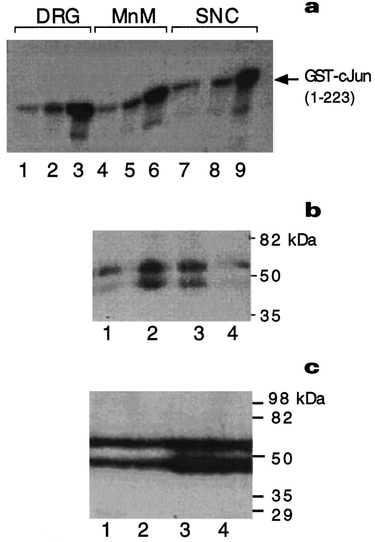
Activation of JNK. a, JNK-1 assay with GST-c-Jun (1–223) as substrate from dorsal root ganglia (DRG) extracts after sciatic nerve cut (lanes 1–3), in MnM after MT transection (lanes 4–6), and in SNC after MFB transection (lanes 7–9). Tissues were isolated from untreated controls (lanes 1, 4, 7), 3 d (lanes 2, 5, 8), or 12 d (lanes 3, 6, 9) after axotomy.b, In-gel kinase assays using GST-c-Jun (1–79) as substrate were performed with nuclear extracts from hippocampus and cortex isolated from untreated rats (lane 1) or 5 min (lane 2), 10 min (lane 3), and 90 min (lane 4) after intraperitoneal injection of pentylenetetrazole. c, In-gel kinase assay using GST-c-Jun (1–79) as substrate from hippocampus or piriform cortex microdissected from untreated rats (lanes 1, 2) or 24 hr after ischemia–reperfusion (lanes 3, 4). The autoradiograms in b and c did not contain additional bands.


