

Article Figures & Data
Figures
- Fig. 1.
Thionine (Nissl)-stained horizontal cryostat sections from stage 28 embryos treated with control antibodies (12-μm-thick section) (A) or with Fab fragments of a monoclonal antibody against N-cadherin (16-μm-thick section) (B). Arrowheads inB point to the borders of morphological changes (rosettes) in the diencephalon and in the tectum induced by N-cadherin blockage. c, Caudal; Di, diencephalon;ml, mantle layer; r, rostral;Tec, tectum; Tel, telencephalon;V, ventricle; vl, ventricular layer. Scale bar, 0.5 mm.
- Fig. 2.
Immunohistochemical characterization of the rosettes. Double-immunostained 12-μm-thick horizontal sections through the tectum of a stage 28 embryo treated with control rat IgG (A, B, E,F, I, K, N,O) or with the monoclonal antibody NCD-2 against N-cadherin (C, D, G,H, L, M, P,R). Double-immunostaining results are shown for N-cadherin (N-cad; A, C) and ZO-1 (B, D), for N-cadherin (E, G) and bromodeoxyuridine (BrdU) (F,H), for BrdU (I, L) and G4 (K, M), and for N-cadherin (N, P) and a radial glia-specific epitope (with the antibody R5) (O, R).Large arrows point to ependymal lining at the center of the rosettes. The small arrow in A points to the ependymal lining (ep). The small arrows in D point to blood vessels that express ZO-1. The small arrows in M point to G4/Ng-CAM-immunoreactive areas in the vicinity of the ventricle. Thesmall arrows in R point to radial glial processes in the mantle layer reaching the pial surface.ml, Mantle layer; v, ventricle;vl, ventricular layer. Scale bars: D, 0.1 mm (A–M); R, 0.05 mm (N–R).
- Fig. 3.
Regional formation of rosettes in the diencephalon. In situ hybridization and thionine (Nissl) staining of 12-μm-thick horizontal sections of a stage 28 embryo treated with control rat IgG (A, C,E, G) or with a monoclonal antibody against N-cadherin (NCD-2) (B, D,F, H). In situhybridization results are shown for N-cadherin (N-cad;A, B), for R-cadherin (R-cad; C, D), for the transcription factor Gbx-2 (Gbx-2; E,F), and for a thionine stain (Thionin; G, H). The arrows in B mark the borders of the morphological changes in the dorsal thalamus. Note that only the dorsal thalamus is severely affected. The arrowheads inG and H mark the borders between the ventral thalamus (vt) and the dorsal thalamus (dt) and between the dorsal thalamus and the pretectum (pt), respectively. c, Caudal;r, rostral; V, ventricle. Scale bar, 0.2 mm.
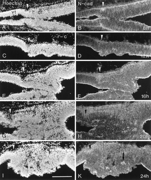
- Fig. 4.
Time course of the rosette formation in the diencephalon. Horizontal 14-μm-thick sections were immunostained for N-cadherin (N-cad; B, D,F, H, K) and counterstained with the nuclear dye Hoechst 33258 (Hoechst; A, C,E, G, I). Embryos were injected with antibody NCD-2 at stage 24 (E4) and allowed to survive for different lengths of time (8 hr, A,B; 12 hr, C, D; 16 hrE, F; 20 hr, G,H; 24 hr, I, K). Note that in some cases, a fusion of the two opposing diencephalic walls is seen (G, H).Arrowheads point to the cell-sparse area of zona limitans intrathalamica at the border between the ventral thalamus (vt in A) and the dorsal thalamus (dt in A). The arrow inK points to the center of a rosette. c, Caudal; r, rostral. Scale bar, 0.2 mm.
- Fig. 5.
A–D, Late effects of N-cadherin blockage on tectal development. Sections through the tectum of an E10–E11 chicken embryo injected at E4 and E5 with control rat IgG (A) or with a monoclonal antibody against N-cadherin (B–D) are shown. Consecutive sections were stained for Nissl substance with thionine (Thio;A, B), hybridized in situwith cRNA probe for N-cadherin (N-cad;C), and immunostained with a monoclonal antibody against axonin-1 (ax-1; D). Note the additional layers between remnants of the ependymal lining (arrowsin B) and the ventricular surface of the NCD-2-injected brains. Small arrowheads in C point to large multipolar N-cadherin-expressing ganglion cells reminiscent of those normally found in layer IV (Redies et al., 1993). In the NCD-2-injected brains, cells with a similar appearance are present in an additional layer close to the ventricle (large arrowheads in C). Small arrowheads in D point to axonin-1 immunoreactivity reminiscent of that normally seen in layer III (Yamagata et al., 1995). Note that in addition, axonin-1 immunoreactivity can also be seen immediately below the ventricular surface of NCD-2-injected tecta (large arrowheads inD). The ventricular surface lacks an ependymal lining (large arrowheads in B).E–J, Late effects of N-cadherin blockage on the development of the dorsal thalamus. Frontal sections through the diencephalon from a noninjected E11 control embryo (E–G) and from an E10–E11 embryo injected with monoclonal antibody NCD-2 against N-cadherin (H–J) are shown. Consecutive sections were Nissl-stained with thionine (Thio;E, H), or immunostained with antibodies against cadherin-7 (cad7; F,I) and R-cadherin (R-cad;G, J). The arrow inG points to the border between the ventral thalamus (VT in E) and the dorsal thalamus (DT in E). This border is marked by the R-cadherin-positive zona limitans (zl inG) (Gänzler and Redies, 1995). The dorsal thalamus, but neither the habenular area of the epithalamus nor the ventral thalamus, is severely distorted by the antibodies against N-cadherin. Arrows in J point to remnants of the ependymal lining, which is missing from most of the ventricular surface of the dorsal thalamus. Note that in the NCD-2-injected brains, gray matter is fragmented and fused at the midline in the dorsal thalamus. Like parts of the dorsal thalamic complex (DCin F), some of the fragments express cadherin-7 and R-cadherin. d, Dorsal; HM, nucleus habenularis medialis; NE, neuroepithelium;SMe, stria medullaris; Tel, telencephalon; tV, tectal ventricle; v, ventral; v3, third ventricle; I-X, tectal layers at E10–E11 (LaVail and Cowan, 1971). Scale bars:D, 0.1 mm (A–D);J, 0.5 mm (E–J).
- Fig. 6.
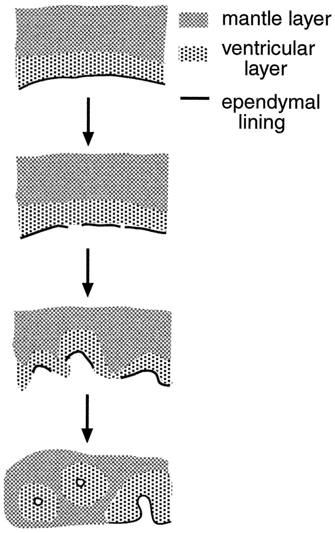
Schematic representation of rosette formation induced by N-cadherin blockage. The top panel represents the normal neuroepithelium, and the three bottom panelsrepresent different time points after antibody injection. Rosette formation starts with a breaking up of the ventricular lining, followed by a folding in of parts of the neuroepithelium, finally resulting in complete rosettes of neuroepithelial tissue. Rosettes are composed of ependymal lining in their centers (solid lines) surrounded by an inner “ventricular” layer of proliferating cells and an outer “mantle” layer of postmitotic neurons (shading).
- Fig. 7.
Hypothetical model to explain early and late morphological changes attributable to N-cadherin blockage. In control brains, neural precursor cells (mitotic cells) proliferate in the ventricular layer. Postmitotic early neurons migrate outward into the mantle layer, presumably along radial glia processes. The end feet of the neuroepithelial cells are tightly connected to each other by N-cadherin-associated adherens junctions, forming the ependymal lining. After N-cadherin blockage, these junctions break up in susceptible regions (see Discussion). Remnants of intact ependymal lining round up and are drawn into the neural tube wall. They become surrounded by proliferating cells. Radial glial processes extend in all directions. Newly generated neurons use these cues to migrate outward in all directions including to the ventricular surface. Consequently, extra gray matter layers are deposited in the vicinity of the ventricle, as seen in the tectum. In the dorsal thalamus, newly generated neurons that normally migrate into the same approximate area of the mantle layer are now also misguided into other directions and aggregate in smaller gray matter fragments.


