

Article Figures & Data
Figures
- Fig. 1.
Generation and characterization ofBcl-xL transgenic mice. A, Schematic presentation of the Bcl-xL transgene construct: RI, EcoRI; N,NotI; S, SacI;Sa, SalI; X,XhoI; Xb, XbaI.B, Southern blot analysis of the different transgenic lines. The 6 kb fragment corresponds to the endogenousBcl-xL gene. The 0.8 kb fragment corresponds toBcl-xL transgene. Lines 7193,7199, and 7194 had the highest copy numbers and were used for further analysis.
- Fig. 2.
The Bcl-xL transgene is expressed at high levels in the facial motor nucleus of line 7193.A, Section through the brainstem of a transgenic mouse hybridized with a probe for Bcl-xL. This probe detects endogenous and transgenic Bcl-xL mRNA, both of which are expressed throughout the brainstem and facial nucleus. B, Adjacent section from the same animal as inA, hybridized with the SV40 probe, which detects only the transgene-derived Bcl-xL mRNA. Note the high levels of expression of SV40 in motor neurons of the facial nucleus (arrows). C, Brainstem section of a wild-type littermate hybridized with the SV40 probe. Note that no signal was detected in the facial nucleus. Scale bar, 0.5 mm.
- Fig. 3.
The Bcl-xL transgene is expressed at high levels in the forebrain of line 7194.A, Section through the forebrain of a transgenic mouse hybridized with a probe for Bcl-xL. This probe detects both the endogenous and transgene-derivedBcl-xL. B, Adjacent section from the same brain as in A, hybridized with the SV40 probe, which detects only transgene-derived Bcl-xLmRNA. Note the high levels of transgene expression in the cerebral cortex (arrows) and thalamic nuclei. Also note the high expression in CA1, CA2 and CA4 regions of the hippocampus, and the minimal expression in the dentate and CA3 regions. Scale bar, 1 mm.
- Fig. 4.
Bcl-xL overexpression protects motor neurons from axotomy-induced cell death. A, Ret-labeled motor neurons in the facial nucleus of a nonaxotomized wild-type mouse. Note the labeling in the medial and lateral (circled) portion of the nucleus. Inset, High-magnification view of Ret labeling in wild-type neurons. B, Ret-labeled motor neurons in the facial nucleus of an axotomized wild-type mouse. Cells in the lateral portion of the nucleus have degenerated (circled), and Ret is no longer detectable except in the medial portion of the nucleus, which is not affected by this lesion paradigm. C, Ret-labeled motor neurons in the facial nucleus of an axotomized transgenic Bcl-xL mouse (line 7193). Many lateral motor neurons survive axotomy (circled), although they are reduced in size.Inset, High-magnification view of Ret labeling in the rescued lateral lateral motor neurons. (Compare the size of motor neurons in the inset in C with the insetin A.) D, Cresyl violet-stained motor neurons in the facial nucleus of an axotomized transgenicBcl-xL mouse. E, High-magnification bright-field view of facial motor neurons 7 d after axotomy in transgenic line 7194. Arrows indicate rescued motor neurons. F, Dark-field view of the same section as in E, labeled with the SV40 probe to detect transgene-derived sequences. Note the high degree of correlation between the rescued motor neurons shown in E and the expression of transgene-derived Bcl-xL shown inF (arrows). Scale bars:A–D, 100 μm; E–F, insets, 25 μm.
- Fig. 5.
Quantification of facial motor neuron rescue in transgenic line 7193. The bar graph shows numbers of facial motor neurons in control and axotomized wild-type andTα1-Bcl-xL transgenic mice. Note the large difference in numbers of facial motor neurons that survive axotomy between wild-type and trangenic animals. Asteriskindicates statistically significant differences (p < 0.05).
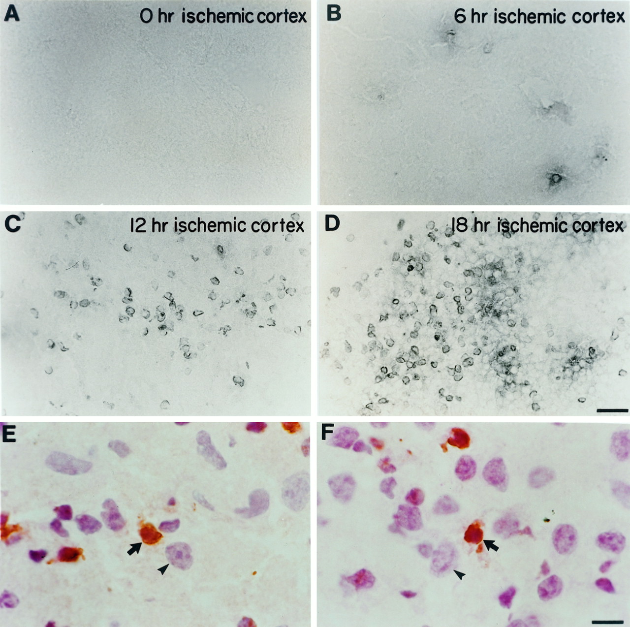
- Fig. 6.
TUNEL labeling after unilateral carotid ligation and exposure to hypoxia. At different time points after hypoxia–ischemia, the cortex ipsilateral to carotid ligation (ischemic cortex) was assessed for the presence of TUNEL-positive cells in P7 mice. A, There were no TUNEL-positive nuclei at 0 hr. B, Occasional TUNEL-positive nuclei began to appear at 6 hr. C, There was an increase in TUNEL labeling at 12 hr, which reached a peak at ∼18 hr (D). Higher-power photomicrographs counterstained with hematoxylin and eosin demonstrate that TUNEL labeling is nuclear. E–F, Arrows point to shrunken TUNEL-positive nuclei (brown) that are also stained with hematoxylin in the cortex 12 hr after hypoxia–ischemia. The arrowheads point to larger, normal-appearing neuronal nuclei adjacent to the smaller, TUNEL-positive nuclei. Scale bars: A–D, 30 μm; E–F, 7.5 μm.
- Fig. 7.
Electron microscopy reveals evidence of apoptosis after hypoxic–ischemic injury. A, Example of a cell from the cortex of a P7 mouse brain ipsilateral to carotid ligation, 12 hr after exposure to 8% oxygen. There is condensed chromatin (arrows) within the nucleus. B, There is a normal-appearing neuronal nucleus in the P7 mouse cortex contralateral to carotid ligation 12 hr after exposure to 8% oxygen. Scale bar, 5 μm.
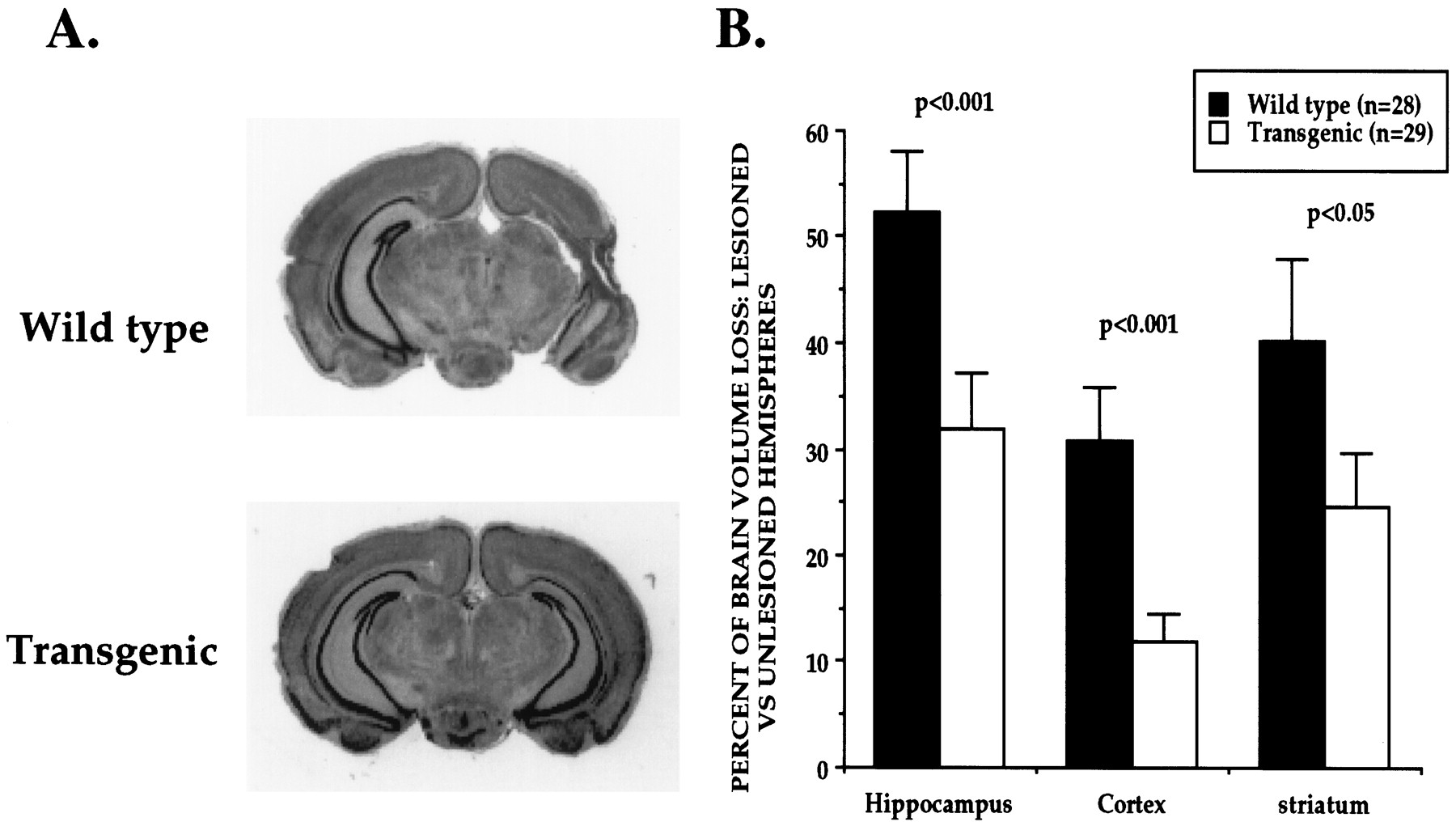
- Fig. 8.
Overexpression of Bcl-xL protects the neonatal mouse brain from hypoxic–ischemic insults. A, Coronal sections of P14 mouse brains 1 week after unilateral (left) carotid ligation and exposure to hypoxia for 1 hr at P7. There was significantly less tissue damage in Bcl-xLtransgenic brains (line 7194) compared with wild-type littermates.B, Quantitative measures of volume loss in transgenic and wild-type animals. The volume of tissue loss in each brain region ipsilateral to carotid ligation (lesioned hemisphere) was compared in each animal with the volume of tissue remaining in the matching brain regions contralateral to carotid ligation (unlesioned hemisphere). The percent volume loss in each structure was determined in each animal, and data are presented as the mean ± SEM.


