

Article Figures & Data
Figures
- Fig. 1.
Neuronal distribution of the novel 4.1 homolog.In situ hybridization was performed on a 4-d-old whole-mount mouse and on adult mouse brain using a digoxigenin-labeled antisense probe corresponding to 285 bp of the novel 4.1 clone (aa 428–523 of m4.1N). A, There is marked enrichment of the novel 4.1 clone in the brain compared with elsewhere in the body.B, The mRNAs are found in almost all neurons of adult mouse brain. C, No signal was evident when sections were incubated with the corresponding sense cRNAs. Scale bars:A, 2.7 mm; B, C, 1.25 mm.
- Fig. 2.
Amino acid sequence of m4.1N. m4.1N is an 879 amino acid protein with sequence homology to 4.1R and 4.1G in the defined membrane-binding, spectrin-actin–binding, and C-terminal domains (domain boundaries are indicated by arrows). The amino acid alignment highlights identical sequence shared by the three proteins (boxed and shaded) and residues with conservative amino acid changes (shaded only). Sequence identities shared by 4.1R and 4.1G are boxedand shaded, whereas sequence identities specific to 4.1G and 4.1N are boxed only. Whereas there is additional m4.1R sequence between the MBD and SABD, the region between the SABD and CTD is expanded in m4.1N. The recently identified Ca2+-dependent calmodulin (CD-CaM)– and Ca2+-independent calmodulin (CI-CaM)–binding sites of 4.1R are located within the MBD. The original m4.1N PCR product obtained from mouse brain cDNA (start site marked by an arrowhead) did not contain two stretches of sequence present in the full-length clone (marked by + symbols and asterisks). m4.1N and h4.1N (accession number 2224617) share 95% amino acid identity. m4.1N sequence data are available from GenBank (accession number AF061283).
- Fig. 3.
Northern analysis of h4.1N. A, A multiple tissue Northern blot was incubated with a probe derived from the 3′-untranslated DNA of h4.1N. The most intense band was a 7.5 kb species identified in brain, which also contained 8.8 and 4.1 kb bands. h4.1N mRNAs were also detected in the heart, kidney, and pancreas, with lesser levels in the placenta, lung, and skeletal muscle; no hybridization was evident in the liver. Whereas the 7.5 kb band is enriched in brain, the 4.1 kb species predominates in peripheral tissues. B, A ubiquitin probe was used as a control for mRNA integrity.
- Fig. 4.
Localization of m4.1N mRNAs in the brain. A–D, High-power views of m4.1N in situ hybridization performed on adult mouse brain reveal robust neuronal labeling in the granule cell layer (GCL) of the cerebellum [note that the molecular layer (ML) is negative] and in deep cerebellar nuclei (DCN) (A), in all layers of the cerebral cortex (B), in the dentate gyrus (DG) and CA1–CA3 regions of the hippocampus (C), and in the striatum [note that the white matter tracts (indicated by black arrowheads) are negative] (D). E, Interestingly, no m4.1N mRNAs are detected in the thalamus except for neurons in the reticular thalamic nucleus (RTN).F, There is intense labeling of olfactory neurons in the olfactory epithelium (black arrow). G, m4.1N mRNAs are detected in enteric neurons (black arrowhead) throughout the gastrointestinal tract.H, The major non-neuronal localization of m4.1N mRNAs is in the kidney, where transcripts are detected in the convoluted and collecting tubule epithelia (white, black arrowheads, respectively). Scale bars:A–F, 200 μm; G, 50 μm; H, 100 μm.
- Fig. 5.
Distribution of m4.1N RNAs in the cerebral cortex during development. A, By embryonic day 11.5 (E11.5), m4.1N mRNAs are detected in the first group of postmitotic neurons that reach the subcortical plate (SP); proliferating progenitor cells in the ventricular zone (VZ) are negative. B, As more postmitotic neurons reach the subcortical plate byE13.5, the thickness of the cell layer increases, and there is likewise more m4.1N labeling. C,D, By E17 (C) andE18.5 (D), successive layers of neurons migrate beyond the subcortical plate to form the cortical plate (CP); whereas m4.1N mRNAs are abundant in the differentiating neurons of the cortical plate, the migrating cells of the intermediate zone (IZ) are negative.E, F, Sections of cortex from 4-d-old (P4; E) and 21-d-old (P21;F) mice reveal m4.1N mRNAs in neurons of all six cortical layers; the white matter of the corpus callosum (CC) is completely negative.
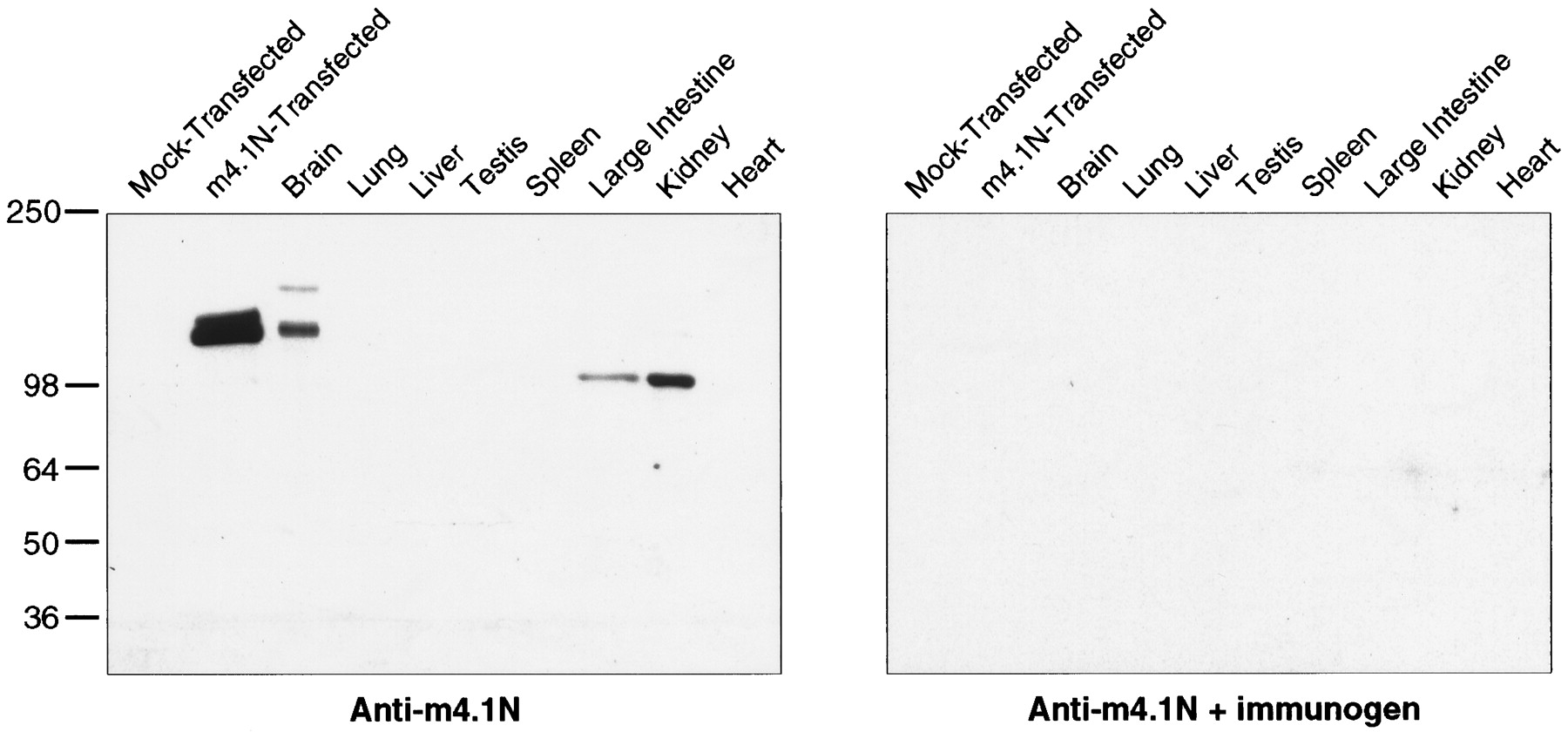
- Fig. 6.
m4.1N Western analysis. A polyclonal antibody was generated against a fusion protein containing the unique N terminal of m4.1N. Left, Western blot analysis of COS-7 cells transfected with the full-length m4.1N clone revealed a major band of ∼135 kDa. No proteins were recognized in control lysates from mock-transfected cells. In the brain, there is a faint 165 kDa band and a major 135 kDa band that comigrates with the expressed protein. Bands of 100 kDa are detected in the kidney and large intestine.Right, When a parallel blot is incubated with antibody preabsorbed with the m4.1N fusion protein, no bands are evident.
- Fig. 7.
Immunolocalization of m4.1N protein. Immunoperoxidase and indirect immunofluorescence staining was performed on floating 40 μm sections of adult rat brain using the m4.1N antibody; immunohistochemistry was also conducted on select peripheral tissues. A, Mitral cells of the olfactory bulb exhibit intense staining of the cell bodies and dendritic processes (black arrowhead); the nuclei are distinctly negative. Among the m4.1N-positive dendritic processes, discrete varicosities exhibit robust staining. B, The olfactory bulb is devoid of staining when incubated with antibody preabsorbed with the m4.1N fusion protein. C, An indirect immunofluorescence confocal image of the same region of the olfactory bulb demonstrates the dendritic labeling of mitral cells with areas of speckling.Inset, Profiles of dendrites in cross section indicate that m4.1N is found at the outer rim of the processes, consistent with a juxta-membrane localization. D, The granule cell layer (GCL) of the cerebellum exhibits a punctate pattern of immunostaining; there is also robust labeling of the molecular layer (ML). E, No signal is detected in the cerebellum when the preabsorbed antibody is used. F, An indirect immunofluorescence confocal image of the cerebellar granule cell layer demonstrates discrete spots of immunofluorescence in the synaptic glomeruli; a white arrow points to one of the punctate structures. G, In the hippocampus, there is a prominent reticular pattern seen in the granule cell layer of the dentate gyrus (DG) and among the pyramidal cells of CA1–CA3; there is also homogeneous speckling of the molecular layer of the dentate and CA3.H, No immunostaining is detected in the hippocampus with the preabsorbed antibody. I, An indirect immunofluorescence confocal image of the granule cell layer of the dentate gyrus reveals discrete spots of immunofluorescence outlining the cell bodies; a white arrow points to one of the punctate structures. The molecular layer (white arrowheads) exhibits a densely speckled pattern of immunofluorescence. J, Within the retina, there is prominent labeling of the apical dendrite and the cell body of bipolar cells. K, Between the smooth muscle layers of the gastrointestinal tract, there is specific immunostaining of the enteric neurons (black arrowheads). Inset, The punctate pattern of labeling is evident at high magnification. L, In the kidney, there is intense immunostaining of the convoluted tubule epithelia; the collecting tubule epithelia stain in a “checkerboard” pattern, such that alternating cells are positive. The cells of the glomeruli are completely negative (asterisks). Inset, The robust immunostaining of a tubular epithelial cell is shown at higher magnification. Scale bars, A, B,K, 40 μm; C, F, 10 μm;D, E, G, H, 160 μm; I, 12.5 μm; J, 16 μm;L, 80 μm.
- Fig. 8.
Enrichment of m4.1N at sites of synaptic contact in primary hippocampal cultures. Double-labeling indirect immunofluorescence of primary hippocampal cultures was performed using the m4.1N antibody and PSD95 (postsynaptic marker) and GluR1 (excitatory postsynaptic marker) antibodies. B,E, m4.1N is enriched at discrete foci along the neuronal dendrites. A–F, An overlay of the FITC-m4.1N immunolabeling (B, E) with the Texas Red GluR1 (A) and PSD95 (D) labeling reveals colocalization of m4.1N with the synaptic proteins at these sites (yellow foci in C,F). Scale bar, 5.3 μm.


