

Article Figures & Data
Figures
- Fig. 1.
Gephyrin, GlyR, and GABAAR distribution on motoneurons cultured alone. A, Immunoperoxidase showing the nuclear staining of the embryonic motoneuronal marker Islet1. B, Double-staining of the dendritic protein MAP2 (green) and the axon-enriched 155 kDa neurofilament protein (red).C–E, Presence of clusters (arrows) of gephyrin (C, red), GlyRα/β (D, red), or GABAARβ3 (E, green) on MAP2-IR somato-dendritic compartment (green in C, D;red in E). GlyRα/β clusters are also accumulated at the level of axon-hillock (arrowhead inD). F, G, Confocal visualization of Geph-IR (F1–2) and GlyRα/β-IR (G1–2), respectively. Discontinuous Geph-IR (arrows) at the cell-to-substrate contact (F1) and at a distance from the coverslip (F2). GlyRα/β forms clusters at the cell-to-substrate contact (arrows in G1) and occasionally displays a continuous labeling (arrows in G2) on sections passing through the nucleus. H, I, Double-immunofluorescence showing that Geph-IR clusters (H1, I1) accumulate in front of synapsin-IR boutons (arrowheads in H2) displaying ChAT-IR (arrowheads in I2). Geph-IR clusters (H1, I1) are also detected at nonsynaptic sites (arrows). In contrast, GlyRα/β (J1) and GABAARβ2/3 (K1) do not concentrate at synaptic sites (arrowheads in J1–2 andK1–2, respectively). Insets inH2, I2, J2, and K2 show superimposed images of H1–H2, I1–I2, J1–J2, andK1–K2, respectively. ChAT, Choline acetyltransferase;GABAARβ2/3, GABAARβ2/3 subunits-IR;GABAARβ3, GABAARβ3 subunits-IR; Geph, gephyrin-IR;GlyRα/β, GlyRα/β subunits-IR;MAP2, dendritic marker; NF, NF155 Kda-IR;Syn, synapsin-IR. Scale bar: A,B, 50 μm; C–K2, 10 μm.
- Fig. 2.
Simultaneous detection of gephyrin-IR (A1, A3, A5) and GlyRα/β-IR (A2, A4, A6) on motoneurons cultured alone for 7 DIV. Motoneurons display large and small, round-shaped Geph-IR clusters. Large Geph-IR clusters do not colocalize with GlyRα/β-IR (crossed arrows). Most (arrowheads) but not all (arrows) small Geph-IR clusters colocalize with GlyRα/β-IR clusters.Geph, Gephyrin-IR; GlyRα/β, GlyRα/β subunits-IR. A1–2, Pairs of digitized images acquired with CCD camera (A1, FITC channel;A2, TRITC channel). A3–4, A5–6, Higher magnification of the plain and dotted outlined regions in A1–2, respectively. Scale bar:A1–2, 10 μm; A3–4,A5–6, 1.2 μm.
- Fig. 3.
Comparison of the functional and pharmacological properties of GlyRs in cultures of purified motoneurons and all spinal neurons. A–C, Concentration–response curves in both types of cultures (purified motoneurons: A,C, bars labeled MN; cultures of all spinal neurons: B, C, bars labeledSN). The internal solution and membrane potential were solution A and −20 mV (A–C, bars labeledMN2 and SN) or solution B and −70 mV(C, bars labeled MN1). The left traces in A or B show superimposed current recordings obtained during successive applications of glycine at increasing concentrations (10–160 μm). Theright graph in A or B(obtained from the corresponding left traces) shows for each cell the fit of the concentration dependence of the peak response by a Hill equation: y =Imax/(1 + (EC50/x)nH). The values and corresponding errors given by the computer for EC50, nH, andImax for each of these cells were, respectively, 46.7 ± 3.4 μm, 1.92 ± 0.16 and 0.744 ± 0.035 nA in A, and 47.7 ± 0.9 μm, 2.16 ± 0.06 and 0.833 ± 0.011 nA inB. C, Mean values and SD for these three parameters, derived from several such experiments performed in different cells under identical conditions. D, Voltage dependence of the responses of purified motoneurons to glycine (internal solution A). Glycine (10 μm) was applied for 2 sec at different test potentials, using long voltage jumps from the holding potential (−20 mV) toward the test potential (the voltage jump beginning 0.8 sec before each glycine application). Left graphs,Normalized I–V curves for three different motoneurons, obtained by dividing each glycine response by the response of the corresponding cell at −60 mV. Right traces, Records obtained in one of these cells during voltage jumps to −80 and +80 mV.E, Strychnine sensitivity of glycine responses (internal solution A, holding potential −20 mV). Left panel, Mean values of the percentage of inhibition of the peak glycine responses by strychnine (50 or 500 nm applied with preincubation) in cultures of purified motoneurons (white bars) and in cultures of all spinal neurons (hatched bars). The glycine concentration was either 40 μm (close to the EC50) or 100 μm. Middle panel, Effect of strychnine applied with preincubation first at 50 nm, then at 500 nm, on the response to glycine (40 μm) of a purified motoneuron (MN). Right panel, Effect of 500 nm strychnine, applied first without preincubation, then with preincubation, on the response to glycine (100 μm) in a culture of all spinal neurons (SN).
- Fig. 4.
Gephyrin and GABAAR but not GlyR form postsynaptic clusters on motoneurons cocultured with DRG neurons. In all pairs of images, motoneurons are identified by the nuclear Islet1 staining. A, B, Geph-IR is concentrated in front of synapsin-IR boutons (arrowheads in A1–2) displaying GAD-IR (arrowheads in B1–2).C, D, GABAARβ2/3 accumulate at synaptic (arrowheads in C1–2) and nonsynaptic loci (crossed arrow in C1–2). Geph-IR clusters colocalize with GABAARβ3 clusters (arrowheads in D1–2). E, F, GlyRα/β (E1, F1) form clusters (arrowheads) that are not adjacent to synapsin-IR (E1–2, crossed arrows) or GAD-IR (F1–2, crossed arrows) boutons.GABAARβ2/3, GABAARβ2/3 subunits-IR;GABAARβ3, GABAARβ3 subunits-IR; GAD, GAD67-IR;Geph, gephyrin-IR, GlyRα/β, GlyRα/β subunits-IR; Isl, Islet1-IR;Syn, synapsin-IR. A1–A2, B1–B2, C1–C2, D1–D2, E1–E2, F1–F2, Pairs of digitized images acquired with CCD camera (A1, B1, C1, D1, E1, F1, TRITC channel;A2, B2, C2, D2, E2, F2, FITC channel). Scale bar, 10 μm.
- Fig. 5.
Gephyrin, GlyR, and GABAAR distribution on motoneurons cocultured with spinal neurons. In all pairs of images, motoneurons are identified by the nuclear Islet1 staining. A–C, Accumulation of Geph (A2), GlyRα/β (B2), and GABAARβ2/3 (C2) in front of most (arrowheads) but not all (crossed arrows) synapsin-IR boutons (A1, B1, C1). D–F, Double-staining of GAD (D1, E1, F1) and Geph (D2); GlyRα/β (E2) or GABAARβ2/3 (F2) show closely apposed signals (arrowheads). Some Geph and GlyRα/β spots are not adjacent to GAD-IR boutons (crossed arrows inD1–2, E1–2, respectively). G, H, Double-immunofluorescence showing that most GABAARβ3-IR spots (arrowheads in G1, H1) are associated with Geph-IR (G2) and GlyRα/β-IR (H2) clusters. Few Geph-IR clusters do not colocalize with GABAARβ3-IR (crossed arrows inG1–2).GABAARβ2/3, GABAARβ2/3 subunits-IR;GABAARβ3, GABAARβ3 subunits-IR; GAD, GAD67-IR;Geph, gephyrin-IR; GlyRα/β, GlyRα/β subunits-IR; Isl, Islet1-IR;Syn, synapsin-IR. A1–A2, B1–B2, C1–C2, D1–D2, E1–E2, F1–F2, G1–G2, H1–H2, Pairs of digitized images acquired with CCD camera (A1, B1, C1, D1, E1, F1, G1, H1, FITC channel; A2, B2, C2, D2, E2, F2, G2, H2, TRITC channel). Scale bar, 10 μm.
- Fig. 6.
Comparison of GluR1-IR with gephyrin- or GlyRα/β-IR clusters on spinal interneurons. Spinal neurons immunolabeled at 11 DIV for GluR1 (A1, B1) and Geph (A2) or GlyRα/β (B2). Most Geph- or GlyRα/β-IR (arrowheads) and GluR1-IR (crossed arrows) do not colocalize. Few Geph- and GlyRα/β-IR clusters colocalized with GluR1-IR (arrows).Geph, gephyrin-IR; GluR1, glutamate receptor subunit GluR1; GlyRα/β, GlyRα/β subunits-IR. A1–A2, B1–B2, Pairs of digitized images acquired with CCD camera (A1, B1, FITC channel;A2, B2, TRITC channel). A3–4, B3–4,Higher magnification of a region outlined in A1–2, B1–2, respectively. Scale bar, A1–2, B1–2, 10 μm; A3–4, B3–4, 2.5 μm.
- Fig. 7.
Relationships during in vitromaturation of gephyrin, GlyR, and GABAAR with synaptic terminals on motoneurons cultured alone. Motoneurons double-stained for synapsin and gephyrin (A1–2, B1–2, C1–2), synapsin and GlyRα/β (D1–2, E1–2, F1–2), and synapsin and GABAARβ2/3 (G1–2, H1–2, I1–2) at 3, 7, and 11 DIV (left, middle, andright columns, respectively). A–C, At 3 DIV, Geph-IR clusters accumulate in front of most but not all synaptic boutons. Numerous Geph-IR clusters are also detected at nonsynaptic loci. At 7 and 11 DIV, the number of postsynaptic Geph-IR clusters increased, and the number of nonsynaptic Geph-IR clusters decreased.D–F, At all stages, most GlyRα/β clusters are detected at nonsynaptic sites. G–I, At 3 and 7 DIV, few GABAARβ2/3 clusters are in front of synapsin-IR terminals. Their number increases at 11 DIV. Arrows, Apposed synapsin and postsynaptic markers (Geph, GlyRα/β, or GABAARβ2/3); crossed arrows, presence of synapsin-IR but not of the above-mentioned postsynaptic markers;arrowheads, presence of Geph-, GlyRα/β-, or GABAARβ2/3-IR clusters without adjacent synapsin-IR.GABAARβ2/3, GABAARβ2/3 subunits-IR; Geph, gephyrin-IR;GlyRα/β, GlyRα/β subunits-IR;Syn, synapsin-IR. A1–A2, B1–B2, C1–C2, D1–D2, E1–E2, F1–F2, G1–G2, H1–H2, I1–I, Pairs of digitized images acquired with CCD camera (A1, B1, C1, D1, E1, F1, G1, H1, I1, FITC channel; A2, B2, C2, D2, E2, F2, G2, H2, I2, TRITC channel). Scale bar, 10 μm.
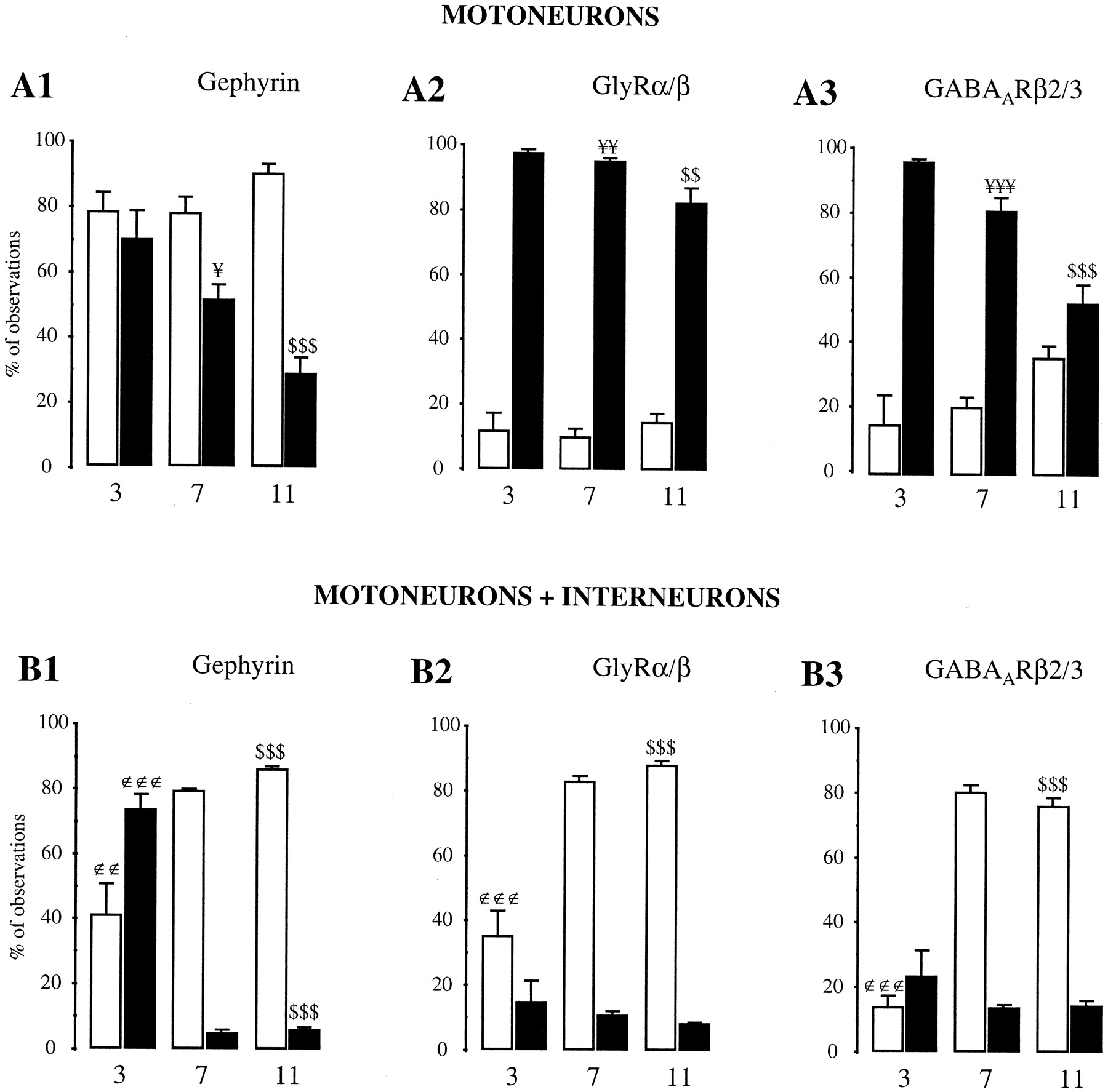
- Fig. 8.
Quantifications of the synaptic localization of gephyrin, GlyR, and GABAAR on motoneurons cultured alone (A1–3) or with spinal interneurons (B1–3). In each case, the open bars give the proportion of synapses with the indicated postsynaptic IR (Gephyrin, GlyRα/β, orGABAARβ2/3), and the filled bars give the proportion of nonsynaptic clusters per cell. Gephyrin, GlyRα/β, and GABAARβ2/3 clusters were classified as synaptic when adjacent to synapsin-IR. Results are means (± SEM) from 10–18 cells. The levels of significance (ANOVA, Scheffé F test) are indicated by one (p < 0.05), two (p < 0.01), or three (p < 0.001) symbols. ∉, Significance between 3 and 7 DIV; ¥, significance between 7 and 11 DIV; $, significance between 3 and 11 DIV.


Tables
% of cells with Motoneurons Motoneurons + interneurons GlyRα/β GABAARβ3 Gephyrin GlyRα/β GABAARβ3 Gephyrin Clusters 71.0 ± 4.6 56.2 ± 13.1 68.2 ± 3.7 63.6 ± 1.9 90.8 ± 3.7 93.1 ± 1.2 Diffuse 22.0 ± 3.4 26.3 ± 0.3 n.d. n.d. n.d. n.d. Total 90.0 ± 3.0 76.2 ± 2.5 68.2 ± 3.7 63.6 ± 1.9 90.8 ± 3.7 93.1 ± 1.2 The number of motoneurons displaying gephyrin-, GlyRα/β-, and GABAARβ3-IR was computed on motoneurons cultured for 7 DIV alone or in the presence of interneurons. Results expressed as percentages (mean ± SEM) were obtained from three independent experiments. The IR could be either diffused or clustered. Totals indicate the percentage of cells with IR independent from its pattern; n.d., IR not detected. Statistical analysis (ANOVA, SchefféF-test) indicates that the presence of interneurons in the culture decreases the number of motoneurons displaying GlyRα/β-IR (p < 0.01), whereas it increases the number of motoneurons with gephyrin-IR (p < 0.001) and GABAARβ3-IR (p < 0.05).
Motoneurons Motoneurons + interneurons Interneurons GlyRα/β 0.251 ± 0.004∉∉∉ 0.360 ± 0.009¥ 0.388 ± 0.007$$$ GABAARβ3 0.337 ± 0.010∉∉∉ 0.502 ± 0.015¥¥¥ 0.659 ± 0.020$$$ Gephyrin 0.311 ± 0.008∉∉∉ 0.423 ± 0.009¥¥¥ 0.479 ± 0.012$$$ Neurons were stained at 7 DIV for gephyrin, GlyRα/β, or GABAARβ3 subunit. For each type of culture and antibody, quantifications were performed on maximum-intensity confocal projections as described in Materials and Methods. Thirty cells were obtained from three independent experiments that were shown to be statistically equivalent (ANOVA, Scheffé F-test). Values are averages ± SEM. The levels of significance (ANOVA, Scheffé F-test) are indicated by one (p < 0.05) or three (p< 0.001) symbols. ∉, Significance between motoneurons cultured alone and motoneurons cultured with interneurons; ¥, significance between interneurons and motoneurons cultured with interneurons; $, significance between interneurons and motoneurons alone.
- Table 3.
Quantifications of the mean numbers of synapses and of gephyrin, GlyR, and GABAAR clusters per cell duringin vitro maturation
Number per cell Motoneurons Motoneurons + interneurons 3 DIV 7 DIV 11 DIV 3 DIV 7 DIV 11 DIV Synapses 5.3 ± 0.8 16.1 ± 2.0∉∉∉ 24.9 ± 2.7¥ 9.6 ± 0.9 50.5 ± 2.8∉∉∉ 63.7 ± 2.4¥¥¥ Gephyrin 24.4 ± 3.8 28.9 ± 4.7 37.7 ± 4.6 10.8 ± 1.7 40.5 ± 3.1∉∉∉ 67.4 ± 3.7¥¥¥ GlyRα/β 22.1 ± 3.1 24.7 ± 3.2 23.8 ± 3.3 4.2 ± 1.1 49.1 ± 4.0∉∉∉ 54.4 ± 3.9 GABAARβ2/3 14.5 ± 2.8 22.7 ± 3.3 16.7 ± 2.0 2.6 ± 0.8 45.2 ± 5.9∉∉∉ 53.3 ± 4.3 Neurons were stained at indicated stages for synapsin, gephyrin, GlyRα/β, or GABAARβ2/3 subunits. Values are averages ± SEM of 10–18 cells. The levels of significance (ANOVA, Scheffé F test) are indicated by one (p < 0.05), two (p < 0.01), or three (p < 0.001) symbols. ∉, Significance of differences between 3 and 7 DIV; ¥, significance between 7 and 11 DIV.


