
Main menu

  • HOME
  • CONTENT
    • Early Release
    • Featured
    • Current Issue
    • Issue Archive
    • Collections
    • Podcast
  • ALERTS
  • FOR AUTHORS
    • Information for Authors
    • Fees
    • Journal Clubs
    • eLetters
    • Submit
  • EDITORIAL BOARD
  • ABOUT
    • Overview
    • Advertise
    • For the Media
    • Rights and Permissions
    • Privacy Policy
    • Feedback
  • SUBSCRIBE

User menu

  • Log in
  • My Cart

Search

  • Advanced search
Journal of Neuroscience
  • Log in
  • My Cart
Journal of Neuroscience

Advanced Search

Submit a Manuscript
  • HOME
  • CONTENT
    • Early Release
    • Featured
    • Current Issue
    • Issue Archive
    • Collections
    • Podcast
  • ALERTS
  • FOR AUTHORS
    • Information for Authors
    • Fees
    • Journal Clubs
    • eLetters
    • Submit
  • EDITORIAL BOARD
  • ABOUT
    • Overview
    • Advertise
    • For the Media
    • Rights and Permissions
    • Privacy Policy
    • Feedback
  • SUBSCRIBE
PreviousNext
ARTICLE

Ca2+-Induced Deprotonation of Peptide Hormones Inside Secretory Vesicles in Preparation for Release

Weiping Han, Danqing Li, Amy K. Stout, Koichi Takimoto and Edwin S. Levitan
Journal of Neuroscience 1 February 1999, 19 (3) 900-905; DOI: https://doi.org/10.1523/JNEUROSCI.19-03-00900.1999
Weiping Han
1Department of Pharmacology, University of Pittsburgh, Pittsburgh, Pennsylvania 15261
  • Find this author on Google Scholar
  • Find this author on PubMed
  • Search for this author on this site
Danqing Li
1Department of Pharmacology, University of Pittsburgh, Pittsburgh, Pennsylvania 15261
  • Find this author on Google Scholar
  • Find this author on PubMed
  • Search for this author on this site
Amy K. Stout
1Department of Pharmacology, University of Pittsburgh, Pittsburgh, Pennsylvania 15261
  • Find this author on Google Scholar
  • Find this author on PubMed
  • Search for this author on this site
Koichi Takimoto
1Department of Pharmacology, University of Pittsburgh, Pittsburgh, Pennsylvania 15261
  • Find this author on Google Scholar
  • Find this author on PubMed
  • Search for this author on this site
Edwin S. Levitan
1Department of Pharmacology, University of Pittsburgh, Pittsburgh, Pennsylvania 15261
  • Find this author on Google Scholar
  • Find this author on PubMed
  • Search for this author on this site
  • Article
  • Figures & Data
  • Info & Metrics
  • eLetters
  • PDF
Loading

Article Figures & Data

Figures

  • Fig. 1.
    • Download figure
    • Open in new tab
    • Download powerpoint
    Fig. 1.

    Depolarization-induced proANF-EGFP fusion protein release is accompanied by an increase in fluorescence. InA and B, cells were superfused first in normal saline and then switched to 100 mmK+ saline (arrows). The fluorescence decrease, indicative of peptide release, was preceded by either a delay (A) or an increase (B) in fluorescence. C, A double-pulse depolarization stimulus (S, arrow), from a holding potential of −80 to 10 mV for 500 msec with a 5 sec interpulse, was delivered to the cell via a patch pipette in whole-cell configuration. Note the the increase in fluorescence was evident immediately after the double pulse. D, A similar fluorescence increase was observed in response to the double-pulse stimulus (S,arrow), without a net fluorescence decrease.
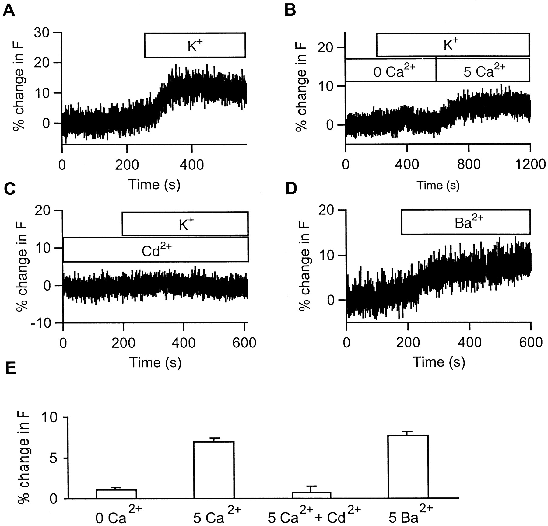
  • Fig. 2.
    • Download figure
    • Open in new tab
    • Download powerpoint
    Fig. 2.

    The depolarization-induced fluorescence change does not depend on exocytosis but requires extracellular Ca2+. Cells were pretreated with 0.2 mmNEM to block peptide release. A, Cells were superfused with normal saline and then with 100 mmK+ saline (bar). Note the apparent and uncontaminated increase in fluorescence. B, Cells were superfused with Ca2+-free normal saline, then with Ca2+-free 100 mmK+ saline, and finally with 100 mmK+ saline containing 5 mmCa2+. The fluorescence increase induced by 100 mm K+ saline occurred only when Ca2+ was present. C, Cd2+ prevented fluorescence increase caused by Ca2+. Cells were superfused with normal saline containing 0.2 mm Cd2+ and then with 100 mm K+ saline containing 0.2 mm Cd2+. D, Ba2+ mimicked Ca2+ in causing the fluorescence increase. Cells were initially superfused with normal saline and then with Ca2+-free Ba2+ containing 100 mmK+ saline. E, Quantification of divalent dependence of the effect of depolarization after NEM treatment. n = 4, 4, 6, and 4 for 0 Ca2+, 5 mm Ca2+, 5 mm Ca2+ plus 0.2 mmCd2+, and 5 mm Ba2+, respectively.
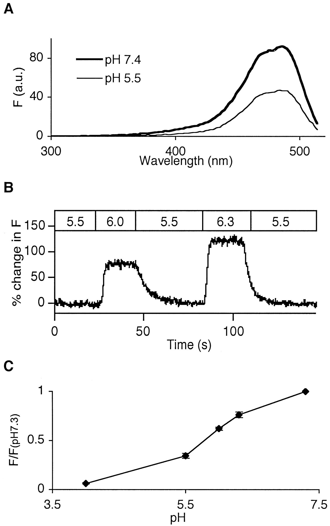
  • Fig. 3.
    • Download figure
    • Open in new tab
    • Download powerpoint
    Fig. 3.

    Fluorescence of EGFP-tagged peptide hormone is sensitive to pH. A, Fluorescence excitation spectra of proANF-EGFP fusion protein at two different pH values.B, Microfluorimetric recordings of the responses of proANF-EGFP fusion protein expressed in cells to solutions at various pH levels. The response of the fluorescence of proANF-EGFP to changing pH was quick and reversible. C, Titration curve for the relative fluorescence of proANF-EGFP.

  • Fig. 4.
    • Download figure
    • Open in new tab
    • Download powerpoint
    Fig. 4.

    The depolarization-induced increase in intravesicular fluorescence requires a pH gradient. Cells were initially superfused with normal saline containing a pH-collapsing agent [1 μm monensin (A), 1 μm nigericin (B), or 1 μm FCCP (C)] and then with 100 mm K+ saline containing the same pH-collapsing agent. Note that fluorescence decrease occurred almost immediately after 100 mmK+ application. D, Comparison of delay from the application of 100 mm K+to the onset of fluorescence decrease in cells without pretreatment (C) and those pretreated with monensin (M), nigericin (N), or FCCP (F). n = 11, 8, 5, and 5 for control, monensin, nigericin, and FCCP, respectively. *p < 0.01 versus control.

  • Fig. 5.
    • Download figure
    • Open in new tab
    • Download powerpoint
    Fig. 5.

    Fluorescence of proANF-Sapphire fusion protein is insensitive to pH. A, Fluorescence excitation spectra of proANF-Sapphire GFP fusion protein at two different pH values.B, Microfluorimetric recordings of the responses of proANF-Sapphire fusion protein to bath superfusion of normal saline at various pH levels. C, Titration curve for the relative fluorescence of Sapphire-tagged proANF.

  • Fig. 6.
    • Download figure
    • Open in new tab
    • Download powerpoint
    Fig. 6.

    The apparent delay after application of 100 mm K+ saline is no longer observed when using proANF-Sapphire. A, A cell was initially superfused with normal saline and then with 100 mmK+ saline. Note that fluorescence decrease occurred almost immediately after 100 mm K+application. Also, no increase in fluorescence was observed with proANF-Sapphire. B, Comparison of delay from the application of 100 mm K+ saline to the onset of fluorescence decrease in cells transfected with proANF-EGFP (EGFP) and those transfected with proANF-Sapphire (Sph). n = 11 and 12 for proANF-EGFP and proANF-Sapphire, respectively. *p < 0.01 versus EGFP.

  • Fig. 7.
    • Download figure
    • Open in new tab
    • Download powerpoint
    Fig. 7.

    NEM increases fluorescence in cells that are transfected with proANF-EGFP but not those with proANF-Sapphire.A, A cell expressing proANF-EGFP was initially superfused with normal saline, then with normal saline containing 0.2 mm NEM, and finally with normal saline containing 1 μm monensin. Note that fluorescence increased gradually after the start of NEM superfusion and that fluorescence increased very quickly when superfusion was switched to monensin (M). B, A cell expressing proANF-Sapphire was initially superfused with normal saline and then with normal saline containing 0.2 mm NEM. NEM did not have any effect on fluorescence of proANF-Sapphire. C, A proANF-Sapphire-expressing cell was pretreated with NEM and DTT. After initial superfusion with normal saline, the solution was switched to normal saline containing 1 μm monensin.

Back to top

In this issue

The Journal of Neuroscience: 19 (3)
Journal of Neuroscience
Vol. 19, Issue 3
1 Feb 1999
  • Table of Contents
  • Index by author
Email

Thank you for sharing this Journal of Neuroscience article.

NOTE: We request your email address only to inform the recipient that it was you who recommended this article, and that it is not junk mail. We do not retain these email addresses.

Enter multiple addresses on separate lines or separate them with commas.
Ca2+-Induced Deprotonation of Peptide Hormones Inside Secretory Vesicles in Preparation for Release
(Your Name) has forwarded a page to you from Journal of Neuroscience
(Your Name) thought you would be interested in this article in Journal of Neuroscience.
CAPTCHA
This question is for testing whether or not you are a human visitor and to prevent automated spam submissions.
Print
View Full Page PDF
Citation Tools
Ca2+-Induced Deprotonation of Peptide Hormones Inside Secretory Vesicles in Preparation for Release
Weiping Han, Danqing Li, Amy K. Stout, Koichi Takimoto, Edwin S. Levitan
Journal of Neuroscience 1 February 1999, 19 (3) 900-905; DOI: 10.1523/JNEUROSCI.19-03-00900.1999

Citation Manager Formats

  • BibTeX
  • Bookends
  • EasyBib
  • EndNote (tagged)
  • EndNote 8 (xml)
  • Medlars
  • Mendeley
  • Papers
  • RefWorks Tagged
  • Ref Manager
  • RIS
  • Zotero
Respond to this article
Request Permissions
Share
Ca2+-Induced Deprotonation of Peptide Hormones Inside Secretory Vesicles in Preparation for Release
Weiping Han, Danqing Li, Amy K. Stout, Koichi Takimoto, Edwin S. Levitan
Journal of Neuroscience 1 February 1999, 19 (3) 900-905; DOI: 10.1523/JNEUROSCI.19-03-00900.1999
del.icio.us logo Digg logo Reddit logo Twitter logo CiteULike logo Facebook logo Google logo Mendeley logo
  • Tweet Widget
  • Facebook Like
  • Google Plus One

Jump to section

  • Article
    • Abstract
    • MATERIALS AND METHODS
    • RESULTS
    • DISCUSSION
    • Footnotes
    • REFERENCES
  • Figures & Data
  • Info & Metrics
  • eLetters
  • PDF

Keywords

  • GFP
  • peptide hormone
  • exocytosis
  • alkalinization
  • secretory vesicle
  • Ca2+

Responses to this article

Respond to this article

Jump to comment:

No eLetters have been published for this article.

Related Articles

Cited By...

More in this TOC Section

  • Cytoskeletal and Morphological Alterations Underlying Axonal Sprouting after Localized Transection of Cortical Neuron AxonsIn Vitro
  • Developmental Increase in Vesicular Glutamate Content Does Not Cause Saturation of AMPA Receptors at the Calyx of Held Synapse
  • Aberrant Chloride Transport Contributes to Anoxic/Ischemic White Matter Injury
Show more ARTICLE
  • Home
  • Alerts
  • Visit Society for Neuroscience on Facebook
  • Follow Society for Neuroscience on Twitter
  • Follow Society for Neuroscience on LinkedIn
  • Visit Society for Neuroscience on Youtube
  • Follow our RSS feeds

Content

  • Early Release
  • Current Issue
  • Issue Archive
  • Collections

Information

  • For Authors
  • For Advertisers
  • For the Media
  • For Subscribers

About

  • About the Journal
  • Editorial Board
  • Privacy Policy
  • Contact
(JNeurosci logo)
(SfN logo)

Copyright © 2023 by the Society for Neuroscience.
JNeurosci Online ISSN: 1529-2401

The ideas and opinions expressed in JNeurosci do not necessarily reflect those of SfN or the JNeurosci Editorial Board. Publication of an advertisement or other product mention in JNeurosci should not be construed as an endorsement of the manufacturer’s claims. SfN does not assume any responsibility for any injury and/or damage to persons or property arising from or related to any use of any material contained in JNeurosci.