
Main menu

  • HOME
  • CONTENT
    • Early Release
    • Featured
    • Current Issue
    • Issue Archive
    • Collections
    • Podcast
  • ALERTS
  • FOR AUTHORS
    • Information for Authors
    • Fees
    • Journal Clubs
    • eLetters
    • Submit
    • Special Collections
  • EDITORIAL BOARD
    • Editorial Board
    • ECR Advisory Board
    • Journal Staff
  • ABOUT
    • Overview
    • Advertise
    • For the Media
    • Rights and Permissions
    • Privacy Policy
    • Feedback
    • Accessibility
  • SUBSCRIBE

User menu

  • Log out
  • Log in
  • My Cart

Search

  • Advanced search
Journal of Neuroscience
  • Log out
  • Log in
  • My Cart
Journal of Neuroscience

Advanced Search

Submit a Manuscript
  • HOME
  • CONTENT
    • Early Release
    • Featured
    • Current Issue
    • Issue Archive
    • Collections
    • Podcast
  • ALERTS
  • FOR AUTHORS
    • Information for Authors
    • Fees
    • Journal Clubs
    • eLetters
    • Submit
    • Special Collections
  • EDITORIAL BOARD
    • Editorial Board
    • ECR Advisory Board
    • Journal Staff
  • ABOUT
    • Overview
    • Advertise
    • For the Media
    • Rights and Permissions
    • Privacy Policy
    • Feedback
    • Accessibility
  • SUBSCRIBE
PreviousNext
ARTICLE, Behavioral/Systems

Control of Response Selection by Reinforcer Value Requires Interaction of Amygdala and Orbital Prefrontal Cortex

Mark G. Baxter, Amanda Parker, Caroline C. C. Lindner, Alicia D. Izquierdo and Elisabeth A. Murray
Journal of Neuroscience 1 June 2000, 20 (11) 4311-4319; https://doi.org/10.1523/JNEUROSCI.20-11-04311.2000
Mark G. Baxter
1Department of Psychology, Harvard University, Cambridge, Massachusetts 02138,
  • Find this author on Google Scholar
  • Find this author on PubMed
  • Search for this author on this site
Amanda Parker
2School of Psychology, University of Nottingham, Nottingham NG7 2RD, United Kingdom,
  • Find this author on Google Scholar
  • Find this author on PubMed
  • Search for this author on this site
Caroline C. C. Lindner
3Laboratory of Neuropsychology, National Institute of Mental Health, Bethesda, Maryland 20892, and
  • Find this author on Google Scholar
  • Find this author on PubMed
  • Search for this author on this site
Alicia D. Izquierdo
3Laboratory of Neuropsychology, National Institute of Mental Health, Bethesda, Maryland 20892, and
4Department of Psychology, The George Washington University, Washington, DC 20052
  • Find this author on Google Scholar
  • Find this author on PubMed
  • Search for this author on this site
Elisabeth A. Murray
3Laboratory of Neuropsychology, National Institute of Mental Health, Bethesda, Maryland 20892, and
  • Find this author on Google Scholar
  • Find this author on PubMed
  • Search for this author on this site
  • Article
  • Figures & Data
  • Info & Metrics
  • eLetters
  • PDF
Loading

Article Figures & Data

Figures

  • Tables
  • Fig. 1.
    • Download figure
    • Open in new tab
    • Download powerpoint
    Fig. 1.

    Schematic of testing sequence for the reinforcer devaluation study. Control subjects received no surgery and were given a period of rest equivalent to that provided for the operated monkeys. Note that surgery 2 completes the disconnection of amygdala and orbital prefrontal cortex in all operated subjects.

  • Fig. 2.
    • Download figure
    • Open in new tab
    • Download powerpoint
    Fig. 2.
    • Download figure
    • Open in new tab
    • Download powerpoint
    Fig. 2.

    Intended lesion and plots of the orbital prefrontal cortex and amygdala lesions in the four operated cases (Op1–Op4), shown on ventral surface views (top) and coronal sections (bottom) from a standard rhesus monkey brain. The intended lesion is shown in the leftmost column. The thick black line and small rectangle between the hemispheres in this and other ventral views indicates the extent of the section of the corpus callosum and anterior commissure, respectively; positions of the stereotaxic levels illustrated in the coronal sections are also indicated. The ventral views for Op1–Op4 show reconstructions of the extent of the orbital prefrontal cortex lesions, and are reversed to aid in matching to the individual sections (i.e., the left hemisphere is on theleft). The numbers to theleft of the coronal sections indicate the distance in millimeters from the interaural plane. Compare and contrast with Figure3.

  • Fig. 3.
    • Download figure
    • Open in new tab
    • Download powerpoint
    Fig. 3.

    Photomicrographs of Nissl-stained coronal sections from case Op3. A, Coronal section ∼30 mm rostral to the interaural plane. The extent of the orbital prefrontal cortex lesion in the left hemisphere is marked by the black arrows. The section of the corpus callosum is also apparent at this level and is marked by the white arrowhead.B, Coronal section 16 mm rostral to the interaural plane. Note the marked loss in volume of the amygdala in the right compared with the left hemisphere. The section of the corpus callosum is marked by the white arrowhead. Therectangles over the left and right temporal lobes show the approximate locations of the regions shown at higher power inC and D, respectively. C, Photomicrograph of the intact (left) amygdala. D, Photomicrograph of the right amygdala ∼9 months after injection of ibotenic acid, at the same magnification used in C. The marked neuronal cell loss and gliosis are characteristic of excitotoxic lesions.
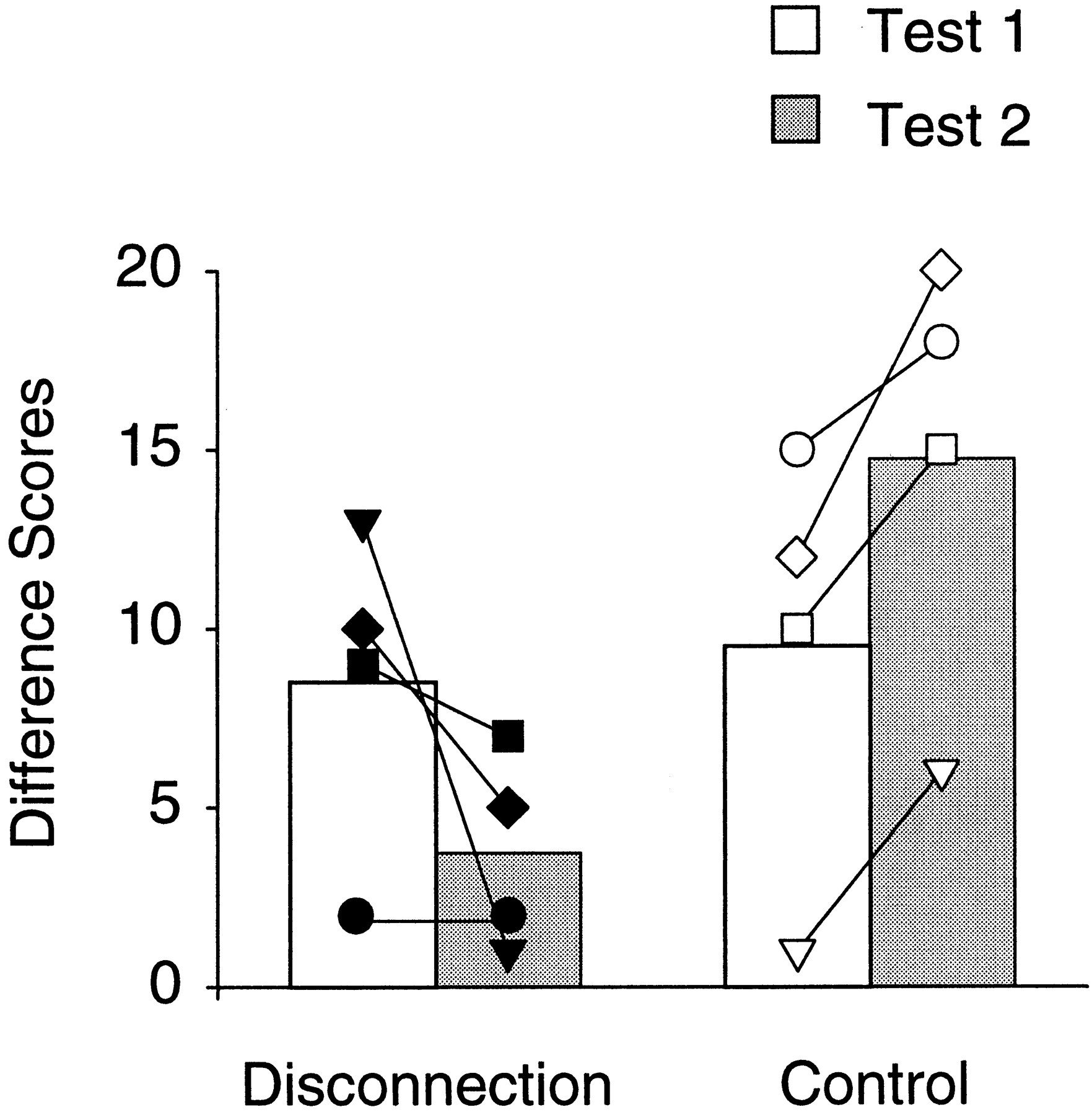
  • Fig. 4.
    • Download figure
    • Open in new tab
    • Download powerpoint
    Fig. 4.

    The effects of reinforcer devaluation performed before (Test 1) and after (Test 2) the surgical disconnection had been completed. Difference scores (devaluation − baseline) for the control and disconnection group means are shown as bars, with the symbols representing scores of individual monkeys. Normal monkeys showed an enhancement of the devaluation effect in the second test, whereas operated monkeys showed a significant reduction of this effect in the second test. Op1,Filled diamonds; Op2, filled squares; Op3, filled inverted triangles; Op4, filled circles; Con1, open diamonds; Con2, open squares; Con3, open inverted triangles; Con4,open circle.

  • Fig. 5.
    • Download figure
    • Open in new tab
    • Download powerpoint
    Fig. 5.

    Results of the devaluation procedure (selective satiation) on progressive ratio testing. Baseline data shown are the mean, for each subject, of the maximum number of responses emitted (i.e., maximum ratio attained) during all four baseline sessions. Satiation data are scores for the single satiation session. Group means are shown as bars, with the symbols representing scores of individual monkeys. All monkeys, regardless of group membership, decreased responding during the test session in which the reinforcer was devalued by selective satiation. Symbols as in Figure 4.

Tables

  • Figures
    • View popup
    Table 1.

    Percent damage to amygdala and orbital prefrontal cortex in the four operated subjects

    CaseAmygdalaOrbital prefrontal
    HemispherePercent damageHemispherePercent damage
    Op1L (first)42.9R (second)65.5
    Op2L (second)34.9R (first)73.3
    Op3R (first)95.3L (second)57.1
    Op4R (second)28.4L (first)41.1
    Mean50.459.3
    • The hemisphere in which each lesion was placed (left, L; right, R) and the order in which the surgeries were conducted are indicated. For example, case Op1 received a neurotoxic amygdala lesion in the left hemisphere before beginning behavioral testing and a lesion of the orbital prefrontal cortex in the right hemisphere, with forebrain commissurotomy in the same surgery, to complete the disconnection. See Figure 1 for a schematic of the behavioral testing sequence.

    • View popup
    Table 2.

    Results of reinforcer devaluation testing

    CaseTest 1Test 2Test 2-test 1 difference
    Baseline F1:F2Satiation F1 F1:F2Satiation F2 F1:F2Difference score (sum)Baseline F1:F2Satiation F1 F1:F2Satiation F2 F1:F2Difference score (sum)
    Con122:814 :1626 :41223.5:6.57 :2327 :3208
    Con214:1612 :1822 :81019.5:10.513 :1728 :2155
    Con327.5:2.528:229 :1120:1013 :1719 :1165
    Con421.5:8.57 :2322 :81515.5:14.56 :2424 :6183
    Mean9.514.755.25
    Op118.5:11.516 :1426 :41025:523 :728 :25−5
    Op224.5:5.517 :1326 :4929.5:0.523 :730 :07−2
    Op320.5:9.512 :1825 :51323.5:6.521 :922 :81−12
    Op4 2.5:27.50 :302 :2820:300 :302 :2820
    Mean8.53.75−4.75
    • The number of food 1 (F1) and food 2 (F2) objects chosen in the baseline sessions (mean of two sessions) and each of the two satiation sessions (preceded by satiation with food 1 or food 2), as well as the difference score between the satiation sessions and the baseline sessions (summed for the two satiation sessions), are given for reinforcer devaluation test 1 and test 2 for each of the eight monkeys (Con1–Con4, control monkeys; Op1–Op4, operated monkeys).

Back to top

In this issue

The Journal of Neuroscience: 20 (11)
Journal of Neuroscience
Vol. 20, Issue 11
1 Jun 2000
  • Table of Contents
  • Index by author
Email

Thank you for sharing this Journal of Neuroscience article.

NOTE: We request your email address only to inform the recipient that it was you who recommended this article, and that it is not junk mail. We do not retain these email addresses.

Enter multiple addresses on separate lines or separate them with commas.
Control of Response Selection by Reinforcer Value Requires Interaction of Amygdala and Orbital Prefrontal Cortex
(Your Name) has forwarded a page to you from Journal of Neuroscience
(Your Name) thought you would be interested in this article in Journal of Neuroscience.
CAPTCHA
This question is for testing whether or not you are a human visitor and to prevent automated spam submissions.
Print
View Full Page PDF
Citation Tools
Control of Response Selection by Reinforcer Value Requires Interaction of Amygdala and Orbital Prefrontal Cortex
Mark G. Baxter, Amanda Parker, Caroline C. C. Lindner, Alicia D. Izquierdo, Elisabeth A. Murray
Journal of Neuroscience 1 June 2000, 20 (11) 4311-4319; DOI: 10.1523/JNEUROSCI.20-11-04311.2000

Citation Manager Formats

  • BibTeX
  • Bookends
  • EasyBib
  • EndNote (tagged)
  • EndNote 8 (xml)
  • Medlars
  • Mendeley
  • Papers
  • RefWorks Tagged
  • Ref Manager
  • RIS
  • Zotero
Respond to this article
Request Permissions
Share
Control of Response Selection by Reinforcer Value Requires Interaction of Amygdala and Orbital Prefrontal Cortex
Mark G. Baxter, Amanda Parker, Caroline C. C. Lindner, Alicia D. Izquierdo, Elisabeth A. Murray
Journal of Neuroscience 1 June 2000, 20 (11) 4311-4319; DOI: 10.1523/JNEUROSCI.20-11-04311.2000
Twitter logo Facebook logo Mendeley logo
  • Tweet Widget
  • Facebook Like
  • Google Plus One

Jump to section

  • Article
    • Abstract
    • MATERIALS AND METHODS
    • RESULTS
    • DISCUSSION
    • Footnotes
    • REFERENCES
  • Figures & Data
  • Info & Metrics
  • eLetters
  • PDF

Keywords

  • amygdala
  • crossed-lesion technique
  • decision-making
  • orbital prefrontal cortex
  • reinforcer devaluation
  • representation
  • rhesus monkey

Responses to this article

Respond to this article

Jump to comment:

No eLetters have been published for this article.

Related Articles

Cited By...

More in this TOC Section

ARTICLE

  • Neural Correlates of Competing Fear Behaviors Evoked by an Innately Aversive Stimulus
  • Distinct Developmental Modes and Lesion-Induced Reactions of Dendrites of Two Classes of Drosophila Sensory Neurons
  • Functional Dissociation among Components of Remembering: Control, Perceived Oldness, and Content
Show more ARTICLE

Behavioral/Systems

  • Appetitive and Aversive Olfactory Learning in Humans Studied Using Event-Related Functional Magnetic Resonance Imaging
  • Electroconvulsive Seizure Behavior in Drosophila: Analysis of the Physiological Repertoire Underlying a Stereotyped Action Pattern in Bang-Sensitive Mutants
  • Spinal Inhibitory Neurons that Modulate Cutaneous Sensory Pathways during Locomotion in a Simple Vertebrate
Show more Behavioral/Systems
  • Home
  • Alerts
  • Follow SFN on BlueSky
  • Visit Society for Neuroscience on Facebook
  • Follow Society for Neuroscience on Twitter
  • Follow Society for Neuroscience on LinkedIn
  • Visit Society for Neuroscience on Youtube
  • Follow our RSS feeds

Content

  • Early Release
  • Current Issue
  • Issue Archive
  • Collections

Information

  • For Authors
  • For Advertisers
  • For the Media
  • For Subscribers

About

  • About the Journal
  • Editorial Board
  • Privacy Notice
  • Contact
  • Accessibility
(JNeurosci logo)
(SfN logo)

Copyright © 2025 by the Society for Neuroscience.
JNeurosci Online ISSN: 1529-2401

The ideas and opinions expressed in JNeurosci do not necessarily reflect those of SfN or the JNeurosci Editorial Board. Publication of an advertisement or other product mention in JNeurosci should not be construed as an endorsement of the manufacturer’s claims. SfN does not assume any responsibility for any injury and/or damage to persons or property arising from or related to any use of any material contained in JNeurosci.