

Article Figures & Data
Figures
- Fig. 1.
Levels of organization in the auditory midbrain. The central nucleus of the IC (CNIC) is one nuclear subdivision that consists of tonotopically (LF, low frequency; HF, high frequency) arranged fibrodendritic laminae. Together, disk-shaped cells tuned to similar frequencies and intermingling afferent layers (purple) make up fibrodendritic laminae. Afferent bands (red andblue) occupy specific sublayers within a given lamina. Afferents that terminate within restricted zones of a target sublayer are termed patches (green). D, Dorsal; V, ventral; M, medial;L, lateral.
- Fig. 2.
Summary figure illustrating the normal developmental progression of the crossed DNLL input to the IC before hearing onset. A–C, Pattern of anterogradely filled DNLL fibers within the right IC at P0, P4, and P12, respectively. Dashed contours represent the ventromedial border of the IC. White arrowheads inB denote the earliest indication of contralateral DNLL bands forming within alternating sublayers of the central nucleus of the IC. By onset of hearing (C), adult-like afferent patches are readily apparent (paired arrowheads). D–F, Retrograde transport from a midline dye placement in the commissure of Probst labels contralaterally projecting DNLL cells (right DNLL is depicted). In the present study, unilateral cochlear ablations were performed at P2, before any evidence of afferent banding. Rat pups were then reared to P12 to determine the effect of unilateral cochlear ablation before experience on the development of the crossed DNLL projection.D, Dorsal; V, ventral;M, medial; L, lateral. Scale bars: A–E, 100 μm; F, 150 μm.
- Fig. 3.
Low-magnification photomicrograph of a coronal section through the auditory midbrain of a unilateral cochlear ablation case (dorsal is up). DiI placement in the commissure of Probst (black arrow) results in labeling of the contralateral projection to the IC from each DNLL. Thus, afferent fibers within the IC ipsilateral to the ablation arise from the contralateral DNLL, and vice versa. Note the marked asymmetry in labeling of the retrogradely filled DNLLs, as well as the contralaterally projecting DNLL fibers within the ICs. Such bilateral asymmetry was characteristic in all of the ablation cases and was striking even at very low magnification. Dashed contoursrepresent the ventromedial borders of the ICs. Scale bar, 500 μm.
- Fig. 4.
Photomicrographs illustrating the crossed DNLL projections to the IC in a P12 control animal. A,B, DiI-labeled DNLL fibers terminating within the IC, left and right, respectively. Note the symmetry between the two sides in both termination pattern and relative density of the input. Such balance was typical of all control cases. The most lateral band inA and B is evidence of a developing projection from the contralateral DNLL to the deep layer of the external cortex of the IC. The vast majority of fibers, however, terminate within the central nucleus of the IC, forming an adult-like pattern of afferent patches. The approximate thickness (ventromedial–dorsolateral dimension) of patches measures 75 μm.D, Dorsal; V, ventral; M, medial; L, lateral. Dashed contoursrepresent the ventromedial borders of the ICs. C,D, Retrogradely filled cells in left and right DNLL, respectively. As was evident for IC, labeling within the DNLL on the two sides appeared to be symmetric. Scale bars, 150 μm.
- Fig. 5.
Photomicrographs illustrating the crossed DNLL inputs to the IC at P12 after unilateral cochlear ablation.A, B, DiI-labeled DNLL fibers terminating within the IC, left and right, respectively. Note the asymmetry between the two sides in the termination pattern and relative density of the input. Afferents within the IC ipsilateral to the ablation (those arising from the deprived DNLL) terminate within appropriate subdivisions of the IC (i.e., as a band in deep layer of external cortex with the majority of the termination within the central nucleus). The heaviest labeling within the central nucleus in this and all experimental cases, ipsilateral and contralateral to the ablation, occupies the correct dorsomedial–ventrolateral position for DNLL patches. The normal pattern of afferent patches within this area of the central nucleus, however, is not evident (A). Although qualitatively fewer, afferents were visible at low magnification in the IC opposite the ablation (B). It is apparent that these fibers not only project to the appropriate region within the central nucleus, but also segregate into distinct afferent bands or patches within this target area. D, Dorsal; V, ventral;M, medial; L, lateral. Dashed contours represent the ventromedial borders of the ICs.C, D, Retrogradely filled cells in the left and right DNLL, respectively. As in all cochlear ablation cases, fewer cells are labeled in the DNLL contralateral to the ablation relative to the DNLL on the side of the ablation. Scale bars, 150 μm.
- Fig. 6.
Quantification of DNLL afferent band features within the central nucleus of the IC. A, Brightness profile of control case illustrated in Figure 4. Stippled curve corresponds to brightness profile of banding in the left IC; solid curve corresponds to brightness profile for the right IC. Periodicity, band width, and peak-to-trough amplitude were not significantly different between the two sides. Offset to theright are linear regression plots that show a best fit sine wave (solid line) for the left andright profiles. B, Brightness profile of unilateral ablation case shown in Figure 5. Stippled curve represents the brightness profile of labeling in the IC ipsilateral to the ablation (left); solid curve reflects brightness profile for the IC contralateral to the ablation (right). Afferent band features (periodicity, band width, amplitude) in the ipsilateral IC were significantly different from controls, whereas features in the contralateral IC were comparable with controls. Offset to theright are linear regression plots that show a best fit sine wave (solid line) for the left andright profiles. Note the lack of periodicity as evidenced by the poor fit for the left side(R2 = 0.06).
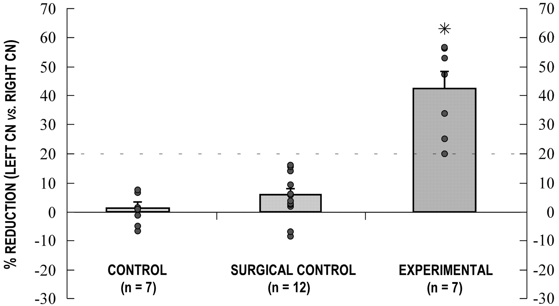
- Fig. 7.
Graph of CN volumes. Each data point represents the percentage difference between the left and right CN volumes for each individual case. Positive data points reflect a reduction of the left CN relative to the right CN. Histogrambars indicate mean percentages, and accompanying error bars represent the SEM for each group. A nonparametric ANOVA was significant (p < 0.001), and a multiple comparison post-test between groups revealed a significant difference (asterisk) between the mean of the experimental group and the means of both the control and the surgical control groups (p < 0.001 and p < 0.01, respectively). A statistical comparison between the control and surgical control means was not considered significant. Thedashed line at the 20% reduction value denotes the CN volume criterion for ablation cases.
- Fig. 8.
Comparison of CN in a normal animal and after a left cochlear ablation. Low-magnification photomicrographs of comparable regions of the anteroventral cochlear nucleus (AVCN) in a control (A,B) and a unilateral ablation case (C,D) in Nissl-stained sections. Dashed contours delineate the boundaries of the AVCN and demarcate its border with the granule (GRAN) cell region. InC, note the drastic reduction in size of the AVCN ipsilateral to the cochlear ablation relative to that on the side opposite the ablation (D). Scale bars, 150 μm.
- Fig. 9.
Cochlear histology of control (A, A', B,B') and unilateral ablation cases (C, D, and E,F). A', B', Low-magnification photomicrographs of the left and right cochleas in a control case. Dashed inset boxes are shown at higher magnification in A and B.SV, Scala vestibuli; SM, scala media;ST, scala tympani; SG, spiral ganglion;StV, stria vascularis. C,D, Photomicrographs of the ablated (C) and intact side (D) in an experimental case. Note the atrophied stria vascularis, the absence of Reissner's membrane (RM), and the lack of hair cells on the ablated side. The spiral limbus (SL) was left intact, and there was no evidence of degeneration in the spiral ganglion. E, F, Photomicrographs of the ablated (E) and intact side (F) in an experimental case with a more complete ablation. Despite abundant debris in the cochlear spaces, remnants of the spiral limbus were distinguishable. Considerable degeneration of the spiral ganglion was apparent on the ablated side in this and similar cases. Scale bars: A′, B′, 500 μm; A–F, 100 μm.
- Fig. 10.
Schematic diagram summarizing the effects of a unilateral cochlear ablation on the development of the crossed DNLL projections to the IC before onset of hearing. Afferent termination within the IC is markedly less dense on both sides when qualitatively compared with P12 control cases. Moreover, in contrast to control cases, all seven ablation cases exhibited striking asymmetry in both distribution pattern and relative density of DNLL afferents. The crossed input from the deprived DNLL to the IC ipsilateral to the ablation always appeared heavier but less organized into bands and patches than the input arising from the nondeprived DNLL to the IC contralateral to the ablation.
- Fig. 11.
Theoretical model of conditions induced by a unilateral cochlear ablation. The “X” indicates ablation of the left cochlea. Arrows represent hypothesized changes in the levels of intrinsic activity of brainstem auditory nuclei known to send patterned inputs to the IC. No arrow is indicated for the CN contralateral to the ablation because it receives monaural input from the unaffected cochlea. Segregation of the crossed DNLL projections into afferent bands appeared to be dependent on afferent activity, whereas the density of the input appeared to be determined by the postsynaptic environment. The ability of the input to locate the appropriate DNLL position or patch location within the central nucleus was unaffected by the ablation.


Tables
Experimental groups Age at experimental manipulation Tracer placement in fixed tissue (P12) Incubation period n = number of animals included in results Control No manipulation DiI in commissure of Probst 2–3 months n = 7 Surgical control (P2) DiI in commissure of Probst 2–3 months n = 12 Unilateral cochlear ablation1-a (P2) DiI in commissure of Probst 2–3 months n = 7 ↵F1-a Ablation cases met the following criteria: (1) showed a 20% greater reduction in CN volume on the side of the ablation relative to the unaffected side and (2) exhibited some degree of cochlear damage as evidenced by temporal bone histology.
Amplitude Period Band width Mean and SD for control group 0.67 ± 0.20 156.35 ± 40.33 82.40 ± 16.18 Mean and SD for experimental group ipsilateral to ablation (left) 0.26 ± 0.222-160 111.21 ± 44.54* 57.60 ± 22.902-160 Mean and SD for experimental group contralateral to ablation (right) 0.74 ± 0.72 129.85 ± 28.2 68.08 ± 21.69


