

Abstract
Genes that control the specification and differentiation of the functionally specialized areas of the mammalian neocortex are likely expressed across the developing neocortex in graded or restricted patterns. To search for such genes we have performed a PCR-based differential display screen using RNAs from rostral neocortex, which included the primary motor area, and caudal neocortex, which included the primary visual area, of embryonic day 16 rats. We identified 82 differentially expressed gene fragments. Secondary screening byin situ hybridization confirmed that five fragments, representing four genes, are differentially expressed across developing rat neocortex. Two of the genes, chick ovalbumin upstream transcription factor I (COUP-TFI) andclose homolog of L1 (CHL1), have been cloned previously, but their differential expression in cortex has not been reported. Sequences from the other two fragments suggest that they represent novel genes. The expression patterns include graded, restricted, and discontinuous expression with abrupt borders that might correlate with those of areas. The differential expression patterns of all four genes are established before the arrival of thalamocortical afferents, suggesting that they are independent of thalamic influence, and could direct or reflect arealization. In addition,COUP-TFI and CHL1 exhibit dynamic expression patterns that undergo substantial changes after thalamocortical afferents invade the cortical plate, suggesting that thalamic axons may influence their later expression. Postnatally,COUP-TFI is most prominently expressed in layer 4, in both rats and mice, and CHL1 is expressed in layer 5.COUP-TFI expression in cortex, and in ventral telencephalon and dorsal thalamus, suggests several possible causes for the loss of layer 4 neurons and the reduced thalamocortical projection reported in COUP-TFI knock-out mice.
The cerebral cortex is divided into several major regions, each composed of distinct areas. The most prominent region, the neocortex, is divided into numerous areas that share a basic organization but nonetheless are characterized by their unique architecture, connections, and function. An important issue is to define the mechanisms that control the specification of areas and their differentiation from the cortical plate. Recent evidence suggests that differential gene expression across the embryonic neocortex has a primary role in regulating arealization (Bishop et al., 2000) and is established by mechanisms intrinsic to it (Miyashita-Lin et al., 1999;Nakagawa et al., 1999). Extrinsic influences on arealization, most notable being thalamocortical afferents (TCAs), the principal input to neocortex, likely operate within the context of this molecular framework to promote the later differentiation of certain areal specializations (Rakic, 1988; O'Leary, 1989; Chenn et al., 1997;Gitton et al., 1999b).
Genes reported to be differentially expressed across the embryonic neocortex include several transcription factor genes: the homeobox genes Emx1 and Emx2 (Gulisano et al., 1996;Mallamaci et al., 1998), the paired-box gene Pax6 (Walther and Gruss, 1991; Stoykova and Gruss, 1994), the LIM-homeodomain gene Lhx2 (Porter et al., 1997; Nakagawa et al., 1999),retinoid Z receptor β (RZRβ), a nuclear melatonin receptor (Becker-Andre et al., 1994; Park et al., 1997;Schaeren-Wiemers et al., 1997), Tbr1, a T-box gene, andId2, a helix–loop–helix gene (Bulfone et al., 1995;Rubenstein et al., 1999). Other differentially expressed molecules include the NGF receptor p75 (Mackarehtschian et al., 1999), some EphA receptors and ephrin-A ligands (Donoghue and Rakic, 1999a;Mackarehtschian et al., 1999), and the cadherins 6, 8, and 11 (Suzuki et al., 1997; Inoue et al., 1998; Nakagawa et al., 1999).
To identify other differentially expressed genes that might be involved in regulating arealization, we used differential display PCR (ddPCR) that used total RNAs from the rostral and caudal neocortex of embryonic day 16 (E16) rats; rostral pieces were from frontal cortex and included the primary motor area, and caudal pieces were from occipital cortex and included the primary visual area. We chose E16 as a compromise in the timing of key developmental phenomena. This age is approximately midway through cortical neurogenesis; marginal zone and subplate neurons, as well as most neurons that will form layers 6, 5, and 4, have been generated (Bayer and Altman, 1991). In addition, TCAs are just beginning to reach the cortex and have not yet invaded the cortical plate (Catalano et al., 1991, 1996; De Carlos et al., 1995). Therefore, differential gene expression at this age would be established independent of TCAs and likely intrinsic to the neocortex and could have a role in the development of area-specific TCA projections.
We have identified four genes differentially expressed across the developing neocortex in a manner consistent with their possible involvement in arealization. In addition, they exhibit layer-specific expression consistent with a role in defining unique properties of subsets of cortical neurons. Two of these genes have been cloned previously: the orphan nuclear receptor chick ovalbumin upstream transcription factor I (COUP-TFI) (Jonk et al., 1994; Qiu et al., 1994) and the cell adhesion molecule close homolog of L1 (CHL1) (Holm et al., 1996). However, their differential expression in the cortex has not been described. The other two gene fragments appear to represent novel genes.
MATERIALS AND METHODS
Animals. Animals used for this study were obtained from timed pregnant Sprague Dawley rats and ICR mice from Harlan Sprague Dawley (Indianapolis, IN). The day of vaginal plug detection is designated E0, and the day of birth is postnatal day 0 (P0).
ddPCR. Pregnant mothers were anesthetized with an overdose of Nembutal (100 mg/kg of body weight). E16.5 rats were used to prepare total RNA. The brains were dissected out from E16.5 rats, and the meninges were removed from the brain surface. Two 500 × 500 μm pieces of cortex (from the surface of the marginal zone to the ventricle) were dissected out from the caudal (occipital) and rostral (frontal) portions of the neocortex (see Fig.1A). These tissues were washed in L15 media and then put directly into lysis buffer (Qiagen RNeasy kit) for total RNA extraction.
For both the rostral and caudal dissections, three separate RNA samples were prepared from two different litters to reduce false positives. Five embryos were used for each RNA preparation, yielding ∼6–7 μg of RNA. The RNA samples were digested with DNase I to remove genomic DNA. The RNA (250 ng) was reverse transcribed with SUPERSCRIPT Preamplification System for First Strand cDNA Synthesis (Life Technologies; 18089-011), priming with downstream primers (Operon Technology). The detailed protocol for reverse transcription and PCR analysis was performed as described by M. Gesemann, E. D. Litwack, and D. D. M. O'Leary (unpublished observations). Each sample of caudal and rostral DNA was amplified with 400 combinations of arbitrary upstream and downstream primers (Operon Technology) by PCR. The PCR products were separated on a 6% denaturing polyacrylamide gel. The differences in the intensity of the bands were recognized by visual inspection. More than 10,000 DNA fragments were analyzed.
In situ hybridization. The DNA fragments obtained from the differential display were used as templates for making riboprobes. These DNA fragments were subcloned into pBluescript. The linear templates used for in vitro transcription were generated either by restriction digest of the plasmids or by amplifying the fragments using a primer containing the T7 RNA polymerase promoter sequence AAAAATGTAATACGACTCACTATAGGGCCCACCGCGGTGGCGGCCGCTCTAGA. All templates were gel-purified before being included in the in vitro transcription reaction in the presence of [35S]-UTP.
The protocol for in situ hybridization was modified from that described by Goulding et al. (1993). Embryos were either immersion fixed or perfused transcardially with 4% paraformaldehyde. Either whole embryos or brains were cryoprotected in 30% sucrose and sectioned at 20 μm on a cryostat. Brains from a minimum of two animals were used per gene for each age analyzed. Sections were secondarily fixed in 4% paraformaldehyde and then pretreated with acetic anhydride and dehydrated in a series of ethanol baths. Hybridization was done overnight at 55°C. After hybridization, the sections were treated with ribonuclease A (20 μg/ml) at 37°C for 30 min and then washed at high stringency in 0.2× SSC at 55°C for 30 min and 0.1× SSC at 55°C for 30 min. Sections were dipped in Kodak NTB2 nuclear track emulsion and stored for 2 d to 2 weeks in the dark at 4°C. The sections were developed with Kodak D-19, fixed with Kodak fixer, and counterstained with 4′,6-diamidino-2-phenylindole (DAPI) or thionin. The sections were again dehydrated in a series of alcohols and xylenes, air-dried, and coverslipped with DPX mountant. Sections were photographed under dark-field or UV fluorescence on a Nikon Microphot microscope. For each montage, all adjustments to contrast and brightness were equally applied. None of the sense control probes generated signals above background level (data not shown).
RESULTS
Identification of genes differentially expressed across the developing neocortex by the use of ddPCR
ddPCR was used to search for differences in gene expression between rostral and caudal parts of the developing neocortex. For each experiment, three independent preparations of total RNA were generated from 500 μm2 pieces of rostral (i.e., frontal cortex, which included the primary motor area) and caudal (i.e., occipital cortex, which included the primary visual area) neocortex dissected from E16 rats (Fig.1A) and used to make first-strand cDNA using arbitrary primers. We used 400 different primer sets and screened >10,000 gene fragments, 148 of which are differentially amplified in at least two of the three independent RNA preparations used. Figure 1B shows two examples of differentially expressed gene fragments: 31v1 exhibits greater amplification from caudal neocortex RNA in two of the three RNA preparations, whereas 36m1 is preferentially amplified from rostral neocortex RNA in all three of the RNA preparations. We were able to reamplify 90 of the 148 differentially amplified gene fragments. Sequence analysis reveals that they represent 82 different gene fragments. Of these, 37 are preferentially amplified from caudal neocortex, and 45 are from rostral neocortex RNA. Table1 summarizes the general categories of the identified genes based on their sequences.

Identification of genes differentially expressed in developing neocortex by the use of ddPCR. A, The dorsal view of an E16 rat brain shows the location of the rostral and caudal pieces of neocortex dissected out for total RNA extraction. Scale bar, 500 μm. B, Examples of ddPCR gels are shown. PCR products amplified from three independent pools of total RNA from rostral and caudal neocortex were separated on a denaturing polyacrylamide gel. Left, 31v1 message is preferentially amplified in two of the three RNA sample preparations from caudal cortex (band marked by an asterisk). This gene fragment was later identified as rat COUP-TFI.Right, 36m1 was preferentially amplified from three samples of RNA from rostral cortex (band marked by anasterisk) but not caudal cortex. The identity of 36m1 is unknown. C, Caudal; R, rostral.
Summary of differential display PCR results
To confirm the differential expression of the amplified fragments and to determine their cortical expression patterns, in situhybridization was performed on E16 rat brain sections using the amplified gene fragments as templates to generate antisense riboprobes.In situ hybridization was successful for 40 of the gene fragments, of which 5 exhibited strong differential expression along the rostral–caudal axis of the neocortex. Three of these gene fragments represent previously identified genes. Fragments 31v1 and 7v2 are identical to the rat orphan nuclear receptor COUP-TFI, originally cloned in mouse (Jonk et al., 1994; Qiu et al., 1994). The 31v1 sequence encodes amino acids Gly87 to Gly184, and 7v2 contains 190 nucleotides of sequence identical to part of the 3′-untranslated region of COUP-TFI. The other fragment, 1v1, is 91% identical and 97% similar to the mouse CHL1from amino acids 581 (Y) to 637 (Y). CHL1 is a member of the L1 family of neural cell adhesion molecules (Holm et al., 1996). The sequences of the remaining two gene fragments do not match any known genes in the database and thus could represent novel genes. Fragment 2m3 contains 200 bp and is 88% identical to human EST AA 771960 at the nucleotide level. Fragment 36m1 has 180 bp and is 100% identical to rat EST AI 145639.
Below we present detailed in situ hybridization analyses ofCOUP-TFI and CHL1 in rat neocortex from E12, when neurogenesis begins, to P7, ∼10 d after it ceases and areas and layers are readily defined (Bayer and Altman, 1991). We also describe the differential expression of the two novel genes in E16 rat cortex and COUP-TFI in postnatal mouse neocortex.
Graded and layer-specific expression of COUP-TFI in developing neocortex
COUP-TFI was reported previously to be expressed in embryonic mice in the optic stalk, the dorsocaudal part of the telencephalon, the diencephalon, the midbrain, and the hindbrain (Jonk et al., 1994; Qiu et al., 1994). Our expression analyses in rats and mice extend these findings.
In E12 rat, we find that COUP-TFI is expressed in the dorsal telencephalon with highest expression in the neuroepithelium that will give rise to cortical structures caudal to the neocortex. The level of expression exhibits a graded caudal-to-rostral decline from the presumptive hippocampus through caudal neocortex (Fig.2A). No expression is detected in rostral neocortex. A strong caudal-to-rostral graded expression in the neocortical neuroepithelium persists throughout the period of neurogenesis (Fig. 2B–D). At E14,COUP-TFI expression is highest in the hippocampal anlage and caudal neocortex. The expression in caudal neocortex is present in both the ventricular zone and preplate (Fig.3A,A′). However, in rostral neocortex, expression above background levels cannot be detected. The expression at E16 is higher than that at E14 but maintains a similar strong high-caudal-to-low-rostral graded pattern (Fig. 2C), which begins in cortical regions caudal to the neocortex and progressively declines across the neocortex. Expression is highest in the ventricular zone and lower in the intermediate zone and cortical plate (Fig. 3B,B′). Expression declines abruptly in the putative somatosensory area (Fig. 2C, arrow). There is also strong expression in the medial ganglionic eminence (Fig.2C). Because this differential expression ofCOUP-TFI is evident before the arrival of TCAs, it is likely to be independent of thalamic influences and established by mechanisms intrinsic to the telencephalon.
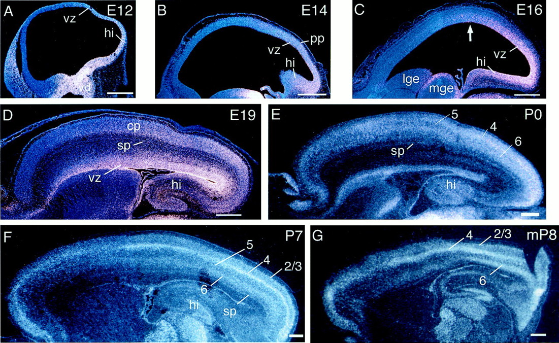
Differential expression of COUP-TFIacross the rostral–caudal axis of developing neocortex. In situ hybridizations using S35-labeled riboprobes on sagittal sections of rat (A–F) or mouse (G) forebrain are shown. A, At E12, COUP-TFI is expressed in a graded manner in the dorsal telencephalon, with higher levels in the future hippocampus decreasing to lower levels in the ventricular zone of the future neocortex. B, A similar gradient ofCOUP-TFI expression is seen at E14, in the ventricular zone and preplate. C, At E16, COUP-TFIexpression is still strongly graded, with expression highest in the ventricular zone. The high expression appears to drop abruptly in the presumptive, future somatosensory area (arrow).D, The high-caudal-to-low-rostral gradient is apparent in all layers at E19, with the strongest expression in the ventricular zone. E, At P0, the ventricular zone has thinned, andCOUP-TFI expression is seen in the cortical plate and subplate. Expression is still highest caudally and in developing layer 4. F, COUP-TFI expression at P7 is still highest caudally in all layers of the cortical plate. Layer 4 shows the strongest expression, which declines abruptly at the presumptive rostral border of visual cortex and exhibits another sharp decrease at the presumptive rostral border of somatosensory cortex.G, A sagittal section of P8 mouse brain shows aCOUP-TFI expression pattern similar to that seen in P7 rat. The highest expression is in layers 4 and 6 of the cortical plate; the two layers exhibit strong, but differing, caudal-to-rostral gradients of expression. A–D are simultaneous exposures using both dark field to show silver grains and UV illumination to show DAPI staining; E–G are only dark-field exposures. Rostral is to the left, and dorsal is up.cp, Cortical plate; hi, hippocampus;lge, lateral ganglionic eminence; mge, medial ganglionic eminence; pp, preplate;sp, subplate; vd, ventral diencephalon; vz, ventricular zone. Arabic numerals indicate differentiated layers of the cortical plate. Scale bars, 500 μm.

COUP-TFI and CHL1are expressed in layer-specific patterns in developing neocortex.In situ hybridizations using S35-labeled riboprobes on sagittal sections of rat forebrain are shown. The left panel in each pair shows a dark-field image of the silver grains indicating gene expression. The right panel in each pair shows the identical field imaged either with a UV fluorescence for DAPI nuclear staining (A′, B′, F′, G′) or with bright-field illumination for thionin Nissl staining (C′–E′, H′–J′). Ages are indicated at thetop. A, A′–E, E′,COUP-TFI shows early expression in the ventricular zone of caudal cortex at E14 (A, A′) and E16 (B, B′). At E19 (C, C′) and P0 (D, D′), expression is seen in the cortical plate and subplate as well as the ventricular zone and subventricular zone. At P7 (E, E′), when all cortical layers have formed,COUP-TFI expression is highest in layer 4 but is also significant in all layers of the cortical plate and the subplate.F, F′–J, J′, In contrast to COUP-TFI,CHL1 shows early expression only in the preplate at E14 (F, F′) and later in the intermediate zone and the cortical plate at E16 (G, G′). At E19 (H, H′), it is expressed strongly in the cortical plate and subplate and weakly in the subventricular zone and the intermediate zone. At P0 (I, I′) and P7 (J, J′), expression of CHL1 is still strong throughout the cortical plate but is highest in layer 5. All photos were taken at a rostral–caudal location in the cortex that lies approximately above the hippocampus or just anterior to it. cp, Cortical plate; iz, intermediate zone; mz, marginal zone; pp, preplate; sp, subplate; svz, subventricular zone; vz, ventricular zone. Arabic numerals indicate differentiated layers of the cortical plate. Scale bars, 200 μm.
Analysis of later ages shows that COUP-TFI expression remains graded throughout cortical development and in addition exhibits layer specificity. At E19, near the end of cortical neurogenesis and when TCAs are beginning to invade the cortical plate,COUP-TFI expression remains strongly graded with the highest levels caudally (Fig. 2D). At this time,COUP-TFI is expressed in the ventricular, subventricular, and lower intermediate zones, as well as in the subplate, cortical plate, and marginal zone (Fig. 3C,C′). At P0,COUP-TFI is expressed throughout the radial extent of the neocortex and in a high-caudal-to-low-rostral gradient in all layers except in the diminished ventricular zone (Fig. 2E). Expression in the cortical plate shows laminar differences, being highest in the nascent layer 4 and lowest in layer 5 (Figs.2E, 3D,D′). Similarly, at P7, when all cortical layers have formed, COUP-TFI is expressed in the subplate and all cortical plate layers, and expression remains highest in layer 4 and lowest in layer 5 (Fig. 2F,3E,E′). Expression is still graded with the exception of layer 4, which exhibits a discontinuous differential pattern of expression. In layer 4, expression is highest caudally, in the presumptive primary visual area, moderate in presumptive somatosensory cortex, and low in a domain between the two as well as more rostrally, in the presumptive primary motor area. Expression declines precipitously at what appears to be the rostral border of the primary somatosensory area (Fig. 2F).
Qiu et al. (1994) reported that COUP-TFI expression is strong in mouse neocortex at E14.5 (a stage similar to E16 rat) but is not detected at E18.5 (a stage similar to E20 rat). Because this report sharply contrasts with our finding of persistent COUP-TFIexpression in postnatal rats and because mice deficient forCOUP-TFI exhibit an excessive, postnatal loss of layer 4 neurons (Zhou et al., 1999), we examined COUP-TFI expression in the neocortex of postnatal mice. Indeed, in situhybridization using the 31v1 probe for COUP-TFI reveals an expression pattern in postnatal mice similar to that in rats, including strong expression in layer 4 in P8 mice (Fig. 2G), as well as in P2 mice (data not shown). This result suggests a cell-autonomous mechanism for the death of layer 4 neurons in the COUP-TFImutant.
To determine whether COUP-TFI expression is graded along the medial–lateral axis of embryonic and postnatal rat neocortex, we performed in situ hybridizations on coronal sections. At all ages examined, COUP-TFI is differentially expressed along the medial–lateral axis with a high-lateral-to-low-medial gradient at both rostral and caudal levels (Fig. 4).COUP-TFI is also expressed in the lateral cortical stream (Fig. 4A,D,F), a population of migrating cells that originate from the lateral part of the cortical ventricular zone and have been proposed to populate the ventrolateral cortical plate (Bayer and Altman, 1991) or, alternatively, the claustrum and laterobasal amygdala (Puelles et al., 1999). Interestingly,COUP-TFI is also expressed in premigratory and migrating neural crest (Qiu et al., 1997). At E19, when TCAs are invading the cortical plate, COUP-TFI expression is highest in the superficial part of the lateral cortical plate (Fig.4D,E). At P0, COUP-TFI expression is still present in the lateral cortical stream, as well as graded in the cortical plate (Fig. 4F,G). By P7, the lateral cortical stream is no longer present. Within layers 2/3, 5, and 6,COUP-TFI still exhibits a high-lateral-to-low-medial graded expression; expression in layer 4 appears uniform in the lateral gustatory and somatosensory areas but declines abruptly in more medial frontal cortex (Fig. 4H,I).

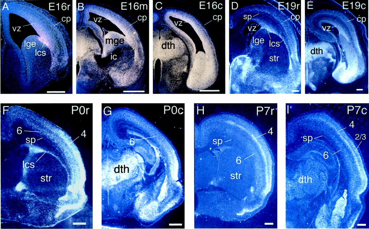
COUP-TFI is expressed in a high-lateral-to-low-medial gradient in developing neocortex, as well as throughout the dorsal thalamus. In situ hybridizations using S35-labeled riboprobes on coronal sections of rat forebrain are shown. A–C, A series of coronal sections of E16 rat brains taken at rostral, mid, and caudal levels along the rostral–caudal axis of the neocortex is shown. At each level, COUP-TFI exhibits a high-lateral-to-low-medial graded expression within the neocortex. A, Rostrally, graded COUP-TFI expression is evident in the cortical ventricular zone. In addition, COUP-TFIexpression is apparent in the lateral cortical stream.B, A mid-level section shows strongCOUP-TFI expression in the dorsal thalamus and graded expression in the cortical ventricular zone. COUP-TFIexpression is also high in the medial ganglionic eminence and within the ventral telencephalon closely associated with the internal capsule, the pathway of TCAs. C, A more caudal section shows very strong expression in the cortical ventricular zone and in the dorsal thalamus, as well as in the thin cortical plate laterally. D, E, By E19, COUP-TFI expression is seen in the subplate and cortical plate in addition to the ventricular zone, lateral cortical stream, and dorsal thalamus. At both rostral (D) and caudal levels (E), the high-lateral-to-low-medial gradient is apparent. F, G, At P0, future layers 4 and 6 exhibit high expression ofCOUP-TFI laterally within the neocortex, and the dorsal thalamus and lateral cortical stream continue to expressCOUP-TFI. H, I, By P7, all cortical layers have formed. Although expression in the dorsal thalamus has declined, cortical expression of COUP-TFI remains robust. Layers 2/3 and 6 exhibit high-lateral-to-low-medial graded expression of COUP-TFI. However, layer 4 shows relatively even expression from the parietal cortex to the rhinal fissure. Images in A–C are simultaneous exposures of dark-field illumination to show silver grains and UV illumination to show DAPI staining; D–I are dark-field images. Dorsal is up, and lateral is to theright. c, Caudal level;cp, cortical plate; dth, dorsal thalamus;ic, internal capsule; lcs, lateral cortical stream; lge, lateral ganglionic eminence;m, mid level; mge, medial ganglionic eminence; r, rostral level; sp, subplate;str, striatum; vz, ventricular zone.Arabic numerals indicate differentiated layers of the cortical plate. Scale bars: A–C, 500 μm;D–I, 1 mm.
Because the majority of TCAs fail to reach the cortex inCOUP-TFI-mutant mice (Zhou et al., 1999), we examinedCOUP-TFI expression relative to the internal capsule, the path of TCAs through the ventral telencephalon, and in the dorsal thalamus, the origin of the TCA projection. At E16, when many TCAs are extending through the internal capsule, COUP-TFI is highly expressed in the medial ganglionic eminence at the level of the internal capsule, as well as in ventral telencephalic cells positioned close to and within the internal capsule (Fig. 4B).COUP-TFI is also highly expressed in the dorsal thalamus at E16 (Fig. 4C), E19 (Fig. 4E), and P0 (Fig.4G), ages that cover much of the period during which TCAs extend from the dorsal thalamus to the neocortex and invade the cortical plate. COUP-TFI expression in the dorsal thalamus declines substantially by P7 (Fig. 4I). These findings suggest that COUP-TFI may regulate TCA development by influencing the differentiation of ventral telencephalic cell groups that direct TCA pathfinding (see Tuttle et al., 1999; Braisted et al., 2000) and of the dorsal thalamic nuclei that give rise to TCAs.
Graded and layer-specific expression of CHL1 in developing neocortex
Previous reports of CHL1 expression are limited (Holm et al., 1996; Hillenbrand et al., 1999). Northern blot analysis ofCHL1 expression shows that it is first expressed at E12 in mouse. Immunohistochemical analysis shows that CHL1 protein is present in subpopulations of neurons, astrocytes, oligodendrocyte precursors, and Schwann cells in mouse and rat. The brief description ofCHL1 expression in neocortex mentions that it is preferentially expressed in layer 5 in postnatal mouse (Hillenbrand et al., 1999).
To better define possible roles for CHL1 in cortical development, we analyzed its expression in E12 to P7 rat forebrain by the use of in situ hybridization. At E12, CHL1 is expressed in postmitotic neurons of the presumptive hippocampus, septum, and ventral diencephalon. CHL1 is not expressed, however, in E12 neocortex (Fig.5A). At E14, CHL1expression extends from the hippocampus into caudal neocortex and is only present in the outermost layer of cells including the preplate (Figs. 3F,F′, 5B). CHL1 expression is graded, being higher in caudal preplate. At E16, CHL1 is expressed in the intermediate zone and at lower levels in the cortical plate of neocortex (Fig. 3G,G′). Interestingly,CHL1 expression shows an abrupt decline in the rostral intermediate zone; this abrupt change in expression is not evident in the marginal zone and cortical plate (Fig. 5C). This differential expression of CHL1 before the arrival of TCAs indicates that it is established independent of their potential influences.

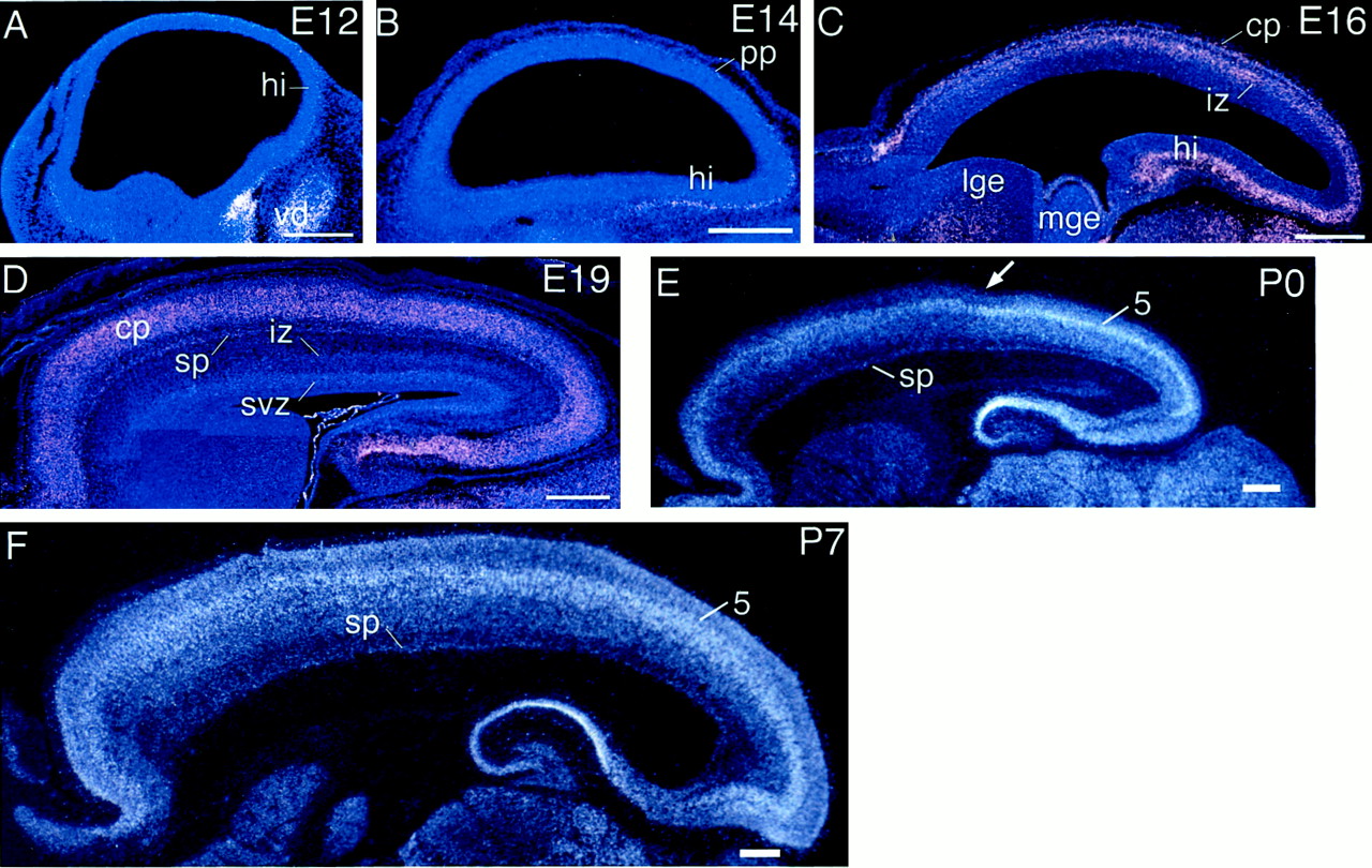
Differential expression of CHL1across the rostral–caudal axis of developing neocortex. In situ hybridizations using S35-labeled riboprobes on sagittal sections of rat forebrain are shown.A, At E12, rat CHL1 is not expressed in the developing neocortex but is present in the hippocampal primordium, the septum, and the ventral diencephalon. B, At E14, within the neocortex, CHL1 is expressed only in the caudal part of the preplate. C, By E16,CHL1 is expressed relatively uniformly rostrocaudally across the cortical plate but exhibits a high-caudal-to-low-rostral graded expression within the intermediate zone that shows a substantial decline rostrally. D, At E19,CHL1 is expressed relatively uniformly rostrocaudally across the cortical plate and subplate; low levels of expression are detected in the intermediate zone and subventricular zone, which is weakly graded with subtly higher levels caudally. E, At P0, CHL1 is expressed in all layers of the cortical plate, but its expression is substantially higher in layer 5. In addition, CHL1 expression is highest in the caudal neocortex, within the presumptive visual cortex, and shows a substantial decline within the presumptive somatosensory cortex (arrow). F, At P7, layer 5 expression is still differential and higher caudally, but the differences in expression levels along the rostral–caudal axis are not as pronounced as at P0. Rostral is to the left, and dorsal isup. A–D are simultaneous exposures of dark-field illumination to show silver grains and UV fluorescence to show DAPI staining; E and F are dark field alone. cp, Cortical plate;hi, hippocampus; iz, intermediate zone; lge, lateral ganglionic eminence;mge, medial ganglionic eminence; pp, preplate; sp, subplate; svz, subventricular zone; vd, ventral diencephalon;5, layer 5. Scale bars, 500 μm.
At E19, CHL1 is highly expressed in the cortical plate, subplate, intermediate zone, and subventricular zone. Expression is not detectable, however, in the marginal zone and ventricular zone (Fig.3H,H′). Although expression in the cortical plate appears to be uniform, expression in the subventricular and intermediate zones is graded with higher expression caudally (Fig. 5D). At P0,CHL1 is expressed throughout the cortical plate, but the highest expression is found in the nascent layer 5 (Fig.3I,I′). The expression in layer 5 is highest in caudal neocortex, including the presumptive primary visual area, and declines abruptly in the presumptive somatosensory area (Fig. 5E,arrow). At P7, CHL1 is expressed in all cortical layers and remains highest in layer 5 (Fig. 3J,J′). Expression in layer 5 is higher caudally than rostrally, although this differential expression is not as obvious as at P0 (Fig.5F).
Examination of CHL1 expression on coronal sections reveals several interesting features. At E16, expression in the cortical plate is graded along the lateral–medial axis at both rostral (Fig.6A) and caudal (Fig.6B) levels. This could reflect the lateral-to-medial maturational gradient of the neocortex. At E19, expression in the cortical plate appears higher in the medial and lateralmost parts of the neocortex than in the parietal cortex interposed between them (Fig.6C). At both P0 (Fig. 6D) and P7 (Fig.6E), layer 5 expression is stronger in retrosplenial and insular cortices than in parietal cortex. This differential layer 5 expression is not observed within the neocortex at levels caudal to the hippocampus (data not shown), which is consistent with the expression observed in sagittal sections (Fig. 5F) showing high layer 5 expression in caudal neocortex. Apart from the cortex,CHL1 is widely expressed in other parts of the forebrain, including thalamus and hypothalamus (Fig. 6B,D), striatal mantle, olfactory tuberculum, septum (Fig.6A), and amygdala (Fig. 6C,D).

CHL1 is differentially expressed along the medial–lateral axis of the developing neocortex. In situ hybridizations using S35-labeled riboprobes on coronal sections of rat forebrain are shown. A, B, Coronal sections of E16 rat brains taken at a rostral (A) and a caudal (B) level along the rostral–caudal axis of the neocortex are shown.CHL1 is expressed in the intermediate zone and the cortical plate. Rostrally, CHL1 exhibits high-lateral-to-low-medial graded expression within the cortical plate but high-medial-to-low-lateral graded expression within the intermediate zone. Caudally, CHL1 appears to exhibit high-lateral-to-low-medial graded expression in both the cortical plate and intermediate zone. C, At E19, CHL1 is present in the cortical plate and subplate and is most highly expressed in more medial and lateral parts of the cortex, with lower levels in between. D, At P0, the pattern of expression is similar to that described for E19, with the notable exception thatCHL1 expression is substantially higher in layer 5 than in other layers. E, By P7, CHL1expression appears lower than at P0. Expression in layer 5 continues to be higher than that in other layers and exhibits the same pattern as at P0, being higher in more lateral and more medial parts of the cortex. Expression in the other layers is low and appears more-or-less uniform.Images shown in A and Bare simultaneous exposures using both dark-field illumination to show silver grains and UV fluorescence to show DAPI staining; those inC–E are dark field alone. c, Caudal level; cp, cortical plate; iz, intermediate zone; lge, lateral ganglionic eminence;r, rostral level; 5, layer 5. Scale bars, 500 μm.
Differential expression of two novel gene fragments across the developing neocortex
In addition to COUP-TFI and CHL1, we isolated fragments, referred to here as 2m3 and 36m1, of two potentially novel genes. In situ hybridization analysis in E16 rat, the age from which RNA preparations were made for the ddPCR screen, confirms that these fragments have differential patterns of expression across the developing neocortex. 2m3 is expressed in the ventricular and the subventricular zones, but little or no expression is detected in the cortical plate (Fig.7A,B). The expression in the ventricular and subventricular zones is graded with higher levels rostrally (Fig. 7A) and ventrolaterally (Fig.7B), where it extends into the basal forebrain, and lower levels caudally and dorsomedially.

Two novel gene fragments identified in the ddPCR screen show graded expression across E16 rat neocortex. In situ hybridizations using S35-labeled riboprobes were performed on sagittal (A, C) and coronal (B, D) sections and imaged using both dark-field illumination to show silver grains and UV fluorescence to show DAPI staining. A, Gene fragment 2m3 is expressed in the ventricular zone in a strong high-rostral-to-low-caudal graded pattern.B, On coronal sections 2m3 appears more highly expressed laterally than medially. The expression continues into the ganglionic eminences of the basal forebrain. C, Gene fragment 36m1 is expressed in the cortical plate in a strong high-rostral-to-low-caudal graded pattern. D, Expression of 36m1 is relatively uniform along the medial–lateral cortical axis, with the exception of a sharp decline laterally. cp, Cortical plate; lge, lateral ganglionic eminence;mge, medial ganglionic eminence; vz, ventricular zone. Scale bars, 500 μm.
In contrast to 2m3, the expression of 36m1 is detected only in the cortical plate (Fig. 7C,D). 36m1 exhibits a strong high-rostral-to-low-caudal graded expression within the neocortex (Fig.7C). Graded expression of 36m1 is less evident along the medial–lateral axis of the neocortex but does appear to decline ventrolaterally in presumptive insular cortex, while remaining strongly expressed in presumptive perirhinal cortex (Fig. 7D). As forCOUP-TFI and CHL1, the finding of early graded expression of 2m3 and 36m1 indicates that their differential expression is established independent of TCAs.
DISCUSSION
This study is the first report of a screen to identify genes differentially expressed across the developing neocortex in patterns suggesting a role in arealization. We have cloned four genes differentially expressed along the rostral–caudal and medial–lateral axes of the developing neocortex, as well as in a layer-specific manner. They include COUP-TFI and CHL1, neither of which has been reported previously to be differentially expressed in cortex, as well as two novel genes.
Roles for COUP-TFI in the development of the TCA projection and the survival of cortical neurons
The COUP-TFs are orphan members of the steroid/thyroid hormone receptor superfamily implicated in regulating signaling pathways involved in development, including those mediated by retinoid acid receptors, retinoid X receptors, the vitamin D3 receptor, thyroid hormone receptors, and hepatocyte nuclear factor-4 (Tsai and Tsai, 1997). Mice have two COUP-TF genes, I andII, both expressed in the developing nervous system (Jonk et al., 1994; Qiu et al., 1994). In vitro studies suggest that COUP-TFI may influence expression of the immediate early response gene NGFI-A (Pipaon et al., 1999), the Purkinje cell-specific gene PCP-2 (Anderson et al., 1998), and the glutamate receptor subunit KA2 (Chew et al., 1999).
COUP-TFI mutant mice have a substantially reduced TCA projection, and TCAs fail to innervate the cortical plate. In addition, subplate and layer 4 neurons undergo excessive cell death (Zhou et al., 1999). Zhou et al. (1999) favor the interpretation that the diminished TCA projection is caused by defects in the putative guidance functions of subplate neurons, which as we show express COUP-TFI. If the diminished TCA projection is secondary to defects in subplate neurons, our finding of a high-caudal-to-low-rostral graded cortical expression of COUP-TFI would suggest that caudal parts of the neocortex, including the visual cortex, would be most severely affected. However, because COUP-TFI is highly expressed throughout development in dorsal thalamus in both mice and rats (Qiu et al., 1994) (present study), the defects in the TCA projection could be autonomous to TCA projection neurons; if so, we would not expect areal differences in the failure of TCAs to invade the cortical plate inCOUP-TFI mutants. In addition, because COUP-TFIis expressed within the ventral telencephalon at the level of the TCA pathway through it, the diminished TCA projection inCOUP-TFI mutants could be caused in part by an aberrant differentiation of ventral telencephalic cell groups and the expression of axon guidance molecules that have been implicated in TCA pathfinding (see Tuttle et al., 1999; Braisted et al., 2000). Zhou et al. (1999)also favor the interpretation that the postnatal loss of layer 4 neurons is caused by their lack of thalamic innervation. Although this is a valid possibility, our finding that layer 4 neurons in both rats and mice highly express COUP-TFI suggests the alternative explanation that their loss is cell autonomous. This would be consistent with the explanation suggested for the death of theCOUP-TFI-expressing neural crest precursors of the ninth cervical ganglion in COUP-TFI mutants (Qiu et al., 1997). Although Zhou et al. (1999) did not analyze COUP-TFIexpression, previous work from the same group (Qiu et al., 1994) reported that COUP-TFI is highly expressed in mouse cortex at E14.5 (a developmental stage similar to E16 rat), but not at E18.5 (similar to E20 rat)—we have no explanation for the discrepancy between these findings of Qiu et al. (1994) and our findings that COUP-TFI continues to be highly expressed in the neocortex of both postnatal rats and mice. If the loss of layer 4 neurons in the COUP-TFI mutant is cell autonomous, we would expect that it would be most pronounced in areas such as visual and somatosensory cortex that most highly express COUP-TFI.
CHL1 in cortical development
The similarity of CHL1 to the L1 family of cell adhesion molecules suggests that CHL1 may have similar functions (Holm et al., 1996). Many of the L1 family members have homophilic or heterophilic interactions and mediate cell–cell (or axon–axon) interactions during development, regeneration, and modification of synaptic activity (Rutishauser, 1993; Schachner, 1997).In vitro, CHL1 promotes neurite outgrowth by heterophilic binding to an unknown ligand. CHL1 is expressed by subpopulations of neurons and glia in the CNS and peripheral nervous system in a pattern that extensively overlaps with other L1 family members (Hillenbrand et al., 1999).
Our results suggest that within the neocortex, CHL1 is only expressed by postmitotic cells. Its graded expression in the intermediate and subventricular zones where neurons are migrating toward the cortical plate suggests that it may regulate cell–cell interactions and neuronal migration differentially along the rostral–caudal axis. The timing of CHL1 expression in the intermediate zone and later its preferential expression in layer 5 suggest that CHL1 may primarily be involved in the migration and process extension of layer 5 neurons.
Novel genes
The two novel gene fragments that we have isolated, 2m3 and 36m1, appear to be partial sequences of novel genes. Both are expressed at E16 in a high-rostral-to-low-caudal graded pattern across the neocortex. However, their expression patterns differ along the medial–lateral axis, with 2m3 exhibiting high-lateral-to-low-medial graded expression, whereas the expression of 36m1 drops off laterally except in the perirhinal cortex. In addition, the two exhibit different laminar expression patterns: 2m3 is expressed in the ventricular zone suggesting that it is expressed by cortical progenitor cells, whereas 36m1 is expressed only in the cortical plate suggesting that is expressed by postmitotic cortical neurons.
Differential gene expression before thalamic input
COUP-TFI and CHL1 exhibit high-caudal-to-low-rostral expression patterns across the neocortex throughout its development, indicating that their graded expression patterns are not caused by the gradients of cortical neurogenesis and maturation but are established by other mechanisms. An issue that has received much attention recently is whether differential patterns of gene expression in the neocortex are established by mechanisms intrinsic to the cortex or by extrinsic influences such as TCAs (Miyashita-Lin et al., 1999; Nakagawa et al., 1999). Because of the timing of development of the TCA projection in rats (Catalano et al., 1991, 1996; De Carlos et al., 1995), differential gene expression evident in the neocortex at E16 or earlier must be established independent of TCAs. We find that COUP-TFI andCHL1 expression is graded in a high-caudal-to-low-rostral pattern as early as E12 and E14, respectively. In addition, the two novel genes 2m3 and 36m1 exhibit strong graded expression at E16, the earliest time that we have examined presently. Thus, the early differential expression of all four genes is established independent of TCAs and likely by a mechanism intrinsic to the neocortex, consistent with a role for them in directing arealization. However, bothCOUP-TFI and CHL1 exhibit substantial changes after the cortical plate is invaded by TCAs. Thus although the early differential expression of these genes is independent of thalamic influence, TCAs may modify their later patterns of expression. This possibility is especially intriguing for COUP-TFI because it is most highly expressed in layer 4, the principal target of TCAs. By P7, COUP-TFI expression in layer 4 is discontinuous, exhibiting abrupt decreases and increases, and appears to be most pronounced in the primary somatosensory and visual areas, which receive a prominent TCA input.
Implications for mechanisms controlling arealization
Two issues relevant to understanding mechanisms that control neocortical arealization are whether area-specific genes exist and whether they are required to differentiate areas. Genes described previously to be differentially expressed across the embryonic neocortex (see introductory remarks), as well as the four genes that we have identified, exhibit, at least initially, graded expression patterns that are not restricted to a single area. Interestingly, none of the 10,000 gene fragments that we examined in the ddPCR screen was expressed exclusively in rostral or caudal E16 rat neocortex; all of the 148 differentially expressed fragments were expressed in both rostral and caudal neocortex. Thus, if area-specific genes are expressed in E16 rat neocortex, they are rare and could have been missed in the screen for a variety of reasons. Our screen did not approach saturation because we did not identify genes shown previously to be differentially expressed in neocortex. In addition, although we screened ∼10,000 bands, some of which are duplicates or different fragments of the same gene, on average a neuron is estimated to express ∼15,000 genes, and our starting RNA samples were from heterogeneous cell populations.
At later stages of development, several genes, includingTbr1, Id2, RZRβ, EphA7,and several cadherins, as well as COUP-TFI as shown here, exhibit expression patterns characterized by relatively abrupt borders that might correlate with those between neocortical areas (Donoghue and Rakic, 1999b; Rubenstein et al., 1999). However, the expression of at least some of these genes, and perhaps all of them, is not limited to a single area. To date, the only example of a genetic marker restricted to a single area is the expression of the H-2Z1 transgene, which marks the granular parts of somatosensory cortex (Cohen-Tannoudji et al., 1994). Although H-2Z1 is not expressed until P2, its area-specific pattern of expression appears to be specified early in embryonic cortical development (Cohen-Tannoudji et al., 1994; Gitton et al., 1999a). Taken together, the available evidence suggests that a neocortical area is primarily defined by the expression of a unique subset of genes, each of which is also expressed in other areas, rather than by the expression of a specific set of genes restricted to that area.
Because neocortical areas have abrupt borders, it is likely (but not required) that the graded expression of genes that regulate arealization is translated in a manner that results in the expression of some downstream genes in patterns with abrupt borders that relate to the borders between areas. For example, the transcription factorsPax6 and Emx2 are expressed in opposing graded patterns across the rostral–caudal axis of the embryonic neocortex (Walther and Gruss, 1991; Stoykova and Gruss, 1994; Gulisano et al., 1996; Mallamaci et al., 1998). An analysis of Pax6 andEmx2 mutant mice suggests that arealization of the neocortex is disproportionately altered in these mutants in opposing manners predicted by their countergradients of expression (Bishop et al., 2000). Studies in Drosophila have shown that gradients of transcription factors can be translated into sharply bordered expression patterns of downstream genes via a thresholding mechanism based on concentration-dependent differences in binding efficacy to promoter and repressor elements (Rusch and Levine, 1996) or the combinatorial action of multiple transcriptional activators and repressors expressed in overlapping graded patterns (Stanojevic et al., 1991; Small et al., 1996).
Interestingly, the graded patterns of gene expression observed in the neocortex often continue beyond it into other regions of the cerebral hemisphere, including the limbic cortex, paleocortex, archicortex, and basal forebrain. These continuous graded expression patterns suggest an intriguing relationship between arealization of the neocortex and other cortical regions, as well as regionalization of the cerebral hemisphere.
Footnotes
↵* Q.L. and N.D.D. contributed equally to this work.
Correspondence should be addressed to Dr. Dennis D. M. O'Leary, Molecular Neurobiology Laboratory, The Salk Institute, 10010 North Torrey Pines Road, La Jolla, CA 92037. E-mail: doleary{at}salk.edu.
This work was supported by National Institutes of Health Grant R01 NS31558 (D.D.M.O.), Cancer Research Fund of the Damon Runyon–Walter Winchell Foundation Fellowship DRG-1544 (N.D.D.), and a National Institute of Neurological Diseases and Stroke National Research Service Award (Q.L.). We thank Matthias Gesemann and David Litwack for technical advice and Yasushi Nakagawa and Rebecca Tuttle for comments on this manuscript.


