

Article Figures & Data
Figures
- Fig. 1.
Temporal and regional patterns of damage after neonatal hypoxic ischemic injury. Dark-field photographs of coronal, Nissl-stained sections at rostral (A, C,E, G) and caudal (B,D, F, H) levels from rats killed at 0 (A, B), 24 (C, D), 48 (E,F), and 168 (G,H) hr after HI. Damaged areas appear as bright objects in a dark field. No injury was apparent at the 0 hr time point (A, B). Clusters of degenerated cells were detected in the cortex and striatum from 24 through 168 hr (7 d) after HI (C–H). Forty-eight hours after HI, lateral and laterodorsal thalamus, medial habenula, and CA1 hippocampus showed damage (F). Scale bar, 100 μm.
- Fig. 2.
Degenerating neurons and caspase-3 immunostaining 48 hr after HI. Dark-field images from 50 μm, Nissl-stained sections of parietal cortex (A), striatum (B), and CA1 hippocampus (C) on the ligated side. A columnar pattern of degenerating neurons was observed in parietal cortex (A). High-magnification images from the penumbra (A1) and ischemic core (A2) showed apoptotic cells with large, round, chromatin clumps (arrows, A1) and necrotic cells with small, punctate chromatin clumps (asterisks,A1, A2). The striatum showed a patchy pattern of clusters of normal and degenerating neurons (B). At higher magnification, apoptotic cells with large, chromatin clumps (arrows, B1) were observed. In CA1 (C), the bright ribbon of degenerating cells contained apoptotic (arrows,C1) and necrotic (asterisks,C1) cells. Caspase-3 immunoreactivity showed a granular and particulate pattern in damaged areas within cerebral cortex (A3), striatum (B2), and CA1 (C2). Noninjured areas, in right parietal cortex 0 hr after HI, at low (D) and high (A4,D1) magnification, showed only diffuse, nonparticulate background staining. Scale bars: A–D, 100 μm;A1–D1, 10 μm.
- Fig. 3.
Degenerating neurons and caspase-3 immunostaining 7 d after HI. Dark-field images of parietal cortex (A), striatum (B), and CA1 hippocampus (C) on the ligated side show a pattern similar to that observed 48 hr after HI. High-magnification bright-field views of the fields in A–C show degenerating cells with varied sizes of chromatin clumps (arrows, A1–C1). Punctate caspase-3 immunoreactivity was distributed in cortex (A2) and striatum (B2) and CA1 (C2) in which apoptotic cells were observed, but the density is less in CA1 at this time point than at 48 hr (compare with Fig. 2C2). Scale bars: A–C, 100 μm; A1–C2, 10 μm.
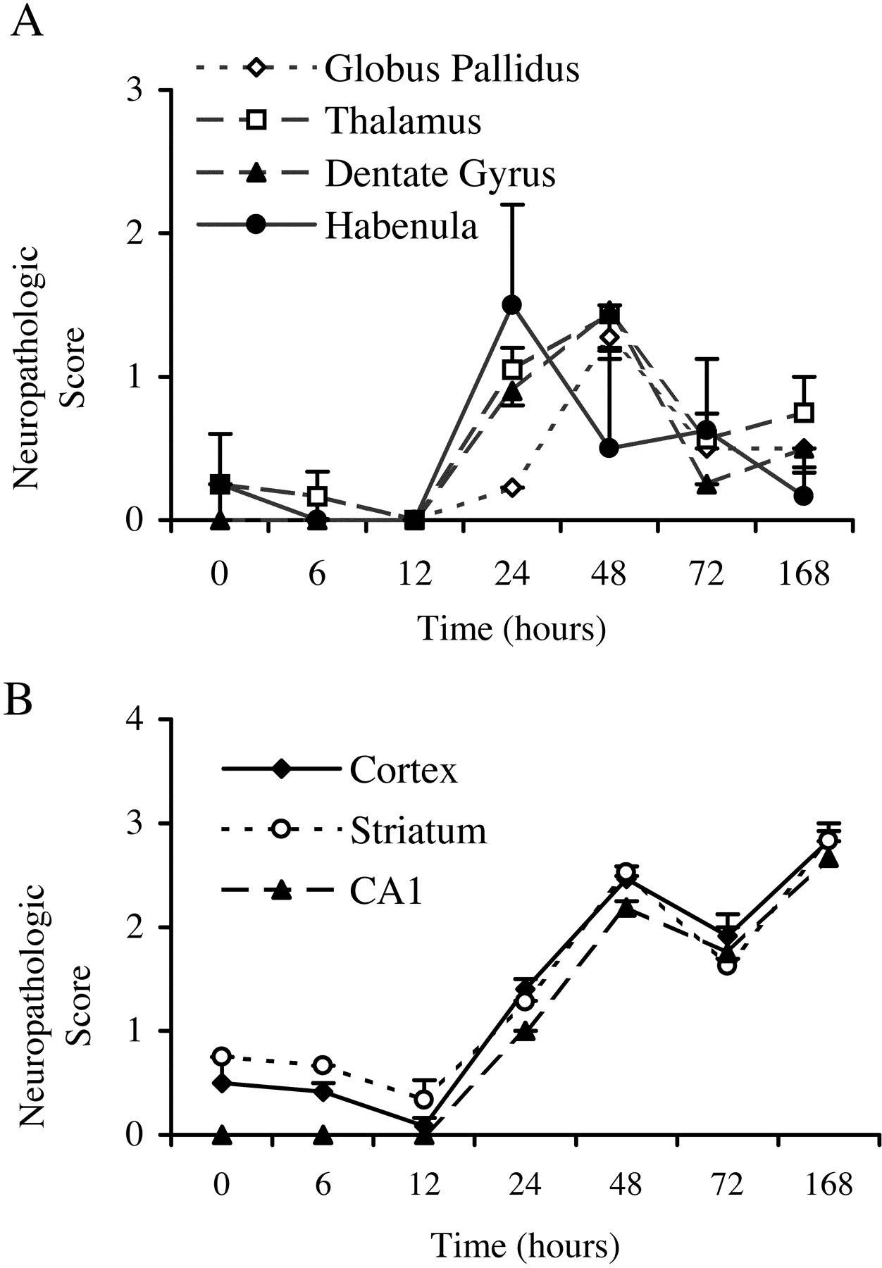
- Fig. 4.
Semiquantitative analysis of regional and temporal patterns of damage. Using the dark-field detection method, the degree of damage, which includes apoptosis, necrosis, and hybrid forms of cell death, was assessed semiquantitatively on a scale of 0 (little damage) to 3 (severe damage) at each time point by two investigators blinded to treatment (n = 2 at 0 and 6 hr;n = 3 at 12 and 168 hr; n = 5 at 24 and 48 hr; n = 4 at 72 hr). The average of the two scores for each animal was used to calculate the group means ± SEM shown here. In globus pallidus, thalamus, dentate gyrus, and medial habenula, the density of damaged cells peaked between 24 and 48 hr after hypoxic ischemic injury and then subsequently declined (A). In cortex, striatum, and CA1, the density of degenerating cells remained elevated through 7 d after HI (B).
- Fig. 5.
Semiquantitative analysis of regional and temporal patterns of necrotic and apoptotic forms of cell death. Examining adjacent Nissl-stained, H&E-stained, and TUNEL-labeled sections, two investigators blinded to treatment semiquantitatively (0 to 3; see Fig.4) assessed the degree of apoptotic and necrotic forms of cell death (n = 2 at 0, 6, 12, and 168 hr;n = 4 at 24 hr; n = 3 at 48 and 72 hr). The average of the two scores for each animal was used to calculate the group means ± SEM shown here. In frontal, parietal, and retrosplenial cortex, caudate putamen, globus pallidus, and CA1, the density of apoptotic cells remained high through 7 d after HI (A–F). In cingulate cortex, CA3, dentate gyrus, subiculum, laterodorsal thalamus, and medial habenula (G–L), the density of apoptotic cells peaked between 24 and 72 hr after hypoxic ischemic injury and then subsequently declined.
- Fig. 6.
TUNEL shows a patchy distribution in the striatum and hippocampus after neonatal hypoxic ischemic injury. Twenty-hour hours after HI, TUNEL-labeled cells form a patchy pattern in the striatum (A). Enlargement of the boxed region in A shows TUNEL-positive cells with round chromatin clumps typical of apoptotic cells (arrows, B); adjacent H&E-stained sections also show neurons with apoptotic features (arrows, C). Seventy-two hours after hypoxic ischemic injury, CA1 hippocampus shows a patchy pattern of TUNEL-positive and counterstained normal cells. Enlargement of theboxed region in D shows TUNEL-positive (E) and H&E-stained cells (F) with apoptotic (arrow,E) and necrotic (asterisks,E, F) morphologies. Scale bars:A, D, 200 μm; B,C, E, F, 10 μm.
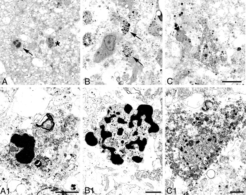
- Fig. 7.
Nuclear changes in apoptotic cells in cortex, 48 hr after HI. Light microscopic photographs of 1 μm, Nissl-stained sections (A–D) and electron micrographs (A1–D1) are shown. Normal neurons are shown inA and A1 for reference. Uniformly condensed, regularly shaped, round chromatin clumps were present in cells at early stages of apoptosis (B,B1). As apoptosis proceeded, neurons showed nuclear and cytoplasmic shrinkage (C, C1). At an advanced stage, phagocytosed apoptotic bodies and debris were observed (D, D1). Scale bars: A–D, 10 μm; A1, B1, D1, 2 μm; C1, 1 μm.
- Fig. 8.
Nuclear changes in apoptotic cells in thalamus and hippocampus, 48 hr after HI. Based on ultrastructural appearance, apoptotic neurons were identified in ligated thalamus (A, B) and hippocampus (C,D). The nucleus of neurons at a middle stage of apoptosis (A, C) showed round chromatin clumps surrounded by an intact nuclear envelope; the cytoplasm was shrunken and condensed but had an intact plasma membrane. Enlargement of the boxed area in A(A1) shows both swollen (asterisks) and normal (arrows) mitochondria, which are typical for cells undergoing apoptotic death. Apoptotic neurons at a more advanced stage (B, D) show more severe nuclear condensation and shrinkage of the cytoplasm. Scale bars:A–C, 2 μm; D, 1 μm;A1, 500 nm.
- Fig. 9.
Nuclear changes in degenerating cells in cortex 48 hr after hypoxia–ischemia. Light microscopic photographs of 1 μm, Nissl-stained sections (A–C) and electron micrographs (A1–C1) are shown. We found an intermediate type of degenerating neurons, hybrid cells (arrow, A, A1) with large, chromatin clumps in the nucleus that were similar in size to those found in cells undergoing apoptosis (see Figs. 7, 8) but that were more irregular in shape. Typical necrotic neurons had smaller clumps of irregularly shaped, condensed chromatin (asterisk inA; arrows in B,B1). Necrotic neurons, which had a relatively homogeneous nucleus with a few irregular chromatin clumps and condensed granular cytoplasm (C, C1) and were typically found in adult ischemic models, were rarely identified in this model. Scale bars: A–C, 10 μm;A1, C1, 2 μm; B1, 1 μm.
- Fig. 10.
Regional and temporal distribution of increased caspase-3 immunoreactivity after neonatal HI. Distribution of caspase-3-immunoreactive protein at the level of the anterior striatum (top) and at the mid-hippocampal level (bottom). Dots show the general distribution of elevated caspase-3 immunoreactivity but do not reflect quantitative values. Intense, punctate caspase-3 immunoreactivity was enhanced in frontal, parietal, and cingulate cortex and in the striatum 24 hr after hypoxic ischemic injury. The density of caspase-3 immunoreactivity in the striatum was higher dorsally than ventrally and decreased slightly by 7 d after HI. In thalamus, increased caspase-3 immunoreactivity was distributed in lateral, laterodorsal, and reticular nuclei with a peak in density at 48 hr after HI. In hippocampus, intense caspase-3 immunoreactivity was present in CA1 and in the dentate gyrus at 48 hr after insult but had nearly disappeared by 7 d after hypoxic ischemic injury.
- Fig. 11.
Regional and temporal differences in caspase-3 protein expression after neonatal HI. Brain tissue lysates (40 μg/lane) were subjected in 10% SDS-PAGE. In the striatum (A) on the ligated right side (R), caspase-3 cleavage product (p17) was expressed from 24 hr through 7 d after HI. In the right hippocampus (B), a faint p17 band was present at 12 hr after HI, and the density of the band increased at 24–48 hr but diminished at 72 hr and 7 d after HI. In the thalamus (C), the p17 band was present at the 72 hr and 7 d time points. In cerebral cortex (D), we did not detect the p17 subunit in either hemisphere at any time point. This experiment was performed using five animals at each time point (4 animals for HI and 1 nontreated control); representative blots are shown. α-Tubulin was used as a protein loading control.L, Left side.


