

Article Figures & Data
Figures
- Fig. 1.
Regional organization of the neurobeachin sequence by alignment with other BEACH-WD40 protein family members.Boxes indicate conserved sequences, andhorizontal lines indicate sequences that are poorly or not conserved. The BEACH domain is symbolized by a filled box, and WD40 repeat units by ovoids.Numbers above sequence regions indicate percentage amino acid sequence identity with the corresponding region of neurobeachin. Positions of RII binding sites in neurobeachin and DAKAP550 regions B are marked by small boxes. Sites of apparent differential splicing in neurobeachin and DAKAP550 are marked bytriangles. Oblique lines indicate that the N-terminal sequences of BGL and DAKAP550 are incomplete. Note that the amino acid scale begins at the common C terminus. The BGL sequence (accession number M83822) was corrected for a frameshift error in region G to achieve a predicted C-terminal sequence homologous to neurobeachin. The CEF10F2.1 sequence was obtained by combining two partial predicted reading frames from overlapping cosmids CEF10F2 and CEF35G12 of the Nematode Sequencing Project (accession numbers Z35598and Z46242) (Wilson et al., 1994). LYST, FAN, YCS2, and LvsA sequences are from Nagle et al. (1996), Adam-Klages et al. (1996), Wicksteed et al. (1991), and Kwak et al. (1999), respectively.
- Fig. 2.
Predicted amino acid sequences of mouse neurobeachin (mNbea), partial chicken neurobeachin (cNbea), and DAKAP550 (DAKAP). The partial DAKAP550 sequence in regions A–D (short variant, DAKAP550S, without the facultative insert in region A) was taken from Han et al. (1997) and completed by us in regions D–G. Approximate borders between regions A and G, based on additional sequence comparisons with BGL, CEF10F2.1, LYST, FAN, YCS2, LvsA, and BEACH-containing ESTs, are indicated above the neurobeachin sequence by diamond symbols and letters. The sequence database accession numbers for mouse and chicken neurobeachin and for the completed DAKAP550 sequence are Y18276–Y18278.
- Fig. 3.
Protein kinase A regulatory subunit binding to neurobeachin measured by surface plasmon resonance (SPR). A, RIIα binds to regions B of neurobeachin and DAKAP550 but not BGL (analyte concentrations, 1.5 μm each). B, SPR tracings of a concentration series of neurobeachin region B binding to RIIα performed for Kd determination.C, Neurobeachin region B binds RIIα and RIIβ but not RIα or RIβ (analyte concentration, 1.5 μm).D, The RII binding peptide from AKAP Ht31 competes with neurobeachin region B for RIIα binding (neurobeachin region B concentration, 1 μm). E, Dissection of neurobeachin region B to delineate the RII binding sequence. Region B comprises amino acids 951–1311. Constructs C1–C3 reach from amino acid 951 to 1225, 1133, and 1041, respectively. Constructs D1–D3 extend from amino acids 1005, 1088, and 1177, respectively, to 1311.Kd values were determined for these constructs by measuring concentration series as in part B and are indicated beside the constructs. As described in Materials and Methods, construct C2 (asterisk) displayed high-affinity binding only if prepared under nondenaturing conditions. D2 and D3 binding were very weak, with approximate Kd values of 5 and 15 μm, respectively, and no binding was detectable with C3 (detection limit, 20 μm). An open frame indicates the sequence region in which the high-affinity RII binding property resides (amino acids 1022–1107). Because known RII binding sites are ∼20 amino acids long, we assume that if the binding site in region B should overlap with the end of a nonbinding construct, C3 or D2, it will do so for a maximum of 20 residues. The candidate core RII binding sequence (amino acids 1081–1099) with high α-helix potential is indicated by a hatched frame in the overview and an open box in the sequence at the bottom. F, Helical representation of amino acids 1081–1099 illustrating the highly amphiphilic nature of this structure. Filled circles symbolize hydrophobic residues, and open circles indicate hydrophilic amino acids.
- Fig. 4.
cAMP-agarose coprecipitates neurobeachin with RII from rat brain cytosol. Immunoblots were developed with antibodies against neurobeachin (Nbea), EEA1(negative control), and RIIα as indicated: cytosol,lane 1; cAMP-agarose beads after incubation in cytosol without (lane 2) or with (lane 3) 10 mm competitor cAMP. γ-Adaptin as a second negative control (data not shown) gave the same result as EEA1. The RIIα band of the cytosol sample is displaced downward by the thick, unlabeled tubulin band migrating above it.
- Fig. 5.
Brain-specific expression of neurobeachin.A, Chicken neurobeachin mRNA [10 μg poly(A+) RNA per lane]. B, Mouse neurobeachin protein (50 μg protein of tissue homogenate per lane). Tissue abbreviations are as follows: A, adrenal gland;B, brain; BS, brain stem;C, cerebellum; FB, forebrain;H, heart; I, small intestine;K, kidney; Li, liver; Lu, lung; M, muscle; O, ovary;Pa, pancreas; Sp, spleen;St, stomach; Te, testis. Smears and minor bands below the main bands are attributed to partial degradation of these very long mRNA and protein molecules.
- Fig. 6.
Immunocytochemical localization of neurobeachin in rat brain at the light-microscopical level.A, Survey photomicrograph of a frontal 50 μm vibratome section exhibits intense staining of perikarya-rich areas, moderate staining of neuropil-rich areas, and weak staining of myelin-rich areas. Views from the parietal cortex (B) and the hippocampal formation (C) illustrate that many different neuronal populations are immunopositive for neurobeachin (cc, corpus callosum; dg, granule cell layer of the dentate gyrus). Note the differential staining of different neuropil-rich areas in C. Views at high magnification from the CA1 area of the hippocampus (D) and the Purkinje cell layer of the cerebellum (E) show the coarsely granular subcellular pattern of neurobeachin in cell bodies at the light microscopical level. Scale bar (shown in C): A, 1.8 mm;B, 210 μm; C, 250 μm;D, 50 μm; E, 55 μm.
- Fig. 7.
Immunoperoxidase electron microscopy of neurobeachin-positive subcellular structures in rat brain neurons.A, The electron-dense neurobeachin immunoreaction product decorates polymorphic tubulovesicular endomembranes (arrowheads) and plasma membrane buds (arrows) of a cerebellar Purkinje cell.B, Neurobeachin immunolabeling of a vesicle cluster next to the concave face of a Golgi stack (arrowhead) in a Purkinje cell body. A multivesicular body (mb) is immunonegative, as generally observed. C, Labeling of the dilated ends of Golgi-like membrane cisternae in a pyramidal neuron of the hippocampus. D, An extensive field of immunopositive tubulovesicular endomembranes surrounded by several Golgi stacks (arrows) in a Purkinje cell.er, Endoplasmic reticulum; n, nucleus. Scale bar (shown in A): A, 1 μm;B, 0.6 μm; C, 0.5 μm;D, 0.8 μm.
- Fig. 8.
Pre-embedding immunogold electron microscopy of neurobeachin-positive endomembranes in rat cerebellar neurons.A, Immunolabeling concentrating at the concave side of a Golgi stack in the cell body of an unidentified neuron in the molecular layer. B, Labeling of the dilated ends of an unidentified endomembrane tubule or cisterna in a Purkinje cell body.C, Labeling of the postsynaptic membrane of a granule cell dendrite in contact with a Golgi cell axon terminal.D, Labeling of small vesicle or tubule cross sections (arrows) in unidentified cell processes in the granule cell layer. a, Axon terminal; d, dendrite; er, endoplasmic reticulum; m, mitochondrion; n, nucleus. Scale bar (shown inA): A, 0.50 μm; B, 0.37 μm; C, 0.26 μm; D, 0.27 μm.
- Fig. 9.
Punctate pattern of neurobeachin immunofluorescence in PC12 cells and effects of AlF4− and BFA. A, AlF4− induces pericentrosomal clustering of perinuclear neurobeachin, concurrent with KDEL receptor (double-immunofluorescence images) and β-COP (data not shown).B, BFA rapidly disperses punctate neurobeachin (left column) close to the nucleus but not in the cell periphery. β-COP (central column; corresponding double-immunofluorescence images) and mannosidase II (right column; representative images from a separate neurobeachin/mannosidase II double immunofluorescence) illustrate the different time courses of the dispersion, by BFA, of a coat protein versus an intrinsic Golgi membrane protein.
- Fig. 10.
Recruitment of neurobeachin from cytosol to Golgi-near membranes is stimulated by GTPγS and antagonized by BFA: immunoblot analysis. Freeze-permeabilized, washed COS-7 cells were incubated for 10 min with cytosol buffer only (buffer), with cytosol only (cytosol), or with cytosol supplemented either with 100 μg/ml BFA or with 0.5 mmGTPγS. For sequential drug treatments, the first 10 min incubation with either BFA or GTPγS was followed by a second 10 min incubation with both reagents combined, all in cytosol. Cells were then washed and pelleted, and proteins bound to them were analyzed by immunoblotting. The first lane (reference) carries a reference aliquot of the cytosol.
- Fig. 11.
Recruitment of neurobeachin from cytosol to Golgi-near membranes is stimulated by GTPγS and antagonized by BFA: double-immunofluorescence analysis. The localization of newly recruited neurobeachin in the presence of BFA versus GTPγS (left/right) is shown in comparison to the reference proteins (top/bottom) giantin (Golgi membranes), γ-adaptin and β-NAP (AP-1 and AP-3 coats), and RIIα (negative control). It can be seen that neurobeachin and the two coat proteins are recruited in a BFA/GTPγS-sensitive fashion to giantin-positive ribbons and, with different relative preferences, additionally to patches in the cell periphery. Recruitment experiments analyzed by immunofluorescence were also performed under the additional incubation conditions of Figure 10 (buffer only, cytosol only, sequential BFA and GTPγS treatments), producing relative recruitment intensities in agreement with Figure 10 (data not shown).
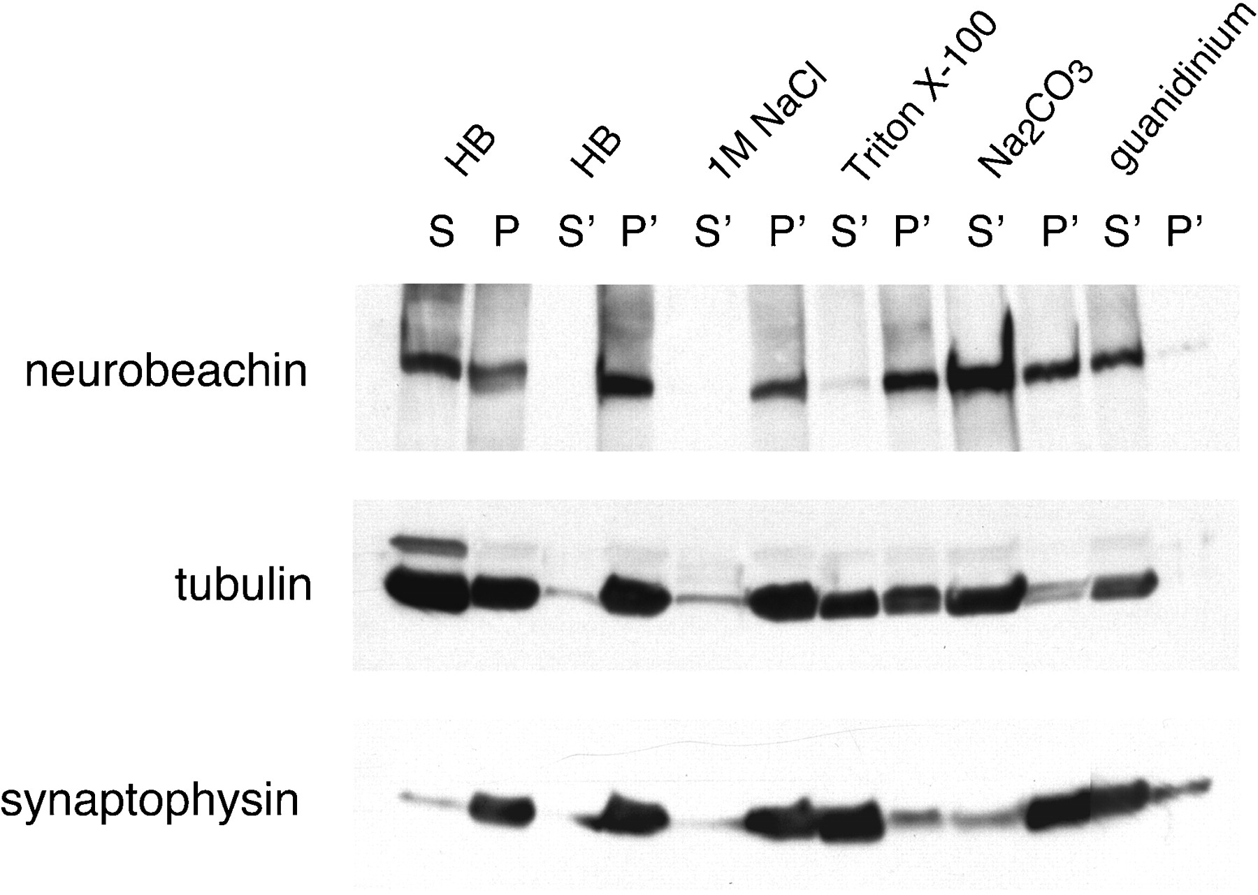
- Fig. 12.
Subcellular fractionation indicates cytosolic and cytoskeletal-associated subpools of brain neurobeachin. Mouse brain homogenate was subjected to 120,000 × gfractionation (S, supernatant; P, washed pellet) in a homogenization buffer (HB) containing 150 mm NaCl as described in Materials and Methods. The pellet fraction P was resuspended in the homogenization buffer (HB) or in various extraction buffers (1 mNaCl in homogenization buffer; 1% Triton X-100 in homogenization buffer without NaCl; 100 mmNa2CO3, pH 11; 6 mguanidinium chloride) and recentrifuged at 120,000 ×g. Supernatant and pellet fractions after recentrifugation are termed S' and P'. Equal aliquots of all fractions were analyzed by SDS-PAGE and immunoblotting with neurobeachin, tubulin, and synaptophysin antibodies. In the experiment shown, extraction was performed at 4°C for 20 min. The same distribution was obtained when extraction was performed at room temperature for 30 min.


