

Article Figures & Data
Figures
- Fig. 1.
TRP is required for the localization of INAD signaling complexes. A, Anti-INAD immunofluorescent staining of cross sections (1 μm thick) of wild-type (wt), inaC209,norpAP41, andtrp343 null mutant photoreceptors. Normal rhabdomeric localization of INAD is seen ininaC209 andnorpAP41 mutants, whereas it is mislocalized severely in trp343mutants. B, INAD, PLC, and eye-PKC also are mislocalized in trp mutants. Shown is immunofluorescent staining of cross sections of trp343 null photoreceptors. Note that Gα, the G-protein protein that shuttles between activated rhodopsin and transducisomes (Bahner et al., 2000), shows normal rhabdomeric localization. C, EM immunogold localization of INAD in wild-type andtrp343 rhabdomeres, demonstrating that ∼25% of the INAD seen in wild-type rhabdomeres still remains intrp mutant rhabdomeres. We cannot exclude the possibility that this small amount of INAD may be binding to TRPL channels in the rhabdomeres.
- Fig. 2.
Mutations that disrupt the INAD–TRP interaction display mislocalization of INAD and TRP. Shown is immunofluorescent staining for INAD and TRP in cross sections (1 μm thick) of wild-type (wt), inaDPDZ3, andtrpC34 transgenic photoreceptors (as indicated). inaDPDZ3 flies express an INAD protein containing three point mutations expected to disrupt the INAD–TRP interaction (see Materials and Methods).trpC34 flies express a truncated TRP protein that is missing its PDZ binding site (C-terminal 34 amino acids deleted). INAD and TRP both are mislocalized severely ininaDPDZ3 andtrpC34 transformants. Given that rhodopsin (Rh1) is not part of the INAD signaling complex (Tsunoda et al., 1997; Huber et al., 1998; B. H. Shieh, personal communication) and that Gα shuttles between Rh1 and the transducisome (Tsunoda et al., 1997; Bahner et al., 2000), we examined the localization of Rh1 and Gα as controls for normal rhabdomeric labeling. Indeed, rhodopsin and Gα are localized normally intrp, inaDPDZ3, andtrpC34 mutant backgrounds (data not shown).
- Fig. 3.
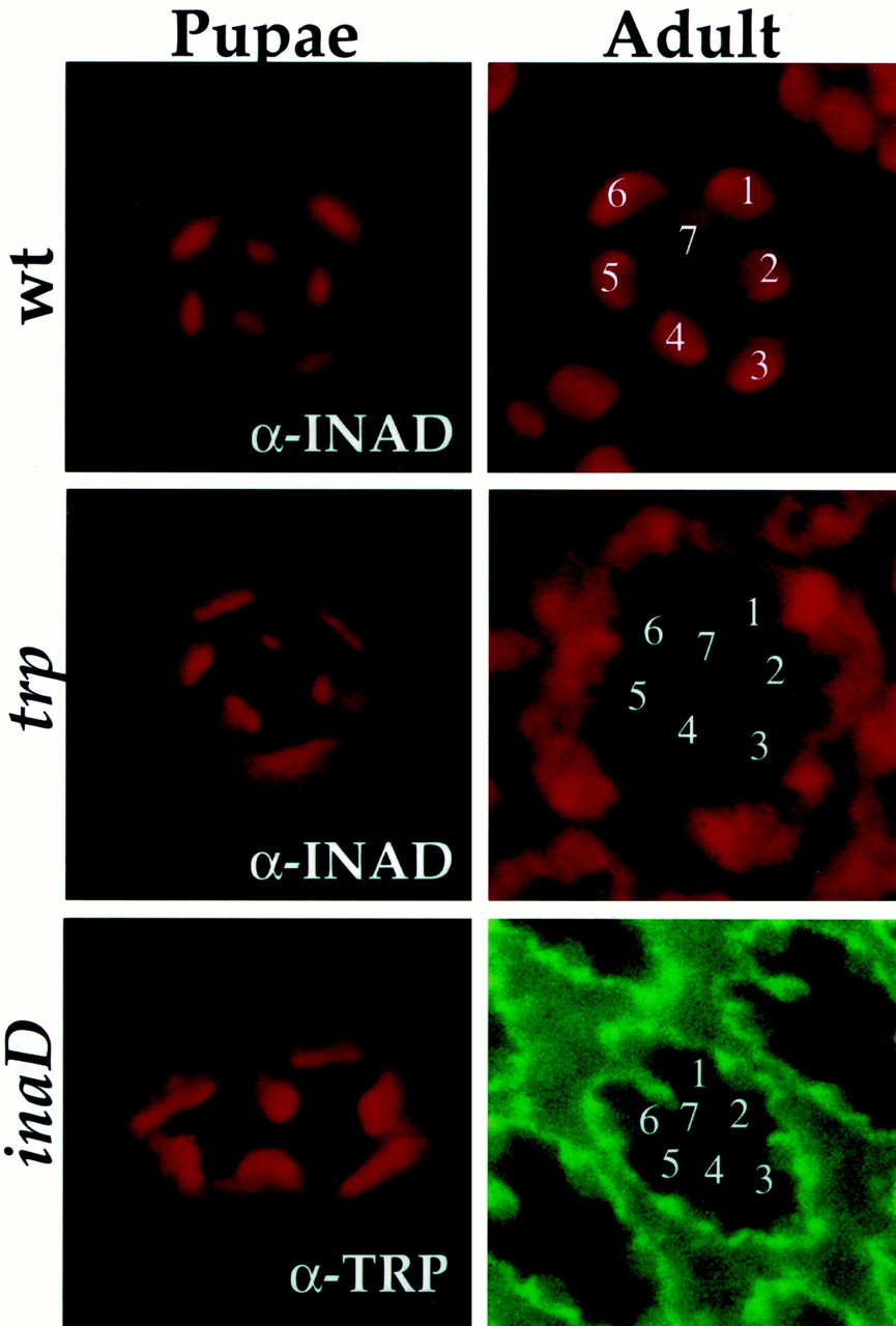
INAD and TRP are targeted independently to the rhabdomeres in pupae but require each other to be maintained in the rhabdomeres. Shown is anti-INAD and anti-TRP (as indicated) immunofluorescent staining of cross sections (1 μm thick) of wild-type (wt),trp343, andinaD1 null photoreceptors from pupae and adult flies. In wild type the INAD (top row) and TRP (data not shown) are targeted to the rhabdomeres in pupae and remain localized in the rhabdomeres of adult photoreceptors. Intrp343 mutants (middle row) INAD is targeted to the rhabdomeres in pupae but becomes mislocalized in adult photoreceptors. Similarly, ininaD1 null mutants (bottom row) TRP is targeted to the rhabdomeres in pupae but becomes mislocalized in adult photoreceptors. Rhabdomeres of individual R1–R7 photoreceptors are indicated by numbers.
- Fig. 4.
PDZ1 is required for localizing INAD at the membrane of CHO cells. A–C, Wild-type and mutantinaD constructs containing point mutations (see Materials and Methods) in PDZ1, PDZ2, PDZ3, PDZ4, or PDZ5 were transfected into CHO cells. Shown are confocal images of immunofluorescently stained CHO cells transfected with wild-typeinaD (INAD; A),inaDPDZ1 (PDZ1;B), and inaDPDZ2(PDZ2; C). Left, Anti-INAD staining. Right, Anti-INAD (green) superimposed with rhodamine-conjugated phalloidin staining (red). INAD shows membrane-associated staining, as seen in A and E, in 72.7% of transfected cells (n = 414), whereas PDZ1 shows membrane-associated staining in only 19.9% of transfected cells (n = 272). In most cells PDZ1 (B, F) is expressed diffusely throughout the cell. The percentage of transfected cells displaying membrane-associated localization was 63.8% for PDZ2 (n = 315), 74.8% for PDZ3 (n = 302), 42.4% for PDZ4 (n = 85), and 40.6% for PDZ5 (n = 256). D–G, Wild-type and mutant INADs redistribute eye-PKC when cotransfected. Shown is immunofluorescent staining of CHO cells cotransfected withinaD (INAD),inaDPDZ1 (PDZ1), orinaDPDZ2 (PDZ2) with eye-PKC. Left, Anti-INAD staining. Right, Anti-PKC staining. PKC transfected alone displays a punctate, perinuclear expression pattern (D). When cotransfected with inaD (E),inaDPDZ1(F), orinaDPDZ2 (G), PKC is redistributed into an expression pattern like that of INAD, PDZ1, or PDZ2, respectively.
- Fig. 5.
PKC and PLC require INAD to be targeted to the rhabdomeres. Shown is anti-PKC and anti-PLC immunofluorescent staining of cross sections (1 μm thick) oftrp343 andinaD1 pupal photoreceptors. Pupae were aged and sectioned at the earliest times of anti-PKC and anti-PLC detection. PKC and PLC are targeted to the rhabdomeres intrp mutants but were mislocalized at similar times ininaD mutants.
- Fig. 6.
Induction of INAD expression in vivo. Shown is immunoblot analysis of transduction proteins in wild-type (wt) and hs-inaD transformant flies. hs-inaD flies without heat shock (no hs) did not express INAD, and levels of TRP, PLC, and eye-PKC were reduced dramatically (Tsunoda et al., 1997).hs-inaD flies were given a 2 hr heat shock (hs) at 37°C, and levels of transduction proteins were assayed at 12 hr intervals after heat shock (hs + 0, hs + 12 hr, etc.). Induction of INAD expression can be seen immediately after heat shock; this pulse of INAD protein expression was followed by a rise in the levels of TRP, eye-PKC, and PLC.
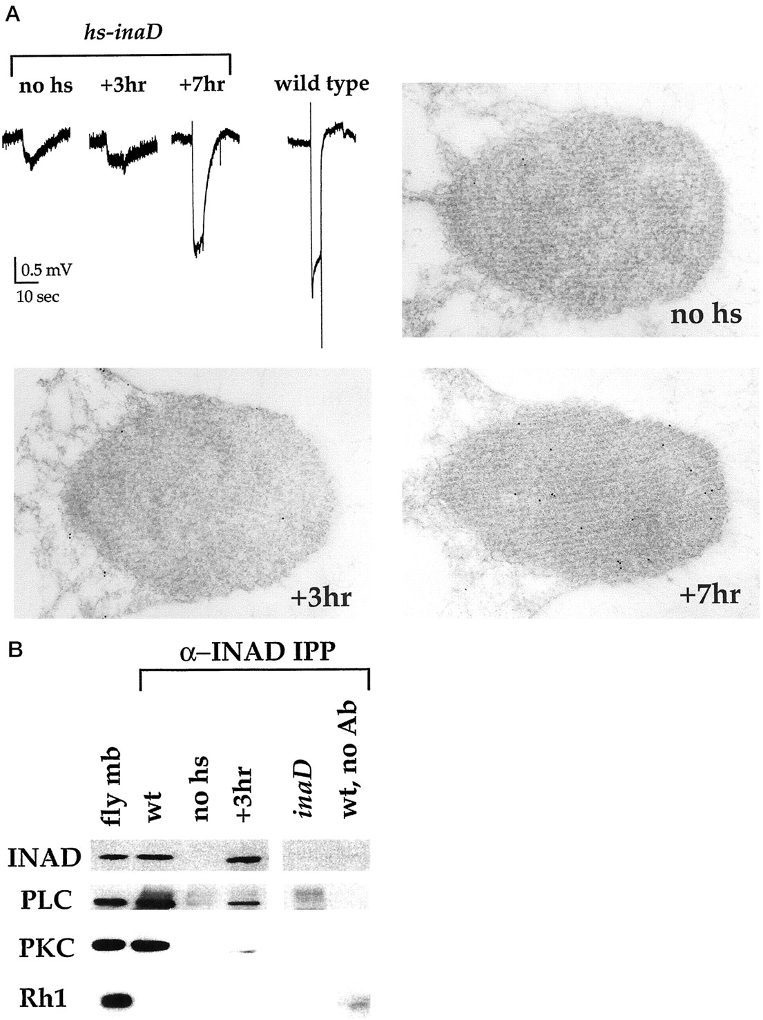
- Fig. 7.
INAD assembles PKC and PLC into complexes before they are targeted to the rhabdomeres.A, Top Left, Electroretinogram (ERG) recordings from wild-type and hs-inaD flies.hs-inaD flies either were uninduced (no hs) or were heat-shocked for 2 hr at 37°C and then recorded at 3 hr (+3hr) or 7 hr (+7hr) after heat shock. The stimulus was a 5 sec pulse of orange light (570 nm long-pass filter). Note the rescue of signaling at 7 hr after heat shock.Top Right to Bottom, Electron microscopic immunogold localization of INAD in hs-inaD rhabdomeres. At 3 hr after heat shock (+3hr) INAD protein had not yet arrived in the rhabdomeres, whereas INAD was present at 7 hr after heat shock (+7hr). Particles per square micrometer that were counted included the following: no heat shock, 1.29 ± 1.90 (n = 28 rhabdomeres); 3 hr, 1.86 ± 2.30 (n = 28); 7 hr, 19.2 ± 5.29 (n = 40). B, Membrane extracts from heads of wild-type (wt), hs-inaD, andinaD1 flies were immunoprecipitated (50 heads for wt and inaD1; 500 heads for hs-inaD) by using an anti-INAD antibody.hs-inaD flies either were uninduced (no hs) or were heat-shocked for 2 hr at 37°C and then assayed at 3 hr after heat shock (+3hr). Immunoprecipitated proteins were separated by SDS-PAGE, transferred to nitrocellulose, and probed with antibodies specific for INAD, PLC, eye-PKC, and rhodopsin (Rh1). Fly mb refers to wild-type membranes before immunoprecipitation. As expected, PLC and eye-PKC coimmunoprecipitated with INAD in wild-type membranes but did not precipitate from inaD null or wild-type membranes incubated without antibody (wt, no Ab). Fromhs-inaD flies, PKC and PLC coimmunoprecipitated with anti-INAD antibodies 3 hr after heat shock, a time when signaling still is not restored and INAD has not yet reached the rhabdomeres (A); it should be noted that it is possible that immunoEM and immunoprecipitation may have different sensitivities. Rhodopsin, which is not a part of the signaling complex, did not coimmunoprecipitate with INAD in any genotype.


