

Article Figures & Data
Figures
- Fig. 1.
Schematic representation of recombinant HSVLatEnk vector and its molecular characterization. Thymidine kinase gene (UL23) in KOS-derived Tk+HSVLatβ-gal DNA [bearing β-galactosidase reporter gene downstream of the Lat-LTR promoter inserted into the gC locus (Antunes-Bras et al., 1998)] was disrupted by homologous recombination with p23d plasmid DNA containing NsiI toSacI deleted HSV TK gene to generate TK-negative HSVLatβ-gal vector (HSVLatβ-gal). HSVLatEnk recombinant was then created as described previously (Antunes-Bras et al., 1998), by inserting Lat-LTR-pEnk transcriptional unit into the gC gene of HSVLatβ-gal by homologous recombination. Purified HSVLatEnk DNA was analyzed by PCR, and subsequent PCR products were separated on ethidium bromide-stained 1.2% agarose gel for the presence of both the TK-deleted gene and the Enk transgene. Positions of respective primers used are indicated on the diagram. Lanes 1 and6 show the Lambda DNA/HindIII-EcoRI and 100 bp molecular weight standards (given in kilobases), respectively. Lane 2, Approximately 1.8 kb PCR product generated by amplification, using primers A/B, of wild-type HSV DNA; lane 3, ∼1.3 kb PCR product generated using the same primers A/B on HSVLatEnk DNA.Lanes 4 and 5 show the PCR amplification products obtained with the set of primers 1/3 and 2/3 on HSVLatEnk DNA, respectively. HSVLatEnk DNA was further analyzed by Southern hybridization. Ten micrograms of HSVLatβ-gal DNA (lane 7), 10 μg of HSVLatEnk DNA (lane 8), and 0.1 μg of pLatEnk DNA (lane 9) were digested withBamHI, applied on 0.8% agarose gel, electrotransferred on nylon membrane, and hybridized with a [32P]-labeled cDNA probe corresponding to the rat pEnk coding region. Whereas no hybridization signal was apparent on HSVLatβ-gal (lane 7), positively labeled DNA fragments of ∼1.35 kb were generated on HSVLatEnk DNA (lane 8) and pLatEnk DNA (lane 9). This size corresponds to the expected size of DNA fragment resulting from theBamHI digestion of both HSVLatEnk and pLatEnk DNA.
- Fig. 2.
Presence and severity of hindpaw joint lesions in control (representing both sham-infected or HSVLatβ-gal-infected polyarthritic rats) (A′) and HSVLatEnk-infected (B′, C′) polyarthritic rats. Individual evolution of polyarthritis-associated joint destruction was assessed by comparing radiograms made 2 weeks after polyarthritis induction (A, B, C), i.e., just before infection with either HSVLatβ-gal or HSVLatEnk, and then 3 weeks later (A′, B′, C′). Osseous lesions of tarsus and metatarsus toe were evaluated bilaterally in both groups of rats (n = 8–10) using a four-degree rating scale. Radiographs were examined by an expert observer, without knowledge of the treatments. The Kolmogorov–Smirnov test was used for statistical analysis of the data. Six of 10 HSVLatEnk-infected rats presented mild lesions (B′), and the remaining four animals had more extensive lesions (C′), which were, however, less important than those of control polyarthritic rats (A′).
- Fig. 3.
HSVLatEnk-infected polyarthritic rats exhibited reduced thermal hyperalgesia and improved spontaneous locomotor activity. Response (paw withdrawal) latencies (A) of controls (n = 9) and HSVLatEnk-infected (n = 15) polyarthritic rats to radiant heating (intensity 7; Ugo Basile) were measured 3 weeks after infection. Rearings (B) and horizontal locomotor activity (C) of control (sham- or HSVLatβ-gal-infected) (n = 12) and HSVLatEnk-infected (n = 15) polyarthritic rats in a red-lighted open field were video monitored and assessed every minute during a 7 min period. Animals were then implanted subcutaneously for 3 d with an Alzet osmotic minipump delivering 3 mg · kg−1 · d−1 of either naloxone (○) or naloxone methiodide (▵) (antago), and thermal hyperalgesia and locomotor activity were assessed. Performances of normal healthy rats are indicated by the horizontal dashed line and gray band in the three behavioral tests (mean ± SEM;n = 6–7). *p < 0.05; **p < 0.01; ***p < 0.001 for control versus HSVLatEnk-infected polyarthritic rats, two-tailed unpaired t test; and for untreated versus naloxone–naloxone methiodide-treated animals, two-tailed pairedt test. Long-term improvement of functional ability (D) in HSVLatEnk-infected polyarthritic rats was evaluated by comparing the locomotor activity in normal healthy rats (n = 5) and in both control (n= 8) and HSVLatEnk-infected (n = 10) polyarthritic rats 3, 5, and 8 weeks after infection. **p < 0.01; ***p < 0.001 for control versus HSVLatEnk-infected rats; two-tailed unpaired ttest.
- Fig. 4.
LATs and PA mRNA expression were detected by in situ hybridization histochemistry. Incubation of 10 μm sections of L4–L5 DRG with digoxygenin-labeled LAT cRNA revealed numerous LAT-expressing neurons (arrowheads) in HSVLatEnk-infected polyarthritic rats (A). In contrast, no LAT-expressing nerve somas could be detected in adjacent L1–L3 DRG (B) or in the spinal cord of the lumbar region (C), suggesting no spread of the vectors to these territories. Every fourth section of L4–L5 DRG from normal healthy rats (D), control (E), or HSVLatEnk-infected polyarthritic rats (F) was incubated with [35S]PA cRNA, and then both unlabeled and labeled neurons were counted (in a total of 30 sections in each group). Approximately 2% of neurons in L4–L5 DRG of healthy rats were labeled with [35S]PA cRNA (D). In contrast, no PA mRNA-expressing neurons were detected in L4–L5 DRG of control (vehicle- or HSVLatβ-gal-infected) polyarthritic rats (E). Infection with HSVLatEnk led to the expression of PA mRNA in ∼12% (110 of a total of 955 neuronal somas) of neurons in L4–L5 DRG (F). The photomicrographs are representative of three to four animals per group, examined 5 weeks after polyarthritis induction (i.e., 3 weeks after vehicle, HSVLatβ-gal, or HSVLatEnk infection). Scale bar:A, B, D–F, 250 μm;C, 390 μm.
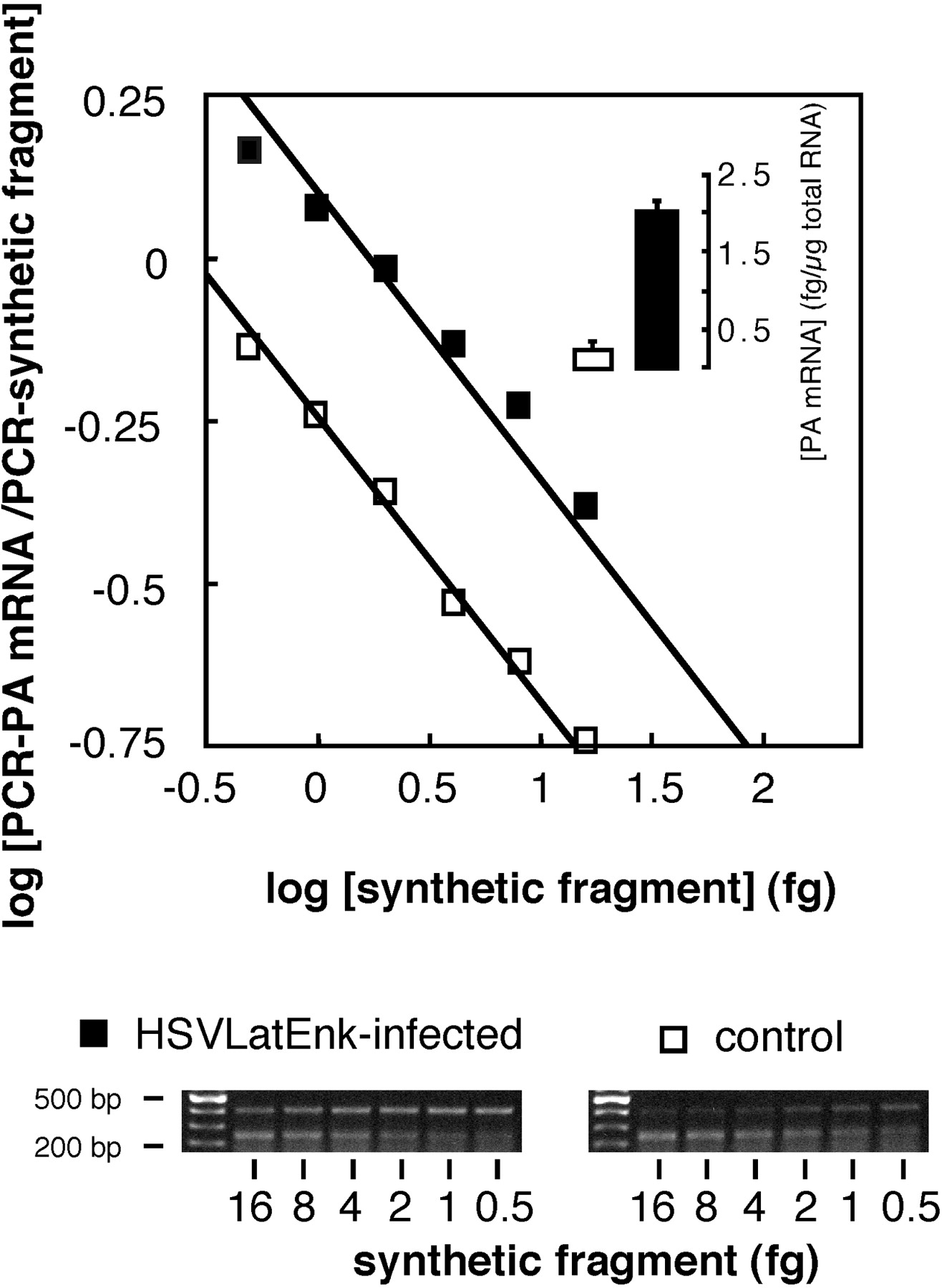
- Fig. 5.
Quantitative RT-PCR measurement of PA mRNA levels in L4–L5 DRG of control (■) or HSVLatEnk-infected (▪) polyarthritic rats. Total RNA was extracted from L4–L5 DRG in both groups of animals (n = 5 for each group) 5 weeks after polyarthritis induction. Five hundred nanograms of total RNA were reverse-transcribed in the presence of six different dilutions of synthetic fragment and amplified for 30 cycles. Measurement of optical density of PCR amplification products of PA mRNA (402 bp) or of the synthetic fragment (241 bp) allowed the drawing of the plot, as described previously (Antunes-Bras et al., 1998). Representative gel analyses of PCR products are shown.
- Fig. 6.
Immunofluorescent detection of Met-enkephalin-like material. Fifteen micrometer sections were stained with monoclonal anti-ME antibody (Valbiotech). No ME staining was detected in DRG neuronal cell bodies (A) and dorsal roots (B) of untreated polyarthritic rats. Only few neuronal processes stained for ME could be visualized in sciatic nerves of these animals (C). In HSVLatEnk-infected polyarthritic rats, numerous ME-stained neuronal soma were present in L4–L5 DRG (D). Scarce neuronal processes were stained in dorsal roots (E), whereas numerous nerve fibers positively labeled for ME were present in sciatic nerves (F) of HSVLatEnk-infected rats. Scale bar:A, D, 250 μm; B,C, E, F, 70 μm.


