

Article Figures & Data
Figures
- Fig. 1.
Expression of SHP1 mRNA and protein in the normal adult rat brain. The dark-field, hematoxylin-stained, light-field micrographs and double-labeling immunohistochemistry for SHP1 and GFAP show coronal brain sections through the hippocampus (A1,A2, A3), through the brainstem (B1, B2, B3), and through the cortical layers II–IV (C1, C2,C3). Note that in the stratum oriens of hippocampus (A3) and in the brainstem in ependymal and subependymal layers lining the fourth ventricle (B3), SHP1 immunoreactivity colocalizes (yellow;arrows) on GFAP-positive astrocytes. In hippocampus (A3) and in cortical layers II–IV (C3), SHP1 immunoreactivity (red; arrowheads) is also located on pyramidal neurons and their apical dendrites.SO, Stratum oriens; SP, stratum pyramidale; SR, stratum radiatum. Scale bars, 100 μm.
- Fig. 2.
Regulation of SHP1 mRNA expression after peripheral (facial, hypoglossal, sciatic) nerve axotomy and direct cortical lesion 3 d after injury. The dark-field micrographs illustrate massive induction of the SHP1 mRNA expression in the axotomized (Ax) facial (A) and hypoglossal (B) motor nuclei. The SHP1 mRNA is also upregulated in ventral and dorsal gray matter of the lumbar spinal cord after sciatic nerve axotomy (C). In response to direct cortical lesion (Dcl), upregulation of SHP1 mRNA is observed only within and around the brain wound (D). In all injury models, weak hybridization signals, slightly above the background, are present on the control, unoperated side (Con). Scale bars, 200 μm.
- Fig. 3.
Time course of SHP1 mRNA and protein expression in the axotomized facial motor nucleus (3–150 d after transection).A, Quantification of SHP1 mRNA using semiquantitative RT-PCR. For each time point the relative increase compared with control facial nucleus was calculated (**p < 0.01 and ***p < 0.001; paired, two-tailed Student'st test; n = 6 per each time point).B, Dark-field micrographs of in situhybridization sections 3, 7, 14, and 28 d after facial nerve axotomy. Note the similarity between the in situhybridization and PCR quantification in the time course of SHP1 regulation. C, Western blot analysis of SHP1 protein in the axotomized facial nucleus. Jurkat cells served as a positive control overexpressing SHP1 protein (68 kDa, arrow). Position of the molecular size marker (in kilodaltons) is indicated on the left. The protein levels of β-actin in cell lysates were also detected as a control for loading variations. Scale bar, 200 μm.
- Fig. 4.
Cellular localization of SHP1 mRNA and protein 3 d after transection of the facial nerve and 1 d after direct cortical lesion. A, Light micrographs of hematoxylin-stained in situ hybridization sections show that silver grains accumulate over small, darkly staining nuclei (red arrows) in the axotomized facial nucleus, whereas motoneurons remain unlabeled. B, C, Facial motor nuclei 3 d after facial nerve transection, triple labeling for Cy3 (anti-SHP1, red), Cy5 (B, microglia, anti-Ox42; green), and DTAF (C, astrocytes, anti-GFAP; green). After transection of the facial nerve, SHP1-IR (red) colocalizes (red and green overlap resulting in yellow) with Ox42-IR on activated microglia (B, arrowheads) and GFAP-IR on astrocytes (C, arrows). D, After direct cortical lesion, invading macrophages identified by anti-ED-1-antibody (green) and a subpopulation of T-lymphocytes identified by anti-CD3-antibody (green) are also positive for SHP1-IR (red, colocalization in yellow).N, Neuron; Con, control, unoperated side;Ax, axotomized side; Dcl, direct cortical lesion side. Scale bars, 100 μm.
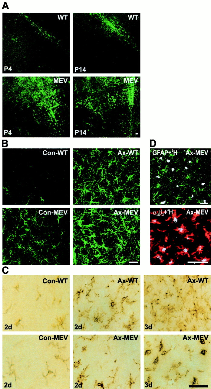
- Fig. 5.
Astrocytic and microglial reaction in the intact brain during adulthood, in the intact (Con) and axotomized (Ax) facial nucleus in normal, wild-type littermates (WT) and SHP1 mutant, moth-eaten viable mice (MEV). A, GFAP-immunoreactivity in the brainstem of WT and MEV mice at postnatal days 4 (P4) and 14 (P14). Note that GFAP-IR is increased in MEV mice compared with the same brain regions of WT littermates. B, GFAP-IR in the brainstem of WT and MEV mice 3 d after facial nerve transection. Note that GFAP-IR is increased on the control, unoperated side in MEV mice (Con-MEV) (as well as in the whole brain) compared with the same region of normal littermates (Con-WT). C, The morphology of microglia, detected by αMβ2-integrin immunoreactivity 2 and 3 d after facial nerve axotomy. Note that after facial nerve axotomy, the microglial cells in MEV mice are characterized by rounded, ameboid morphology with delayed and decreased ramification. D, Combination of [3H]-thymidine autoradiography (white points) with either GFAP-immunoreactivity (green) or αMβ2-integrin-IR (red) 3 d after facial nerve axotomy. Note that the proliferating cells in MEV mice are exclusively αMβ2-integrin-positive microglia. Scale bars, 100 μm.
- Fig. 6.
Quantitative effects of reduced SHP1 activity on astrocytic (A) and microglial reaction (B) in the intact and injured brain as well as on axonal outgrowth (C). A, Quantification of GFAP-IR (pixel number) in the intact brain (random brainstem area, nonaxotomized facial nucleus) and 3 d after facial nerve axotomy in wild-type (WT) littermates and MEV mice. Note that only in the uninjured brain regions there is a statistically significant increase for GFAP-IR in MEV mice compared with WT mice (6 sections per each animal; n = 4 animals). B1, Quantification of αMβ2-integrin-positive microglia in intact brain regions (random brainstem area and nonaxotomized facial nucleus). Note that the number of resting microglia is similar in WT and MEV mice. B2, Reduction in the number of proliferating ([3H]-thymidine-labeled) αMβ2-positive microglia in MEV mice 2 d after direct cortical lesion. B3, Reduction in the number of αMβ2-positive microglia and the proliferation rate ([3H]-thymidine-labeled cells) in MEV mice 2 and 3 d after facial nerve axotomy compared with wild-type littermates. The microglial reaction was always quantified in six sections per animal (n = 4). C, Four days after crush near the foramen stylomastoideum, the facial nerve was cut longitudinally and stained for galanin or CGRP, which accumulate in the terminals of the elongating neurites. The average distance between the most distal-labeled growth cone and the crush side was determined for each axonal marker in five tissue sections per animal (n = 6). Both the galanin- and CGRP-positive axonal populations show the same regeneration distance of ∼6 mm at day 4 in the WT littermates and MEV mice. All statistical analyses were performed using a paired, two-tailed Student's t test. Statistically significant changes are indicated byasterisks (*p < 0.05, ***p < 0.001).
- Fig. 7.
Association of SHP1 with CSF-1-receptor (R) and EGF-receptor (R). A, After 48 hr starvation in serum-free medium, microglial cells were stimulated with CSF-1 (200 ng/ml) for 0, 10, 30, and 60 min at 37°C and lysed in lysis buffer. Cell lysates were immunoprecipitated with an antiserum against SHP1. The proteins were resolved by SDS-PAGE, transferred to membrane, and probed with antibodies against phosphotyrosine (Anti-PTyr), CSF-1-R, or SHP1. The positions for the migration of CSF-1-R and SHP1 are indicated by arrows. Stimulation of microglial cells with CSF-1 induces increase in tyrosine phosphorylation of CSF-1-R and SHP1. Tyrosine phosphorylation of both proteins reached a maximum after 30 min and declined after 60 min. Note that CSF-1-R was detected in SHP1 immunoprecipitates of nonstimulated and CSF-1-stimulated microglial cells. B, Cultured astrocytes were starved in serum-free medium for 48 hr, stimulated with EGF (100 ng/ml) for 0, 10, 30, and 60 min at 37°C, and lysed in lysis buffer. Cell lysates were immunoprecipitated with an antiserum against SHP1. After SDS-PAGE, proteins were transferred to membrane and probed with antibodies against phosphotyrosine (Anti-PTyr), EGF-R, or SHP1. The positions for the migration of EGF-R and SHP1 are indicated by arrows. EGF stimulation induces increases in tyrosine phosphorylation of EGF-R and SHP-1. Note that the amount of EGF-R detected in SHP1 immunoprecipitates was increased in stimulated cells and paralleled the level of EGF-R tyrosine phosphorylation.


