

Article Figures & Data
Figures
- Fig. 1.
Temporal and spatial correlation of astrocyte differentiation and vascularization in the developing optic nerve. Longitudinal cryosections of developing rat optic nerves from E17 (A–C), E18 (D–F), E19 (G–I), E20 (J–L), and P1 (M–O) were stained by a GFAP antiserum (A, D, G,J, M), which is a specific marker of astrocytes. GFAP staining was scarce at E17 but was first detected near the surface of the nerve at E18. The intensity of the staining increased with age, extending more deeply into the nerve. The staining extended throughout the nerve by P1. Optic nerve sections of the same ages were double by two endothelial-specific markers, a VWF antiserum (B, E, H,K, N) and rhodamine-conjugated BSLI (C, F, I,L, O). VWF and BSLI labeling colocalized and displayed a similar time course and pattern to the GFAP staining. Scale bar, 100 μm
- Fig. 2.
GFAP staining in developing optic nerves. Longitudinal cryosections of E18 and E19 optic nerves were stained by an anti-GFAP antibody and observed under higher magnification than that shown in Figure 1. The majority of GFAP staining at E18 is near the surface of the nerve, right underneath the pia (arrows). By E19, the staining beneath the pia has become much more intense and is beginning to extend more deeply into the nerve. Scale bar, 50 μm.
- Fig. 3.
Morphology and immunoreactivity of purified VECs in culture. Phase-contrast micrographs of purified VECs (A) and pial cells (B) after 3 d in serum-free culture. VECs tend to be spindle-shaped (A), whereas the pial cells display a flat sheet-like appearance (B). To confirm the purity of the VECs, they were immunostained immediately after isolation with an anti-Tie2 antibody (C, D). All of the purified cells, indicated in (C) with the 4′,6′-diamidino-2-phenylindole (DAPI) nuclear stain, were Tie2+ (D). Scale bar, 100 μm.
- Fig. 4.
Effects of purified VECs and pial cells on astrocyte differentiation. A, Purified APCs were cultured on a coverslip suspended above a conditioning layer of purified VECs or pial cells, as indicated. APCs cultured alone (none) or with LIF (LIF) were used as negative and positive controls. After 4 d, the APCs cultures were labeled with a GFAP antiserum. Astrocyte differentiation was significantly promoted by VECs purified either from both E17 and P1 optic nerves but not by pial cells purified from nerves of either age. Results represent means ± SD (n = 3).B, Effect of LIF neutralizing antibody on astrocyte differentiation promoted by VECs. VEC-conditioned medium was collected and incubated overnight at 4°C with nothing (VECs), neutralizing antibody against LIF (VECsαLIF), neutralizing antibody against CNTF (VECs αCNTF), or neutralizing antibody against LIF plus excess LIF (VECs αLIF LIF). Purified APCs were then cultured in these conditioned media for 4 d before GFAP immunostaining. The ability of VEC-conditioned medium to induce astrocyte differentiation was significantly reduced by the LIF neutralizing antibody unless excess LIF was added to the medium. Results represent means ± SD (n = 3).
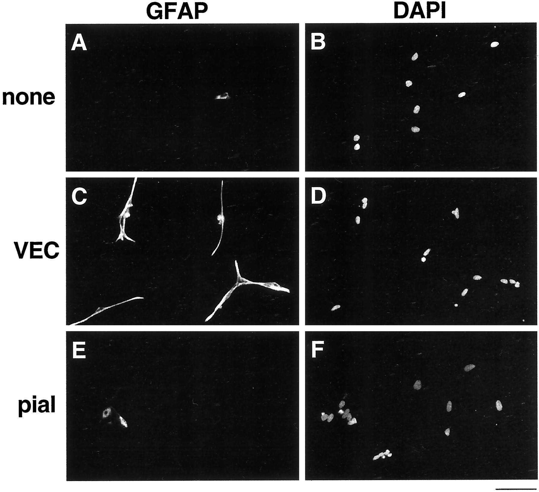
- Fig. 5.
Effect of VECs on astrocyte differentiation. Purified APCs were cultured alone (A, B) or suspended above a conditioning layer of purified VECs (C, D) or pial cells (E,F). After 4 d, the APC cultures were stained with an anti-GFAP antibody (A, C,E), as well as the DAPI nuclear stain (B,D, F). Most APCs remain undifferentiated unless cultured with VECs. Scale bar, 100 μm.
- Fig. 6.
Effect of LIF neutralizing antibody on astrocyte differentiation induced by VECs. Purified APCs were cultured in VEC-conditioned medium treated with anti-LIF antibody (A, B), anti-CNTF antibody (C, D), or anti-LIF antibody plus excess LIF (E, F). After 4 d, the APC cultures were stained with an anti-GFAP antiserum (A, C, E) and the DAPI nuclear stain (B, D,F). Few APCs differentiate into GFAP+ astrocytes in the presence of the LIF neutralizing antibody.
- Fig. 7.
RT-PCR analysis of LIF mRNA in optic nerve and retina. Total RNA was extracted from E17 retinas, purified E17 APCs, and from VECs and pial cells isolated from P1 optic nerves. Subsequently, total RNA was subjected to RT-PCR using LIF-specific primers. RT-PCR or PCR without reverse transcription using GAPDH primers were used as controls. The amplified products were separated on a 1.5% agarose gel and visualized by ethidium bromide.


