

Article Figures & Data
Figures
- Fig. 1.
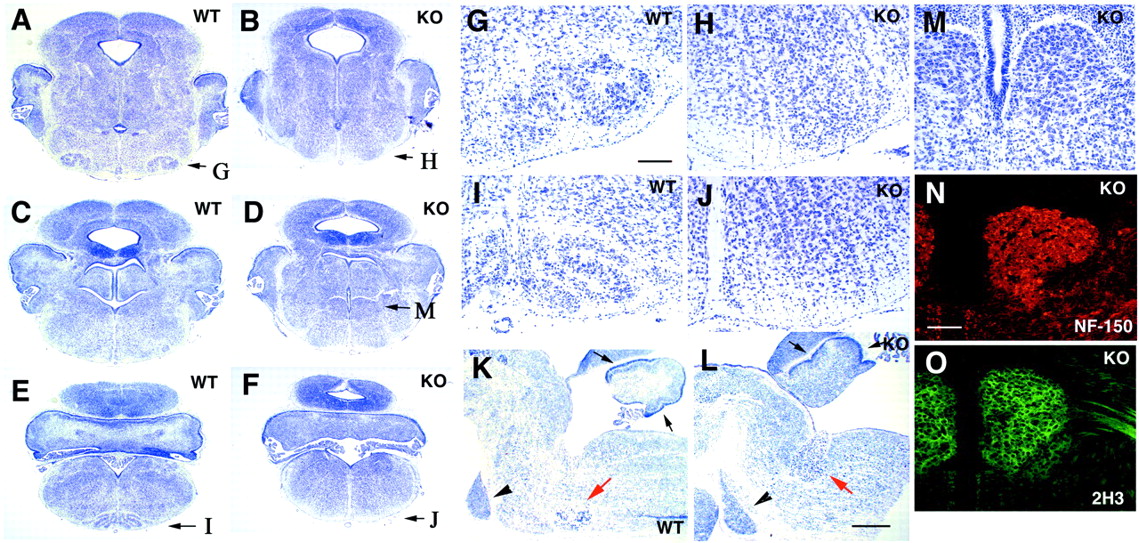
Selective loss of the VIIth motor nucleus and IO in Cdk5−/− mice. Nissl-stained coronal (A–J, M) and sagittal (K, L) sections from Cdk5+/+ (A, C, E, G, I, K) and Cdk5−/− (B, D, F, H, J, M) mice at E18.5. Genotypes are indicated as wild type (WT) (Cdk5+/+) and knock-out (KO) (Cdk5−/−). A–J andM are paraffin sections stained with cresyl violet.K and L are frozen sections stained with toluidine blue. G–J and M are higher magnifications of a specific part of the sections in A, B, E, F, and D, respectively; the magnified area in each section is indicated by a letter on thebottom right corner in A, B, andD–F. A loss of typical structures of the facial nerve nucleus (B, H) and IO (F, J) is observed in the null mice. As seen in the sagittal sections, the appearance and position of the pontine nucleus (arrowheads) are similar in Cdk5+/+ and Cdk5−/− mice (K, L). In Cdk5−/− hindbrain, an abnormal cell mass is seen in L and M. This ectopic mass (M and red arrow inL) consists of postmitotic neurons positively stained with the anti-neurofilament antibodies NF-150 (N) and 2H3 (O). Black arrows inK and L indicate the external granule cell layer of the cerebellum. The red arrow inK indicates the location of VIIth nucleus in Cdk5+/+ mice. Arrowheads in K and Lindicate pontine nucleus. A–F, G–J, andM–O are at the same magnifications, respectively. Scale bars: G, N, 120 μm; L, 700 μm.
- Fig. 2.
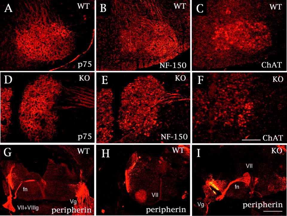
Immunohistochemical characterization of a neuronal mass of facial branchiomotor neurons. Serial coronal sections from Cdk5+/+ (A–C) and Cdk5−/− (D–F) mice at E18.5 were stained with anti-p75 (A, D), anti-NF150 (B, E), and anti-ChAT (C, F) antibodies. Both the facial nucleus in Cdk5+/+ mice and the neuronal mass in Cdk5−/− mice are positive for these antibodies. G–I, Coronal sections from Cdk5+/+ (G, H) and Cdk5−/− (I) mice at E14.5 stained with anti-peripherin antibody. The facial nerves (fn), axonal bundles of FBM neurons, are formed early after the neurons are generated and extend dorsally toward the exit point at the r4 level. The facial nerve bends in parallel to the migration of the cell bodies, forming the “genu” of the facial nerve. Thus, the facial nerve exits at the rostral level (G) from the facial nucleus (VII in H) in Cdk5+/+ mice. In Cdk5−/− mice, the facial nerve extends from the neuronal mass (VII) and exits at the same level (I). These results indicate that the neuronal mass in Cdk5−/− mice consists of migratory-arrested FBM neurons at the site at which they were born. Scale bars: F, 160 μm; I, 400 μm.Vg, Vth ganglia; VII, VIIth ganglia;VII+VIIIg, VIIth and VIIIth ganglia; WT, wild type; KO, knock-out.
- Fig. 3.
In situ hybridization studies for the Cdk5 mutant hindbrain. Sections were probed with either peripherin (A–D, G–J) or Phox2b (E, F, K, L); these sections were from either Cdk5+/+ (A, C, E, G, I, K) or Cdk5−/− (B, D, F, H, J, L) mouse embryos of age E18.5 (A, B), E14.5 (C–F), and E16.5 (G–L). The sections shown are either coronal (A–F) or sagittal (G–L) sections. The ectopic neuronal mass was identified as VIIth motor nuclei because it stained positive for Phox2b as well as peripherin (arrows inB and H). I andJ are higher magnifications of G andH, as indicated with arrows, respectively. The arrow in A indicates the VIIth motor nucleus in Cdk5+/+ mice. E, F, and K, L are serial sections of C, D and I, J, respectively, hybridized with Phox2b probe. In Cdk5−/− mice (N, O) at E18.5, the motor nuclei of the Vth (arrows in O), the Xth (arrowheads in M andN), and the XIIth (arrow inM and N) nuclei are comparable with Cdk5+/+ mice (M), as shown by the expression pattern of peripherin. However, segregation of the Xth (arrowheads) and XIIth (arrow) nuclei was less clear in Cdk5−/− mice (N). Although the VIIth nucleus (arrows) appeared normal in p35−/− mice (P), an elongated shape of the VIIth nucleus (arrows) was observed in p35−/−Cdk5+/− mice (Q) at E18.5, as visualized by peripherin expression. The arrowhead in P indicates motor nuclei of the Vth nerve in p35−/− mice. Scale bars: B, 400 μm; C, 80 μm; N, 100 μm.WT, Wild type; KO, knock-out.
- Fig. 4.
IO neurons are positioned abnormally in Cdk5−/− mice. Using the IO neuronal marker ER81 (A–F) and retrograde labeling with DiI (G, H), IO neurons were identified in Cdk5−/− mice (B, D, F, H). These neurons distribute diffusely in the parenchyma of the medulla at E14.5 (B) and at E18.5 (D, F, H), whereas they form a typical structure in the wild-type neurons at E14.5 (A) and E18.5 (C, E, G).Arrows in E and F indicate ER81-positive neurons in the sagittal sections of Cdk5+/+ (E) and Cdk5−/− (F) mice at E18.5. Crossing fibers of the olivary commissure are clearly seen (arrows in G, H;insets, higher magnifications). In Cdk5−/− mice, the number of crossing fibers is decreased, but commissure fibers run correctly beyond the midline (H). The posterior extramural stream in Cdk5−/− mice (J) is comparable with that in Cdk5+/+ mice (I) at E14.5, as shown in the Nissl-stained sagittal sections (arrows in I and J;insets, higher magnifications). Scale bars:A, 400 μm; C, F, 800μm;inset in I, 160 μm. WT, Wild-type; KO, knock-out.
- Fig. 5.
Reelin signaling in the FBM neuronal migration.A–L, In situ hybridization study in Cdk5+/+ (A–F) and Cdk5−/− (G–L) mice at E14.5. D–F andJ–L are higher magnifications of A–Cand G–I at indicated areas (arrows) in each panel, respectively. The FBM neurons express Dab1 mRNA (B, E, H, K), and Reelin mRNA is expressed in the surrounding area at the migratory termination of FBM neurons in the wild-type mice (A, D). Cadherin-8 (Cad8) expression confirms the identification as FBM neurons in the wild-type (C, F) and Cdk5−/− mice (I, L). Migration-arrested FBM neurons also express Dab1 (O) as well as peripherin (M) and neurofilament (N), as shown by the immunostaining of the coronal serial sections of E18.5 Cdk5−/− brain with anti-Dab1, anti-peripherin, and anti-NF-150 antibodies, respectively. Scale bars:A, 300 μm; D, 80 μm; M, 400 μm. WT, Wild type; KO, knock-out.
- Fig. 6.
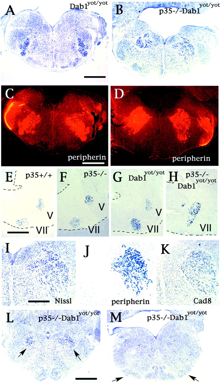
Abnormal FBM neuronal migration in Dab1yot/yot and p35−/−Dab1yot/yot mice. Disturbance of radial migration of FBM neurons in yotari, Dab1yot/yot, mice is shown in Nissl-stained (A) and peripherin-stained (C) coronal serial sections at E18.5. Deterioration of FBM neuronal migration is advanced in p35−/−Dab1yot/yot mice as seen in Nissl-stained (B) and peripherin-stained (D) coronal sections at E18.5. These abnormalities are confirmed by in situ hybridization with a peripherin probe in the sagittal sections from p35+/+ (E), p35−/− (F), Dab1yot/yot (G), and p35−/−Dab1yot/yot (H) mice at P0. Dashed lines indicate the margin of sections. I–K, Serial sections in coronal sections are either Nissl-stained (I) or analyzed byin situ hybridization with peripherin probe (J) and cadherin-8 (Cad8) probe (K). Abnormally located FBM neurons in p35−/−Dab1yot/yot mice are identified by their cadherin-8 mRNA expression (K). L, M, Migration abnormality of the IO neurons is also detected in rostral (L) and caudal IO (M) sections of p35−/−Dab1yot/yot mice with Nissl stain at P0.Arrows in L indicate ectopically located FBM neurons; arrows in M indicate the dorsal accessory olive, which is located laterally in p35−/−Dab1yot/yot mice. Scale bars:A, 300 μm; E, 240 μm; I, 60 μm. V, Vth nucleus; VII, Vth nucleus.
- Fig. 7.
Abnormalities of FBM and IO neurons are rescued by neuron-specific expression of Cdk5 transgene. No abnormality is found in the VIIth motor nucleus and IO complex in Nissl-stained coronal sections from adult Cdk5TgKO mice (B, D) compared with age-matched wild-type mice (A, C). A–Dare at the same magnification. Scale bar, 1 mm.


