

Article Figures & Data
Figures
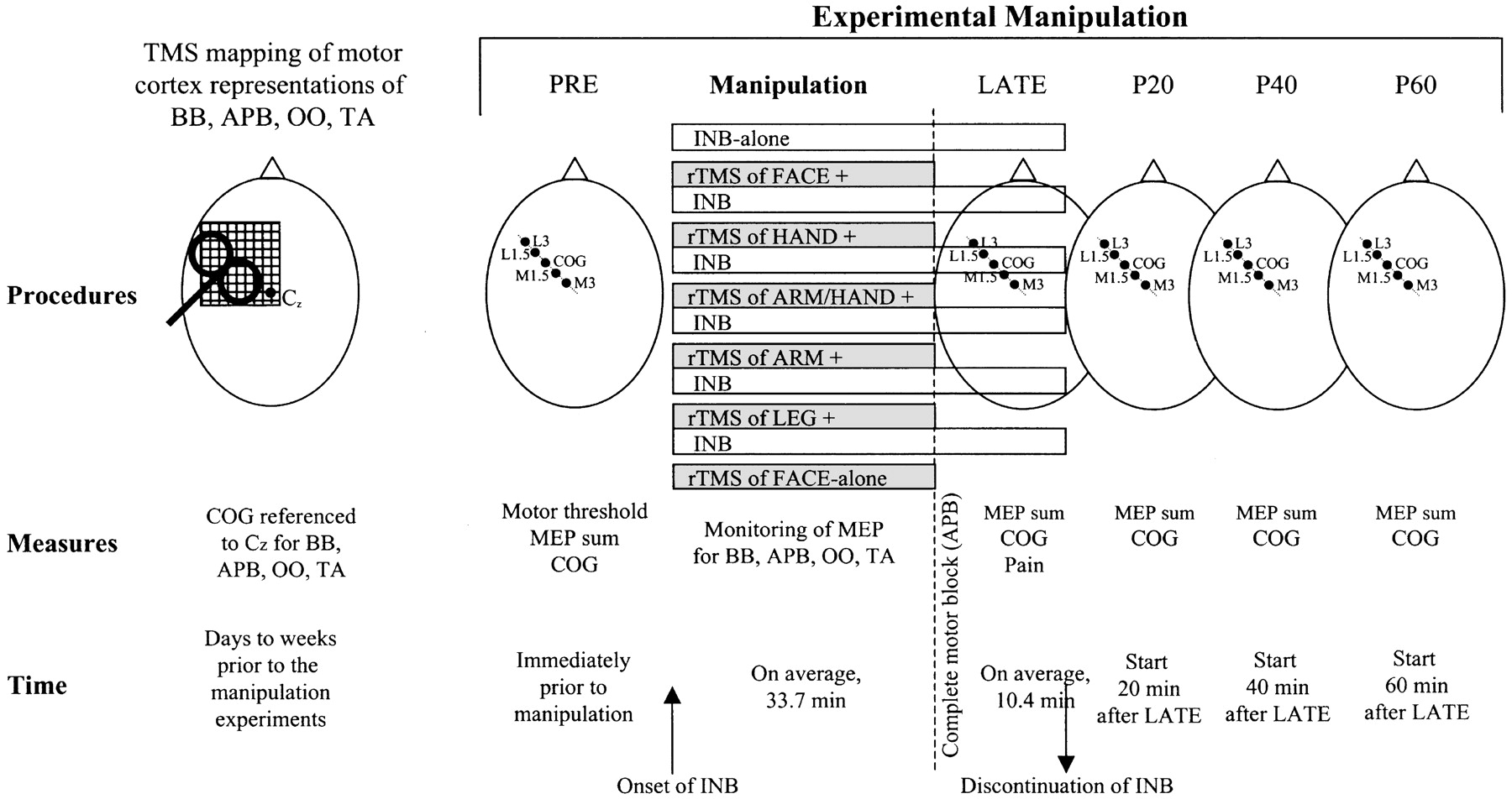
- Fig. 1.
Time line of experimental procedures. First, detailed two-dimensional maps in motor cortex of the BB, APB, OO, and TA were obtained, using an eight-shaped coil that was moved along a 1 × 1 cm grid referenced to the vertex (Cz, left-most panel; subject's head viewed from above). Days later, seven different manipulation experiments separated by at least 1 week were conducted in randomized order. Immediately before each manipulation (PRE), excitability (motor threshold, MEP sum) and location (COG) of the BB map were measured along one axis, testing five grid points 1.5 cm apart (panel beneath PRE). The central point (COG) corresponded to the BB COG obtained in the initial two-dimensional mapping experiment. The manipulations consisted either of ischemic nerve block of the hand (the INB-alone control experiment), focal 0.1 Hz rTMS of the face representation in motor cortex (the FACE-alone control experiment), or the combination of rTMS delivered to one of five different sites (FACE, HAND, ARM, LEG, or ARM/HAND overlap representation in motor cortex) and INB. The onset of INB (inflation of a tourniquet at the forearm) is indicated by the upward facing arrow. MEP in the BB, APB, OO, and TA elicited by rTMS were monitored throughout. Complete motor block to the APB was reached on average after 33.7 min (indicated by the vertical dashed line). At this point (in the FACE-alone experiment after 30 min) rTMS was stopped, and the LATE measurements of the BB map were started following the same protocol as the PRE measurements (panel beneath LATE). Immediately thereafter INB was discontinued (downward facing arrow). Further measurements of the BB map were obtained 20, 40, and 60 min (P20, P40, P60) after the end of the LATE measurements. The data were analyzed by comparing the LATE, P20, P40, and P60 measures with the PRE measures, using two-way repeated measure ANOVA with experimental manipulation and time as the within-subject effects.
- Fig. 2.
Normalized amplitudes of the MEP of the four different target muscles (OO, APB, BB, and TA), as elicited during experimental manipulation by repetitive transcranial magnetic stimulation delivered to different sites of the motor cortex (FACE, HAND, ARM/HAND overlap area, ARM, LEG; for definition, see Materials and Methods). INB, Ischemic nerve block of the hand. Thetop panel refers to the first 30 trials after start of INB (5 min), and the bottom panel to the last 30 trials before reaching complete motor block in the APB. Length of bars (y-axis) reflects the MEP amplitude normalized to the maximum amplitude for a given target muscle and subject (n = 6) across stimulation sites.Numbers indicate the mean maximum MEP amplitudes of the different target muscles (in millivolts).
- Fig. 3.
Changes in MEP amplitude in the biceps muscle of one representative subject induced by six different experimental manipulations (FACE, HAND, ARM/HAND, ARM, LEG = 0.1 Hz focal rTMS of the face, hand, arm/hand overlap, upper arm and leg representations in motor cortex, respectively, combined with INB). All recordings are averages of five trials obtained at the center of gravity of the biceps muscle using stimulation intensity of 30% above biceps motor threshold. Gray curves (top row) indicate MEP before manipulation (PRE), the other curves show MEP late into INB (LATE), and 20 and 60 min after the end of INB (P20, P60). Percentage values indicate the change of MEP amplitude compared with PRE. Note the long-lasting MEP increase with ARM/HAND + INB and ARM + INB compared with the only transient increase with INB-alone. Note further the much weaker transient increase with HAND + INB and FACE + INB compared with INB-alone.
- Fig. 4.
Changes in MEP sum of the biceps muscle (means of six subjects) over time. The thin curves in all diagrams refer to the control experiment (INB-alone). The thick curves refer to the experimental manipulation indicated at thetop of each diagram (for abbreviations, see Materials and Methods). The time of MEP sum measurements is given on thex-axis (for abbreviations, see Fig. 1).Asterisks indicate significant differences between the two curves in a given diagram. p values (paired two-tailed t tests) are also shown. Note that INB-alone resulted in only a transient increase of MEP sum, whereas ARM/HAND + INB and ARM + INB led to a long-lasting increase. In contrast, FACE + INB and HAND + INB canceled the transient increase obtained with INB-alone, and HAND + INB even resulted in a long-lasting decrease of MEP sum. LEG + INB was not significantly different from INB-alone.
- Fig. 5.
A, MEP mapping of the biceps muscle. MEPs were elicited by focal TMS at the COG of the biceps map and at sites 1.5 and 3 cm anterior–lateral (L1.5, L3) and posterior–medial (M1.5, M3) from COG. The different curves refer to the different times of measurement, as indicated in theinset (for abbreviations, see Fig. 1). MEP amplitudes are normalized to the individual maximum MEP during PRE (usually, but not always obtained at COG). The different manipulations are indicated at the top of each diagram (for abbreviations, see Fig.1). B, Changes of the COG along the lateral-to-medial axis. Data are from the MEP maps in A. The thin curve in each diagram refers to the INB-alone (control) condition, and thick curves refer to the experimental manipulation shown at the top of each diagram (for abbreviations, see Fig. 1). The asterisk indicates a significant lateral shift of the COG with HAND + INB compared with INB-alone.
- Fig. 6.
A, MEPs in the OO muscle of the same subject as in Figure 3, measured before (gray curve, PRE), LATE into, and 20 and 60 min after (P20, P60) FACE + INB (top panel) or ARM + INB (bottom panel, for abbreviations see Fig. 1). All MEPs were elicited 6 cm lateral from the COG of the biceps (BB), and are averages of five trials. Numbers indicate the percentage change of MEP amplitude compared with the MEP measured during PRE. Note that ARM + INB resulted in an only transient increase of the OO MEP, whereas the increase with FACE + INB was long-lasting.B, Group data (n = 4) of changes in OO (top panel) and BB (bottom panel) MEP sum induced by two different experimental manipulations, FACE + INB (filled squares) and ARM + INB (open circles). Note that, similar to the single subject data in Figure 6A, FACE + INB resulted in a long-lasting increase of MEP sum in the OO but not BB. Conversely, ARM + INB led to a long-lasting increase of the MEP sum in BB but not OO. C, Group data (n = 4) of changes in the COG of the OO and BB maps induced by two experimental manipulations. Positive values (y-axis, in centimeters) denote shifts of the COG in the medial direction, and negative values shifts in the lateral direction. Otherwise, same arrangement and conventions as in B.


Tables
- Table 1.
Excitability and location of the arm representation in motor cortex before manipulation
INB LEG + INB ARM + INB ARM/HAND + INB HAND + INB FACE + INB FACE alone F p MTCOG 39.7 37.7 38.8 37.7 39.2 38.2 39.6 0.54 NS MTL1.5 46.8 46.0 44.3 41.3 44.2 43.7 47.4 1.37 NS MTL3 63.3 66.7 61.8 58.1 59.5 63.3 62.4 2.12 NS MTM1.5 46.3 43.5 43.5 45.1 45.8 43.3 46.4 0.79 NS MTM3 63.0 64.0 59.7 62.1 65.8 64.7 67.4 0.87 NS MEP sum 2.0 2.6 2.0 2.9 3.6 3.1 3.5 1.13 NS ΔCOG 0.060 0.065 0.077 −0.014 −0.105 −0.036 −0.017 0.16 NS MT, Motor threshold of the biceps muscle (in % of maximum stimulator output) at the different stimulation sites given by the indices; MEP sum is given in millivolts; ΔCOG, deviation from COG (in centimeters, negative values indicate deviation lateral from COG). All values are means of six subjects.
- Table 2.
Time to complete motor block, total duration of INB, and level of INB-induced pain
INB LEG + INB ARM + INB ARM/HAND + INB HAND + INB FACE + INB F p CMB 33.3 34.8 34.2 31.7 33.1 35.0 1.65 NS tINB 42.3 44.7 44.2 42.1 43.6 47.5 4.30 0.006 Pain 10.2 9.7 10.7 9.3 8.8 11.2 0.96 NS CMB, Time to complete motor block (in minutes); tINB, total INB time (in minutes). The level of INB-induced pain is scaled by a standardized psychophysical pain ratio scale (maximum 20). All values are means of six subjects.


