

Article Figures & Data
Figures
- Fig. 1.
Targeted disruption of the murinetn-C gene. a, Diagram of a part of the murine tn-C gene (denoted as tn-C +) covering exons 2–5, the targeting vector, the targetedtn-C gene, and the resulting mutant (denoted astn-C -) allele after Cre recombinase-mediated excisionin vitro. Artificial introduction of aBamHI site combined with the excision of the floxed exon 2 converted a 11.5 kb BamHI genomic fragment indicative of the wild-type allele (tn-C +) to a 4.6 kbBamHI genomic fragment indicative of the mutant allele (tn-C -). Selected restriction enzyme recognition sites are indicated as follows: B, BamHI;Bg, BglII; D,DraI; V, EcoRV. Probe5′A and primers 1A,1B, and 1C are indicated.b, Southern blot analysis of representative tail DNA samples from wild-type (tn-C +/+), heterozygous (tn-C +/−), and homozygous (tn-C−/−) mice. DNA was digested with BamHI and subjected to hybridization using probe 5′A (a). c, Determination of genotypes by PCR. Multiplex PCR using primers 1A,1B, and 1C (a) as performed routinely from representative tail DNA samples.d, Northern blot analysis of total RNA from wild-type (lane 1, 3) and TN-C-deficient mice (lanes 2, 4). RNA was isolated from cerebrum (lanes 1, 2) and cerebellum (lanes 3, 4) of 7-d-old mice. Wild-type samples gave rise to a broad band with a size of ∼6–8 kb. Note that the weak band in lane 4 is shifted to lower molecular mass with respect to the intensive wild-type band inlane 3. Probing blots with a GAPDH-specific probe revealed no differences in RNA amounts loaded. e, RT-PCR analysis of TN-C-specific cDNAs derived from wild-type (lanes 1, 3, 5, 7) and TN-C-deficient mice (lanes 2, 4,6, 8). Reverse transcription was performed on total RNA from cerebrum (lanes 1,2), cerebellum (lanes 3,4), lung (lanes 5,6), and thymus (lanes 7,8) of 7-d-old mice. In the subsequent PCR, primers were used as depicted. Note that the mutant TN-C message lacking exon 2 could be amplified from all TN-C-deficient tissues tested.
- Fig. 2.
Western blot and immunohistological analysis of TN-C-deficient mice. a, Western blot analysis of crude protein extracts from brain, thymus, and lung tissue of 7-d-old mice from the indicated genotypes (tn-C +/+, tn-C +/−, and tn-C −/−) using polyclonal TN-C antibody pK7. In each lane, 10 μg of total protein was loaded. Intense immunoreactive bands specific for TN-C were easily detected in tissues from wild-type mice, but no signals were detectable in samples from TN-C-deficient animals. Heterozygous mice showed a reduced signal intensity of TN-C-immunoreactive bands. b, Quantitation of the TN-C protein content in different genotypes. The indicated amounts of total protein from cerebra of 7-d-old wild-type (tn-C +/+) and homozygously TN-C-deficient littermates (tn-C −/−) were applied and detected after blotting with the polyclonal TN-C antibody pK7. In wild-type animals, TN-C was detectable in as little as 0.1 μg total protein, whereas in mutant littermates no band was detectable even in 200 μg total protein. Molecular weight markers are indicated at the left margins in kilodaltons. c, d, Immunohistological localization of TN-C by indirect immunofluorescence on fresh frozen sections of cerebella of 7-d-old wild type mice (c) and TN-C-deficient littermates (d) using polyclonal antibody KAF 9–2. Intense TN-C immunoreactivity is visible on sections from wild-type mice, whereas no immunoreactivity is detectable in age-matched TN-C deficient littermates. Sections incubated only with secondary antibody showed no immunoreactivity (data not shown). egl, External granular layer; igl, internal granular layer;pcl, Purkinje cell layer. Scale bar (shown ind): c, d, 150 μm.
- Fig. 3.
Morphological and immunohistochemical analysis of TN-C-deficient mice. Light microscopic analysis of the cerebellar cortex (a, b) and retina (c, d) of 2-month-old wild-type (a, c) and TN-C-deficient (b, d) mice demonstrates an apparently normal histoarchitecture of both CNS structures in the mutant. All cerebellar (compare a, b) and retinal (compare c, d) layers of TN-C-deficient animals display a normal thickness, and there is no evidence for ectopically positioned cell types in TN-C-deficient tissues. TN-R immunoreactivity in the cerebellar cortex of wild-type mice (e) is homogeneously distributed in the molecular layer (mol) and internal granule cell layer (igl) and is accumulated in the white matter (wm). A similar distribution and intensity of TN-R positivity is detectable in the cerebellar cortex of TN-C-deficient mice (f). The distribution of oligodendrocytes and myelin in the optic nerve (on) of adult wild-type (g) and TN-C-deficient (i) mice was visualized with MAG antibodies (h and j are the phase-contrast images ofg and i, respectively). MAG immunoreactivity is absent from the retinal end of the optic nerve and from the retina of both genotypes. Arrows ing and i indicate the transition zone from myelinated to nonmyelinated segments of retinal ganglion cell axons. Electron microscopic analysis of optic nerves from TN-C-deficient mice (k) revealed a normal ultrastructure of the meninges and glia limitans (k). Virtually all ganglion cell axons of mutant mice (some labeled with axin k and l) are surrounded by a myelin sheath (some labeled with m in kand l). Analysis at a higher magnification demonstrates the presence of ultrastructurally intact CNS myelin sheaths in TN-C-deficient optic nerves. as, Astrocyte;ax, axons; igl, internal granule cell layer; inl, inner nuclear layer; ipl, inner plexiform layer; m, myelin sheath, mol, molecular layer; on, optic nerve; onl, outer nuclear layer; pcl, Purkinje cell layer;wm, white matter. Scale bars: (shown inb) a, b, 100 μm; (shown in d) c, d, 100 μm; (shown in f) e, f, 200 μm; (shown in j) g–j, 200 μm;k, 2 μm; l, 1 μm.
- Fig. 4.
Hippocampal morphology of TN-C-deficient mice.a, b, TN-C immunoreactivity was detectable in the hippocampal CA1 region of 5-week-old wild-type mice (a) by indirect immunofluorescence using the polyclonal antibody KAF 9–2. Specificity of weak signals is demonstrated by comparison with TN-C-deficient littermates (b). c, d, Nissl staining revealed an apparently normal histology of CA1 through CA3 regions and of the dentate gyrus of TN-C-deficient mutants (c) when compared with wild-type mice (d). e–h, Immunohistochemical localization of the presynaptic marker synaptophysin (e,f) and Timm's staining (g,h) revealed a similar laminated organization of the CA3 subfield in wild-type (e, g) and TN-C-deficient mice (f, h).i–l, GAD (i, j; only CA1 subfield shown) and parvalbumin immunoreactivity (k, l) showed normal distribution and appearance of GAD- and parvalbumin-positive interneurons in TN-C-deficient mice (j, l) when compared with wild-type littermates (i, k). m, n, Immunohistochemistry of WFA lectin-binding sites showing the interneuron-enwrapping perineuronal nets in the CA1 region of wild-type (m) and TN-C-deficient (n) mice. They were not detectably altered in the mutant. o,p, The morphology of dendrites and spines of hippocampal pyramidal cells of wild-type (o) and TN-C-deficient animals (p), visualized with the Golgi method, is indistinguishable between genotypes. Scale bars: (shown in b) a,b, 25 μm; (shown in d) c,d,50 μm; (shown in j) i,j, 25 μm; (shown in l)e–h, k,l, 50 μm; (shown inn) m,n, 25 μm; (shown in p) o,p, 5 μm.
- Fig. 5.
Unaltered learning, spatial memory, and relearning of TN-C-deficient mice. a, Latencies to climb the platform during the visible platform task, the spatial learning acquisition, and the relearning. Each value represents one trial (mean ± SEM). Mice from both genotypes quickly learned to locate the platform in all three phases. b, During the probe trial at the end of the acquisition phase, both genotypes showed a preference for the area surrounding the platform in NE. The preference for the area in NW might be attributable to the fact that animals were started from the west (mean ± SEM).c, After five trials with the platform located at a new position (NW), both genotypes showed a preference for the area around the platform location (NW) as well as for the area where the platform was located during the former acquisition phase (NE). Asterisksindicate a difference to the chance level of 10% at a significance ofp < 0.005 (Wilcoxon signed rank test; mean ± SEM). d, The analysis of the number of trials needed to reach criterion for three successive platform locations during the trial-to-criterion task (days 7–12) revealed no difference between genotypes (mean ± SEM). e, Scheme of the circular water maze (diameter, 155 cm) with different platform locations (platform diameter, 14 cm) surrounded by a circular area equal to 10% of the total area of the maze.
- Fig. 6.
LTP and LTD in the CA1 region are impaired in TN-C-deficient mice. a, Input–output curves for slopes of fEPSPs evoked by stimulation of Schaffer collaterals at different stimulation strengths. No significant difference between genotypes was found. b, Paired-pulse facilitation (PPF) was measured as the ratio between the slopes of fEPSPs evoked by the second and first pulses and plotted for several interpulse intervals. Field EPSPs were recorded at 30% from the subthreshold strength. To measure slopes of overlapping fEPSPs evoked by paired-pulse stimulation (for interpulse intervals <50 msec), fEPSPs evoked by a single-pulse stimulation were subtracted from fEPSPs evoked by paired-pulse stimulation. Examples of fEPSPs evoked by paired-pulse stimulation are shown in c, right panels. No significant difference between genotypes was found.c, TBS of Schaffer collaterals (applied at time point 0) evoked a high increase in the slopes of fEPSPs recorded in the CA1 region of slices from wild-type mice. In slices from TN-C-deficient mice (tn-C −/−), the potentiation appeared lower than in wild-type mice (tn-C +/+). The mean slope of fEPSPs recorded 0–10 min before TBS was taken as 100%. Data represent mean + SEM; n indicates the number of tested slices;N indicates the number of tested mice. Right panels show fEPSPs recorded before and 60 min after TBS. Scale bars, 20 msec and 500 μV. d, Two trains of low-frequency stimulation (1 Hz, indicated by horizontal bars) of Schaffer collaterals reliably decreased the slopes of fEPSPs in slices from wild-type mice (tn-C +/+). In slices from TN-C-deficient mice (tn-C −/−), the slope returned to the baseline. The mean slope of fEPSPs recorded 10 min before the first train was taken as 100%. Data represent mean + SEM;n indicates the number of tested slices;N indicates the number of tested mice. Right panels show fEPSPs in TN-C-deficient (tn-C −/−) and wild-type (tn-C +/+) mice before and 60 min after induction of LTD. Calibration: 10 msec, 500 μV.e, Cumulative plots representing levels of STP and LTP from all experiments. Each symbol represents a single experiment. Cumulative probability at any given value Xis the probability to observe potentiation less than or equal toX. Lower values of LTP (there is no overlap of values measured in young tn-C −/− and tn-C +/+ mice) are evident for TN-C-deficient mutants when compared with wild-type littermates. f, Cumulative plots representing levels of STD and LTD from all experiments. Each symbolrepresents a single experiment. Cumulative probability at any given value X is the probability to observe depression less than or equal to X. Lower values of both STD and particularly LTD are evident for TN-C mutants when compared with wild-type littermates.
- Fig. 7.
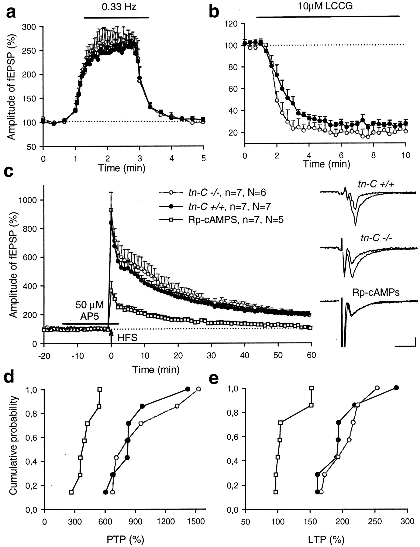
Normal LTP in the CA3 region of TN-C-deficient mice. a, Stimulation of mossy fibers with a frequency of 0.33 Hz similarly increased the amplitudes of fEPSPs in acute slices from both TN-C-deficient (tn-C −/−) and wild-type (tn-C+/+) mice. b, Application of the type II metabotropic glutamate receptor agonist L-CCGI (10 μm) reduced the amplitude of fEPSPs in slices from both TN-C-deficient (tn-C −/−) and wild-type (tn-C +/+) mice to the same level. c, High-frequency stimulation (HFS) of mossy fibers (applied at time point 0) evoked a similar increase in slopes of fEPSPs in slices from TN-C-deficient (tn-C −/−) and wild-type (tn-C+/+) mice, respectively. The potentiation was impaired in wild-type slices treated with a competitive inhibitor for PKA, Rp-cAMPS. The time interval of the application of the NMDA receptor antagonist AP-5 is shown by ahorizontal bar. Mean slope of fEPSPs recorded 0–10 min before HFS was taken as 100%. Right panels show averaged fEPSPs recorded before and 60 min after induction of LTP in TN-C-deficient (tn-C −/−) and wild-type (tn-C +/+) mice. Calibration: 10 msec, 100 μV.d, e, Cumulative plots representing levels of PTP (d) and LTP (e) from all experiments. Eachsymbol represents a single experiment. Cumulative probability at any given value X is the probability to observe a potentiation less than or equal to X. Note similar values of PTP and LTP in TN-C-deficient (tn-C−/−) and wild-type (tn-C+/+) mice and that Rp-cAMPS completely blocked LTP in five of seven experiments. 100% corresponds to the baseline level.
- Fig. 8.
Normal LTP in the dentate gyrus of TN-C-deficient mice. a, b, Short high-frequency stimulation (SHFS) of the medial (a) or lateral (b) perforant pathway (applied at time point 0) evoked a similar potentiation in slices from wild-type (tn-C +/+) and TN-C-deficient mice (tn-C −/−) in the presence of 100 μm picrotoxin. Mean slope of fEPSPs recorded 0–10 min before TBS was taken as 100%. Four points recorded during the period marked by SHFS represent potentiation recorded between five trains (10 sec after a train). Data represent mean + SEM; nindicates the number of tested slices; N indicates the number of tested mice. Right panels show fEPSPs recorded before and 60 min after TBS. Calibration: 10 msec, 250 μV.c, d, Cumulative plots representing levels of LTP measured 50–60 min after beginning of high-frequency stimulation of medial (a) or lateral (b) perforant pathway. Each symbolrepresents a single experiment. Cumulative probability at any given value X is the probability to observe a potentiation less than or equal to X. No significant difference between genotypes was found.
- Fig. 9.
Blockade of GABAA receptor-mediated inhibition does not fully rescue LTP in the CA1 region of TN-C-deficient mice. a, TBS of Schaffer collaterals in the presence of picrotoxin evoked an increase in the slopes of fEPSPs recorded in the CA1 region of slices from wild-type mice (tn-C +/+). In slices from TN-C-deficient littermates (tn-C −/−), potentiation appeared significantly lower than in wild-type mice. Mean slope of fEPSPs recorded 0–10 min before TBS was taken as 100%. Data represent mean + SEM; nindicates the number of tested slices; N indicates the number of tested mice. Right panels show fEPSPs recorded before and 60 min after TBS. Calibration: 20 msec, 500 μV.b, c, Cumulative plots represent levels of STP (b) and LTP (c) from all experiments performed in the presence of picrotoxin. Eachsymbol represents a single experiment. Cumulative probability at any given value X is the probability to observe a potentiation less than or equal to X. Note the overlap of STP values and significant difference in distribution of LTP levels: 9 of 11 values corresponding to TN-C-deficient mutants are lower than the smallest LTP value for wild-type littermates.d, Cumulative plot represents values of the NMDA receptor-mediated component in fEPSPs evoked by single theta bursts. Examples of such fEPSPs recorded without (solid lines) or in the presence (dotted lines) of the NMDA receptor antagonist AP-5 are shown on the right.Horizontal bar indicates the time interval used for measurements of the NMDA receptor-mediated component. No significant difference between genotypes was found.
- Fig. 10.
Voltage-dependent Ca2+channels mediate deficits in synaptic plasticity in TN-C-deficient mutants. a, b, Short high-frequency stimulation (SHFS) in normal ACSF (a) or theta-burst stimulation (TBS) in the presence of 20 μm nifedipine (b) applied at time point 0 evoked a similar potentiation in the CA1 region in wild-type (tn-C +/+) and TN-C-deficient (tn-C −/−) mice. Four points recorded during the period marked by SHFS represent potentiation recorded among five trains (10 sec after a train).c, Bath application of 25 mmtetraethylammonium (applied at time point 0 for 7 min) evoked stronger LTP in the CA1 region of wild-type (tn-C +/+) as compared with TN-C-deficient (tn-C −/−) mice. Ina–c, mean slope of fEPSPs recorded 0–10 min before TBS was taken as 100%. Data represent mean + SEM; nindicates the number of tested slices; N indicates the number of tested mice. Right panels show fEPSPs recorded before and 60 min after induction of LTP. Calibration: 10 msec, 500 μV. d, e, Cumulative plots representing levels of LTP measured 50–60 min after induction of LTP according to protocols given in a and b(d) and c(e), respectively. To facilitate comparison of experiments performed in the presence and absence of nifedipine (Nif), data presented in Figure 6efor young mice are added to d. Eachsymbol represents a single experiment. Cumulative probability at any given value X is the probability to observe LTP less than or equal to X.


