

Article Figures & Data
Figures
- Fig. 1.
Tissue distribution and clearance of infused BDNF. BDNF (2 μg/2 μl, 25 min) was infused into stratum lacunosum-moleculare CA1, immediately above the hippocampal fissure, ∼300 μm above the medial perforant path→granule cell synapses. Cytochrome c (Cyt C) was infused as a protein control. Brains were obtained at 15 min or 1, 3, 6, or 24 hr after infusion, and coronal sections were immunostained for BDNF.A, Schematic depiction of cannula–electrode assembly in the hippocampus. B, C, BDNF immunostaining obtained at 15 min and 1 hr after BDNF infusion, respectively. BDNF was rapidly delivered and cleared from the dentate gyrus. D, Cyt C 15 min control. B–D were taken through the area of the cannula tract. The BDNF antibody titer for the BDNF-infused brains was 1:20,000 (minimizing endogenous staining and facilitating detection of exogenous protein). The antibody titer for Cyt C-infused brains was 1:10,000 (therefore darkerstaining of mossy fibers). Scale bar, 1 mm.
- Fig. 2.
BDNF infusion elicits enhanced synaptic transmission and enhanced E–S coupling at medial perforant path→granule cell synapses. A, Time course plots showing BDNF-LTP of the evoked fEPSP and population spike (Pop Spike). BDNF was infused during the period indicated by the hatched bar. Values are group means ± SEM) expressed as percentage of baseline (n = 6).B, Representative input–output curves obtained during baseline and 2 hr after BDNF infusion. Values are means of four responses. C, E–S plot based on values shown inB. The leftward shift in the E–S curve indicates an increase in granule cell excitability to synaptic input. The regression coefficient was 0.95 in both plots.
- Fig. 3.
BDNF-LTP does not require NMDAR activation.A, Group mean changes in the fEPSP slope and population spike (Pop Spike) amplitude. CPP was injected intraperitoneally (10 mg/kg) 2 hr before BDNF infusion (hatched bar; n = 6). B, Representative plot showing the effect of HFS (arrow) plus BDNF (bar) in the presence of CPP. NMDAR blockade abolished HFS-LTP, but had no effect on BDNF-LTP. C, Field potentials (average of four sweeps) obtained at the times indicated in B. Calibration: 3 mV, 2 msec. D, Mean fEPSP slope obtained 2 hr after BDNF infusion in the CPP-treated specimens and nontreated controls.
- Fig. 4.
BDNF-LTP does not require low-frequency test stimulation. The normal paradigm for monitoring responses involves delivery of low-frequency test responses throughout the experiment at a rate of one per 30 sec. Here, test stimulation was omitted during BDNF infusion (hatched bar) and for 6 hr after infusion. At the end of this time, six responses were collected. Changes in the fEPSP and population spike (Pop Spike) are shown. Values are means ± SEM of four experiments expressed as percentage of baseline. Note that potentiation is seen in response to the first stimulus applied after infusion. The magnitude of the fEPSP increase (41.6 ± 4.1%) was not significantly different from that of controls (47.6 ± 5%; n = 5) receiving continuous test stimulation.
- Fig. 5.
BDNF-LTP requires rapid transcription. The RNA synthesis inhibitor ACD was applied at various time points relative to infusion of BDNF. A–E, Group time course plots. A, BDNF alone (n = 5).B, ACD alone (n = 5). ACD was given 1 hr before BDNF (C; n = 6), immediately before BDNF (D; n = 6), or 2 hr after BDNF (E; n = 5). The periods of ACD infusion (4 μg, 1 μl; black bar) and BDNF infusion (hatched bar) are indicated.F, Summary bar graph of fEPSP changes. All values are group means ± SEM expressed as percentage of baseline. Values for the bar graph were obtained 2 hr after BDNF infusion in the ACD pretreatment group and 4 hr after BDNF (or ACD alone) infusion in the other groups. *Significantly different from BDNF group. The residual potentiation in the ACD + BDNF group was significantly elevated above baseline. Pop Spike, Population spike.
- Fig. 6.
ACD blocks Arc upregulation associated with BDNF-LTP. Western blot assays of Arc were run on aliquoted samples from microdissected dentate gyrus (DG) and hippocampal regions CA1 and CA3 after BDNF-LTP in vivo.A, Group mean + SEM changes in Arc immunoreactivity levels based on densitometric analysis. Optical density values are expressed as a ratio between the treated and nontreated (control) side for each region. BDNF-LTP is associated with enhanced Arc expression at 3 hr (n = 7) but not 15 min (n= 8; data not shown). ACD infusion 1 hr before BDNF blocked BDNF-LTP and the associated increase in Arc expression (n = 5). No changes in Arc expression were detected in the CA1 or CA3 regions. B, Representative immunoblot from the ACD-pretreated group. Infusions were made into the left hippocampus.
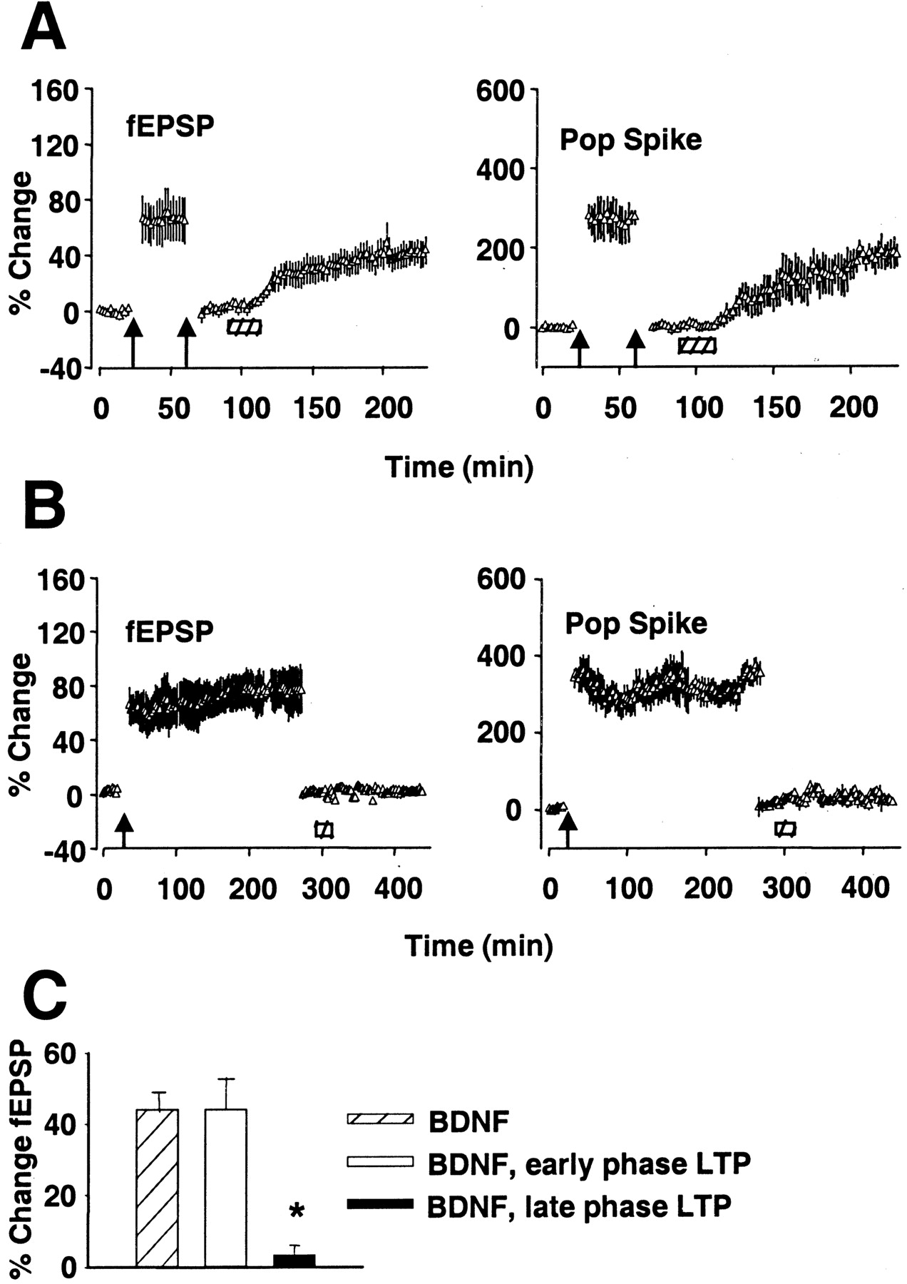
- Fig. 7.
BDNF-LTP is occluded by late phase but not early phase HFS-LTP. A, LTP was induced by three sessions of HFS (400 Hz) and recorded for 30 min. The stimulus intensity was then lowered to reset the fEPSP slope to baseline. A second session of HFS produced no further increase, demonstrating saturation of HFS-LTP. BDNF infusion (hatched bar) 60 min after the first HFS led to increased synaptic transmission (n = 6).B, HFS-LTP was induced and recorded for 240 min and then reset to baseline as in A. BDNF infusion 260 min after HFS had no effect on fEPSP slope or population spike (Pop Spike) amplitude for the duration of recording (n = 6). Values are group means ± SEM expressed as percentage of baseline. C, Summary of fEPSP slope increases obtained in group receiving BDNF after baseline recording (BDNF), 60 min after HFS (BDNF, early phase LTP), or 260 min after HFS (BDNF, late phase LTP). *Significantly different from BDNF group. The magnitude of LTP in the BDNF group and BDNF, early phase LTP group was not statistically different (p < 0.05).


