

Article Figures & Data
Figures
- Fig. 1.
The rate of adaptation increased when apical spiral ganglion neurons were exposed to BDNF, yet remained relatively unchanged when exposed to NT-3. APmax for all apical neurons plotted in a frequency histogram for each of the three conditions: control cultures, cultures exposed to BDNF, and cultures exposed to NT-3. Inset, Example traces from five different apical neurons taken at APmax for each of the three conditions: control cultures (left series of sweeps), cultures exposed to 5 ng/ml BDNF (middle series of sweeps), and cultures exposed to 5 ng/ml NT-3 (rightseries of sweeps).
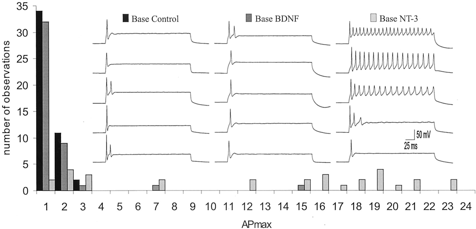
- Fig. 2.
The rate of adaptation decreased when basal spiral ganglion neurons were exposed to NT-3, yet remained relatively unchanged when exposed to BDNF. APmax for all basal neurons plotted in a frequency histogram for each of the three conditions: control cultures, cultures exposed to BDNF, and cultures exposed to NT-3. Inset, Example traces from five different basal neurons taken at APmaxfor each of the three conditions: control cultures (leftseries of sweeps), cultures exposed to 5 ng/ml BDNF (middle series of sweeps), and cultures exposed to 5 ng/ml NT-3 (right series of sweeps).
- Fig. 3.
APmax and latency were systematically altered by addition of neurotrophins to the neuronal cultures. Action potential durations, which were significantly different between apical and basal neurons in control cultures, were relatively unchanged when cultures were supplemented with either BDNF or NT-3. a, The maximum number of action potentials that an individual neuron is capable of firing (APmax) was significantly different between apical and basal neurons in control cultures. Both apical and basal neurons exposed to BDNF showed APmaxvalues similar to the base control neurons. Apical and basal neurons exposed to NT-3, however, showed APmaxvalues similar to or greater than the apical control neurons. **p < 0.01; *p < 0.05, for comparisons between experimental conditions denoted by the solid lines for this and subsequent bar graphs. The following conditions were compared statistically using Student's two-tailedt test: apex control to base control, apex control to apex BDNF, apex control to apex NT-3, base control to base BDNF, and base control to base NT-3. b, A similar pattern exists for action potential latency measurements. The initial difference in action potential latency between the apical and basal neurons in control conditions at threshold was no longer evident in the presence of each neurotrophin. Both apical and basal neurons exposed to NT-3 had latencies more similar to the apical than basal control neurons. Apical and basal neurons exposed to BDNF had latencies more similar to the basal than the apical control neurons. c, Aside from the significant difference between apical and basal neurons in control cultures, neurotrophins appeared to have no effect on action potential duration. The duration showed a tendency to be decreased by treatment with either BDNF or NT-3 when compared with apex controls but the differences failed to achieve statistical significance.
- Fig. 4.
Action potential duration is altered by neurotrophins in rapidly adapting, but not slowly adapting neurons.a, Action potential duration differed in rapidly adapting neurons exposed to low concentrations of neurotrophins (5 ng/ml). BDNF decreased this parameter in apical neurons; NT-3 increased it in basal neurons. **p < 0.01 and *p < 0.05 for comparisons between the experimental conditions denoted by thesolid lines. b, Action potential durations in slowly adapting neurons were brief and remained unaltered when exposed to neurotrophins. In apical neurons no significant effects were noted in this parameter between control, BDNF, and NT-3 conditions. The control basal neurons could not be compared because none of the 47 neurons fell within the slowly adapting category; nevertheless there was no significant difference between the BDNF and NT-3 conditions in slowly adapting basal neurons.
- Fig. 5.
Exposure to BDNF increased KCaantibody staining, whereas NT-3 decreased it. a–l, Representative images of spiral ganglion neurons from the apex and base in control, BDNF, and NT-3 conditions. The top panels(a, c, e, g, i, k) show neurons labeled with FITC-conjugated KCa antibody, and the bottom panels (b, d, f, h, j,l) show the overlay of the green FITC-conjugated KCa antibody and the red TRITC-conjugated NF200 antibody. Because a pure count of the number of stained cells would not accurately represent the obvious difference in label intensity that we consistently observed (compare Fig. 6a,b), these results also illustrate the need for a weighted ranking method of analysis. a–d, Spiral ganglion neurons in control cultures label significantly more with anti-KCa in the base compared with the apex.a, The faint profiles of KCa-labeled neurons are discernable; however, they were considerably lighter than those in the base (compare arrow in a witharrow in c). b, The double exposure shows prominent NF200-positive labeling, indicating that neurons were present in the culture, and demonstrating that the faintly stained cells in a were neurons. Thearrows in a and b indicate an NF200-positive neuron that minimally expressed the protein for KCa, therefore in b it retains most of the red (TRITC) color in the overlay.c, Anti-KCa stained a population of neurons removed from the base of the cochlea. The arrowindicates the round profile of a neuron strongly labeled with the anti-KCa. d, A double exposure of KCa (green) and NF200 (red) staining revealed that the majority of neurons were labeled with both antibodies, as indicated by theyellow color of each of the neurons.e–h, Neurons isolated from the cochlea and grown in culture supplemented with 5 ng/ml BDNF showed an increase in KCa antibody labeling. e, Neurons from the apical cochlea stained similarly to those from the base.f, The double exposure shows yellowneuron cell bodies, verifying that the basal neurons exposed to 5 ng/ml BDNF were labeled with both NF200 and KCa antibodies.g, Neurons from the basal, high-frequency region of the cochlea stained similarly to those from the base control.h, The double exposure shows yellowneuron cell bodies, verifying that the basal neurons exposed to 5 ng/ml BDNF are labeled with both NF200 and KCa antibodies.i–l, Cultures of spiral ganglion neurons supplemented with 5 ng/ml NT-3 demonstrated a decrease in KCa antibody labeling in neurons isolated from the base, while preserving the apex–base differences observed in controls. i, Neurons isolated from the apex of the cochlea lightly labeled with the KCa antibody. j, The double exposure shows that every neuron identified with anti-NF200 weakly labeled with anti-KCa, resulting in a predominance of thered NF200 color. k, Neurons isolated from the base of the cochlea stained significantly darker than the apex; there was, however, significantly less staining than that observed in the base control neurons. l, Double exposure of KCa-positive neurons (green) and NF200-positive neurons (red). For a–l, spiral ganglia were isolated from postnatal day 5 CBA/CaJ mice and maintained for 7 d in vitro. Tissues were incubated in a 1:800 dilution of anti-KCa overnight at 4°C.m, Histogram of the weighted percentage of KCa antibody staining of apical and basal spiral ganglion neurons in each condition (Control, BDNF, andNT-3) for three experiments. The error bar is not shown for the base BDNF condition because all three values were 100%. **p < 0.01 for comparisons between the experimental conditions denoted by the solid lines.
- Fig. 6.
Exposure to BDNF increased Kv1.1 antibody staining, whereas NT-3 was relatively ineffective. a–l, Representative images of spiral ganglion neurons from the apex and base in control, BDNF, and NT-3 conditions. The top panels(a, c, e, g, i, k) show neurons labeled with FITC-conjugated Kv1.1 antibody, and the bottom panels(b, d, f, h, j, l) show the overlay of thegreen FITC-conjugated Kv1.1 antibody and thered TRITC-conjugated NF200 antibody.a–d, Spiral ganglion neuron cultures without exogenously added neurotrophins labeled significantly more with anti-Kv1.1 in the basal neurons compared with the apical ones.a, Spiral ganglion neurons labeled lightly with anti-Kv1.1. b, The double exposure shows prominent NF200-positive labeling, indicating that neurons were present in the culture, and demonstrating that the faintly stained cells were neurons.c, Anti-Kv1.1 stained a population of neurons removed from the base of the cochlea. NF200-positive neurons (d) strongly labeled with the anti-Kv1.1 (c). The intensity of Kv1.1 label, however, revealed heterogeneity because neurons from the same cochlear location were either labeled intensely (arrow) or weakly (arrowhead). d, A double exposure of Kv1.1 (green) and NF200 (red) staining showed that the majority of neurons observed ind labeled with anti-Kv1.1 (compare with neurons inc). e–h, Neurons isolated from the cochlea and grown in culture supplemented with 5 ng/ml BDNF. Kv1.1 staining was significantly greater in apical neurons. e,Neurons from the apex and exposed to BDNF showed anti-Kv1.1 staining comparable with that observed in the base control where some were strongly labeled (arrow) and others weakly labeled (arrowhead). f, Kv1.1-positive neurons,green; NF200-positive neurons, red; double-labeled neurons, yellow. g,Neurons from the base of the cochlea and exposed to BDNF stained similarly to base controls. h, The double exposure shows neuron cell bodies that are yellow in color, verifying that the basal neurons exposed to 5 ng/ml BDNF were labeled with antibodies against both NF200 and Kv1.1 antibodies.i–l, Cultures of spiral ganglion neurons supplemented with 5 ng/ml NT-3 did not show significantly changed distributions of Kv1.1 protein in neurons isolated from either the base or the apex, thus the apex–base differences observed in controls was preserved.i, Neurons isolated from the apex of the cochlea labeled lightly with the Kv1.1 antibody. j, The double exposure shows that some of the neurons identified with anti-NF200 labeled weakly with anti-Kv1.1. k, Neurons isolated from the base of the cochlea showed significantly more staining than neurons from the apex. l, Double exposure of Kv1.1-positive neurons, green; NF200-positive neurons,red. For a–l, spiral ganglia were isolated from postnatal day 5 CBA/CaJ mice and maintained for 7 din vitro. Tissues were incubated in a 1:200 dilution of anti-Kv1.1 overnight at 4°C. m, Histogram of the weighted percentage of Kv1.1 antibody staining of apical and basal spiral ganglion neurons in each condition (Control, BDNF, and NT-3) for four experiments. *p < 0.05 for comparisons between the experimental conditions denoted by the solid lines.
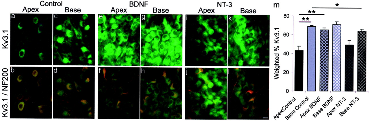
- Fig. 7.
Exposure to BDNF increased Kv3.1 antibody staining, whereas NT-3 had relatively little effect.a–l, Representative images of spiral ganglion neurons from the apex and base in control, BDNF, and NT-3 conditions. Thetop panels (a, c, e, g, i, k) show neurons labeled with FITC-conjugated Kv3.1 antibody, and thebottom panels (b, d, f, h, j, l) show the overlay of the green FITC-conjugated Kv3.1 antibody and the red TRITC-conjugated NF200 antibody.a–d, Spiral ganglion neuron cultures without exogenously added neurotrophins. Neurons isolated from the base showed significantly more anti-Kv3.1 label than neurons from the apex.a, A low percentage of spiral ganglion neurons labeled lightly with anti-Kv3.1. b, The double exposure shows NF200-positive labeling, indicating that neurons were present in the culture, but only a small percentage labeled with the Kv3.1 antibody (green). c, Anti-Kv3.1 stained a population of neurons removed from the base of the cochlea.d, A double exposure of Kv3.1 (green) and NF200 (red) staining revealed that the majority of neurons were labeled with both antibodies, as indicated by the yellow color of each of the neurons. e–h, Neurons isolated from the cochlea and grown in media supplemented with 5 ng/ml BDNF. Apical neurons showed a significant increase in Kv3.1 labeling. e, Neurons from the apical cochlea stained similarly to those from the base.f, Kv3.1-positive neurons, green; NF200-positive neurons, red; double-labeled neurons,yellow. g, Neurons from the base of the cochlea stained similarly to those from the base control.h, The double exposure shows neuron cell bodies that areyellow in color, verifying that the basal neurons exposed to 5 ng/ml BDNF were labeled with antibodies against both NF200 and Kv3.1. i–l, Cultures of spiral ganglion neurons supplemented with 5 ng/ml NT-3. Anti-Kv3.1 staining was reduced in neurons isolated from the base. i, Neurons isolated from the apex of the cochlea labeled lightly with the Kv3.1 antibody.j, The double exposure shows that most neurons identified with anti-NF200 only labeled weakly with anti-Kv3.1.k, Neurons isolated from the base of the cochlea were stained significantly more than neurons from the apex.l, Double exposure of Kv3.1-positive neurons,green; NF200-positive neurons, red. Fora–l, spiral ganglia were isolated from postnatal day 5 CBA/CaJ mice and maintained for 7 d in vitro. Tissues were incubated in a 1:400 dilution of anti-Kv3.1 for 48 hr at 4°C. m, Histogram of the weighted percentage of Kv3.1 antibody staining of apical and basal spiral ganglion neurons in each condition (Control, BDNF, and NT-3) for four experiments. **p < 0.01 and *p < 0.05 for comparisons between the experimental conditions denoted by thesolid lines.
- Fig. 8.
Exposure to NT-3 increased Kv4.2 antibody staining, whereas BDNF had relatively little effect.a–l, Representative images of spiral ganglion neurons from the apex and base in control, BDNF, and NT-3 conditions. Thetop panels (a, c, e, g, i, k) show neurons labeled with FITC-conjugated Kv4.2 antibody, and thebottom panels (b, d, f, h, j, l) show the overlay of the green FITC-conjugated Kv4.2 antibody and the red TRITC-conjugated NF200 antibody.a–d, Spiral ganglion neurons cultured without exogenously added neurotrophins. Apical neurons showed significantly more anti-Kv4.2 label than basal neurons. a, Anti-Kv4.2 stained a population of neurons removed from the apex of the cochlea. The intensity of Kv4.2 label, however, was not uniform. Neurons from the same cochlear location were either intensely labeled (arrow) or weakly labeled (arrowhead).b, A double exposure of Kv4.2 (green) and NF200 (red) staining revealed that not all neurons observed in b labeled with Kv4.2 (compare with neurons in a). c,Spiral ganglion neurons labeled lightly with anti-Kv4.2.d, The double exposure shows NF200-positive labeling, indicating that neurons were present in the culture, however, the intensity of label with the Kv4.2 antibody (green) was lower in basal (c) than apical neurons (a). e–h, Neurons isolated from the cochlea and grown in media supplemented with 5 ng/ml BDNF. Kv4.2 antibody staining was increased in neurons isolated from the basal cochlea but not the apex, therefore eliminating the apex–base difference observed in control cultures. e, The majority of neurons from the apex stained with anti-Kv4.2 with intensities similar to controls where some were strongly labeled (arrow) and others weakly labeled (arrowhead). f, Double exposure of Kv4.2-positive neurons, yellow; NF200-positive neurons,red. g, Neurons from the basal cochlea stained similarly to those from the apex. h, The double exposure shows that not all of the population of neurons identified with NF200 stained with anti-Kv4.2. i–l, Cultures of spiral ganglion neurons supplemented with 5 ng/ml NT-3. The distribution of Kv4.2 protein in neurons isolated from both the base and the apex was significantly increased from controls, and the apex–base difference was preserved. i, Neurons isolated from the apex of the cochlea labeled strongly with the Kv4.2 antibody.j, The double exposure shows that most of the neurons identified with anti-NF200 labeled with anti-Kv4.2. k,The addition of NT-3 significantly increased the amount of Kv4.2 staining in basal neurons compared with controls; however, a large population of neurons remained only lightly labeled. l,The double exposure of Kv4.2-positive neurons (yellow) and unlabeled NF200-positive neurons (red). For a–l, spiral ganglia were isolated from postnatal day 4 CBA/CaJ mice and maintained for 7 din vitro. Tissues were incubated in a 1:400 dilution of anti-Kv4.2 overnight at 4°C. m, Histogram of the weighted percentage of Kv4.2 antibody staining of apical and basal spiral ganglion neurons in each condition (Control, BDNF, and NT-3) for four experiments. **p < 0.01 and *p < 0.05 for comparisons between the experimental conditions denoted by the solid lines.


