

Article Figures & Data
Figures
- Figure 1.
Development of abnormal cluster distribution and axonal branching in MyoD-/- mice. Confocal images of motor axons and AChR clusters in diaphragm muscles from BALB/c and MyoD-/- mice at P1, 14, 28, and 42 (top panels), and E14.5 and E15.5 (bottom panels). Muscles were reacted with a mixture of antibodies against neurofilament and synaptophysin and with an FITC-conjugated secondary antibody to label preterminal and terminal portions of the motor axons (A–F, a–f) and with rhodamine-conjugated α-BuTx to label AChRs (A′–F′,a′–f′). In the BALB/c diaphragms, at all postnatal ages, neuronal branches (A–D) and AChR clusters (A′–D′) are confined to the middle of the muscle (in a discrete endplate band), whereas in the diaphragms of MyoD-/- mice, there is extensive ramification of neuronal branches (a–d) throughout the muscle, extending to the edge of the diaphragm (a, d, arrowheads). AChR clusters are distributed throughout the muscle, corresponding to the distribution of the axon terminals (a′–d′). At E14.5 a discrete band of AChR clusters is found in the middle of the diaphragms of both BALB/c (E′) and MyoD-/- mice (e′). The FITC and rhodamine images were overlapped (E, F, e, f), showing that some terminal axons are not apposed to AChR clusters at E14.5 (E, e, arrows.). By E15.5 motor axons have grown out of the central zone of the muscle fibers, and new AChR clusters are formed at the axon terminals of the MyoD-/- mice (f), resulting in a wider, more diffuse endplate band (f′) than is found in age-matched BALB/c muscle (F′). Scale bars, 100 μm. For each age group, micrographs are at the same magnification.
- Figure 2.
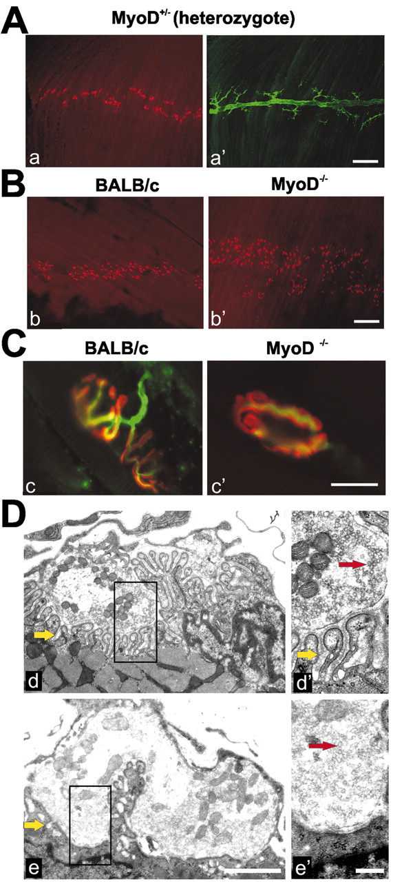
Confocal and electron microscope images of nerve terminals and endplates demonstrating abnormalities in the MyoD-/- mouse. A, Diaphragm muscles of P28 MyoD+/- (heterozygote) mice display an AChR cluster distribution (after reaction with rhodamine-labeled α-BuTx) (a) and nerve branching (after immunostaining with a mixture of antibodies against neurofilament and synaptophysin and an FITC-conjugated secondary antibody) (a′) similar to that in wild-type muscles. Scale bar, 200 μm. B, Intercostal muscles of P28, BALB/c (b), and MyoD-/- (b′) mice reacted with rhodamine-conjugated α-BuTx. The endplate zone in the MyoD-/- intercostal muscle is wider than in the BALB/c muscle. Scale bar, 200 μm. C, Diaphragm muscles of P42 BALB/c (c) and MyoD-/- (c′) mice reacted with antibodies against neurofilament and synaptophysin and an FITC-conjugated second antibody and rhodamine-conjugated α-BuTx. Terminal branches of the axons overlie postsynaptic AChR clusters in both muscles. The branching of the terminal axons is less complex, and the “pretzel-like” appearance of the wild-type AChR clusters is not present in the MyoD-/- mice. Scale bar, 20 μm. D, Electron micrographs of endplates in diaphragms from P42 BALB/c (d, d′) and MyoD-/- mice (e, e′). Yellow arrows indicate subsynaptic folds; red arrows indicate presynaptic vesicles. Boxed regions in d and e are enlarged in d′ and e′, respectively. Endplates with complex, well differentiated synaptic folds are found in the control mice (d, d′). Junctional folds at endplates of MyoD-/- muscles are generally shallower and less complex (fewer synaptic folds). Occasionally, junctional folds are almost absent in regions adjacent to axon terminals (e′). Scale bars, 1 μm.
- Figure 3.
The agrin–MuSK–rapsyn pathway is functional in MyoD-/- muscle, and MyoD-/- mice assemble a complex postsynaptic apparatus. A, MyoD-deficient myotubes form AChR clusters when exposed to neural agrin. Primary cultures of myoblasts derived from leg muscles of newborn BALB/c or MyoD-/- mice were allowed to differentiate into myotubes. Myotubes in both types of cultures displayed few AChR clusters in the absence of agrin, whereas both types of cultures, when treated with 10 nm recombinant neural agrin, displayed numerous AChR clusters. Scale bar, 10 μm. B, Confocal images of cross-sections of the diaphragm of P42 MyoD -/- mice reacted with rhodamine-labeled α-BuTx (a–e) and with an antibody to MuSK (a′) rapsyn (b′), neuregulin (c′), erbB (d′), or utrophin (e′), followed by an FITC-conjugated secondary antibody. All of these proteins colocalize with the AChR clusters at the neuromuscular junctions of the diaphragm of MyoD-/-mice, as they do in wild-type mice (data not shown). Scale bar, 50 μm. C, Lysates from leg muscles of E15–P14 MyoD-/- and BALB/c mice were subjected to SDS-PAGE and immunoblotted with an antibody to MuSK and an HRP-labeled secondary antibody. Lysates from denervated (11 d after denervation) hindlimb muscles served as standard for MuSK (arrow) identification. There were no marked deficits in MuSK levels in MyoD-/- muscles at any stage studied. Both types of muscle demonstrated an age-related decrease in MuSK during the perinatal period, and both were able to upregulate MuSK in response to denervation.
- Figure 4.
Developmental transition from fetal to adult-type AChRs is delayed in the diaphragm of MyoD-/- mice. Partially purified AChR lysates from the diaphragms of P1 –P42 mice were subjected to SDS-PAGE and immunobloted with antibodies specific for the γ-, ϵ-, or β-subunit and HRP-labeled secondary antibodies. Elimination of the γ-subunit protein is delayed by 1 week in MyoD-/- as compared with BALB/c mice. The appearance of theϵ-subunit was also delayed by 1 week in MyoD-/- mice and did not reach control levels for an additional week. Changes in γ- and ϵ-subunit expression were specific, because at any given stage wild-type and MyoD-/- muscles expressed similar amounts of the β-subunit. Standards on the right are in kilodaltons.
- Figure 5.
Prolonged expression of the AChR γ-subunit in legmuscles of the MyoD-/- mice. Confocal images of sections of the tibialis anterior muscle, taken from P1 –P28 BALB/c and MyoD-/- mice, that were incubated with an antibody to theγ-subunit, FITC-conjugated secondary antibody, and rhodamine-labeled α-BuTx. The γ-subunit immunoreactivity diminished after P7 in BALB/c muscle, whereas it persisted at P21 in MyoD-/- mice, as can be seen by the colocalization of weak immunostaining of the γ-subunit (d′, arrow) with α-BuTx (d, arrow). Scale bar, 50 μm.
- Figure 6.
Delayed expression of the AChR ϵ-subunit in leg muscles of the MyoD-/- mice. Confocal images of sections of the tibialis anterior muscle, taken from P1–P28 BALB/c (left panels) and MyoD-/- (right panels) mice, that were incubated with an antibody to the ϵ-subunit, FITC-conjugated secondary antibody, and rhodamine-labeled α-BuTx. The ϵ-subunit is clearly visible by P7 in the BALB/c mice, whereas the ϵ-subunit in MyoD-/- mice is clearly visible by P21. Scale bar, 50 μm.


Tables
E14.5 E15.5 Innervated clusters (%) Uninnervated clusters (%) Endplate zone width (μm) Innervated clusters (%) Uninnervated clusters (%) Endplate zone width (μm) BALB/c 25.4 74.6 53 ± 3 92.5 7.5 81 ± 4 MyoD−/− 24.8 75.2 60 ± 4 93.1 6.9 109 ± 7* The presence or absence of innervation of AChR clusters on diaphragm muscle was evaluated with confocal microscopy of muscles reacted with α-BuTx and antibodies to synaptophysin/neurofilaments (300 clusters; n = 4 muscles for each group at each developmental stage). The width of endplate zone containing AChR clusters represents the mean ± SEM from six animals.
↵* p < 0.05; MyoD−/− significantly different from BALB/c controls.


