

Article Figures & Data
Figures
- Figure 1.
Expression and localization of PSD-93 in the major pain-related regions of the CNS. A, RT-PCR analysis showed that a 571 bp product from PSD-93 was highly detected in the dorsal horn (DH) of spinal cord and forebrain (FB), whereas the mRNA was detected very weakly or not detected in the ventral horn (VH) of spinal cord and dorsal root ganglion (DRG). PCR product was cloned directly into the TA cloning vector and verified as PSD-93 by automatic DNA sequencing. β-Actin mRNA was used as a loading control. B, Western blot analysis showed that PSD-93 protein was expressed abundantly in the PSD fraction of the forebrain and dorsal horn, weakly in ventral horn, and not detected in the dorsal root ganglion. Tubulin was used as a loading control. C, PSD-93 immunoreactivity was distributed mainly in the superficial dorsal horn of wild-type (WT) mice. There was no immunostaining with the PSD-93 antibody in the dorsal horn of PSD-93 knock-out (KO) mice. Scale bar, 125 μm. D, The subcellular localization of PSD-93 in the superficial laminas of dorsal horn (DH) and the anterior cingular cortex (CO) of forebrain. Immunogold labeling of PSD-93 was localized most commonly in the postsynaptic membrane (arrows). Pr, Presynaptic; Po, postsynaptic. Scale bar, 0.2 μm.
- Figure 2.
Identification of a complex assembled by PSD-93 and NMDARs in the spinal cord and forebrain. A, The subcellular localization of NR1 in the superficial laminas of dorsal horn (DH) and that of NR2A/2B in the anterior cingular cortex (CO) of forebrain. Immunogold labeling of NR1 in postsynaptic structures in the superficial laminas was associated almost exclusively with the postsynaptic membrane (arrow). In the CO the immunogold labeling for NR2A/2B was associated mainly with the postsynaptic membrane (arrow) but also was prominent in the postsynaptic spine apparatus in some spines (data not shown). Pr, Presynaptic; Po, postsynaptic. Scale bar, 0.2 μm. B, Similar distribution of PSD-93 and NR2A/2B in the spinal dorsal horn. Scale bar, 125 μm. C, Colocalization of PSD-93 and NR2A/NR2B in the superficial laminas of dorsal horn (DH) and the anterior cingular cortex (CO) of forebrain. Sections were double-labeled with PSD-93 antibody (10 nm gold) and NR2A/2B antibody (5 nm gold). Arrows indicate the postsynaptic membrane. Scale bar, 0.1 μm. D, Binding of PSD-93 and NR2A or NR2B in the spinal cord and forebrain. PSD-93 antibody was able to immunoprecipitate (IP) not only itself but also NR2A or NR2B in the soluble PSD fraction. However, PSD-93, NR2A, and NR2B could not be immunoprecipitated when PSD-93 antibody was preincubated with the PSD-93 fusion protein. The amount of sample loaded for the input was 10% of that for the immunoprecipitation. IB, Immunoblotting.
- Figure 3.
Expression of NR2A and NR2B in the spinal cord and forebrain of PSD-93 knock-out mice. A, Immunoblots of PSD-93, NR2A, and NR2B proteins in total extracts of spinal cord and forebrain of wild-type (+/+), heterozygous (+/-), and knock-out (-/-) mice. PSD-93 protein is undetectable in knock-out mice. There was no significant difference in the density of NR2A or NR2B bands among wild-type, heterozygous, and knock-out mice (p > 0.05; n = 5 for each group). Tubulin was used as a loading control (data not shown). B, The expression and distribution of NR2A/2B immunoreactivity in the spinal cord of wild-type (+/+) and PSD-93 knock-out (-/-) mice. The density and localization of NR2A/2B immunoreactivity in the spinal cord of wild-type mice are similar to those in knock-out mice. Scale bar, 50 μm. C-E, Effect of PSD-93 deletion on surface NR2A and NR2B expression in the cultured neurons of spinal dorsal horn. The amount of sample loaded for the total (T) was 10% of that for the surface (S). C, The top panel depicts a representative Western blot that shows samples of total and biotinylated surface NR2A in wild-type (WT) and PSD-93 knock-out (KO) mice. The bottom panel shows the statistical summary of the densitometric analysis. Average surface NR2A levels in knock-out mice were reduced by 64% of the value in wild-type mice (*p < 0.05). D, The top panel depicts a representative Western blot that shows samples of total and biotinylated surface NR2B in wild-type (WT) and PSD-93 knock-out (KO) mice. The bottom panel shows the statistical summary of the densitometric analysis. Average surface NR2B levels in knock-out mice were reduced by 50% of the value in wild-type mice (*p < 0.05). E, The top panel depicts a representative Western blot that shows samples of total and biotinylated surface GluR1 in wild-type (WT) and PSD-93 knock-out (KO) mice. The bottom panel shows the statistical summary of the densitometric analysis. There was no significant difference in surface GluR1 expression between wild-type and knock-out mice (p > 0.05). F, G, Effect of PSD-93 deletion on surface NR2A and NR2B expression in the spinal cord neurons from adult mice. Surface expression was assayed by using a membrane-impermeable cross-linking agent (BS3). The cross-linked product is unable to enter polyacrylamide gels, so only the intracellular pool is resolved by Western blotting. Changes in surface expression are detected by observing changes in this indirectly detected intracellular pool. F, A representative Western blot that shows samples of total and intracellular NR2A, NR2B, and α-actinin, respectively, in the untreated control and BS3-treated groups of wild-type (WT) and PSD-93 knock-out (KO) mice. G, The statistical summary of the densitometric analysis. Average percentages of intracellular NR2A and NR2B levels in knock-out (KO) mice were increased by 190 and 194%, respectively, of the value in wild-type (WT) mice (*p < 0.05). In contrast, the average percentage of intracellular α-actinin level in knock-out mice was decreased by 0.92% of the value in wild-type mice.
- Figure 4.
PSD-93 deletion reduced NMDAR-mediated mEPSCs from the cultured neurons of spinal dorsal horn. A, mEPSCs were acquired in the presence or absence of 100 μm AP-5. The addition of 100 μm AP-5 to the extracellular solution abolished the slow exponential, whereas the addition of AP-5 plus NBQX (100 μm) completely blocked miniature events. One hundred events were selected before (control ACSF) and during AP-5 perfusion. After AP-5 perfusion the decay time of the fast component was not altered significantly (before AP-5, τfast = 1.98 ± 0.20 msec; during AP-5, τfast = 1.96 ± 0.19 msec; n = 20). Therefore, the amplitude of the second exponential was taken as the NMDAR peak current. The amplitude of the averaged mEPSC was taken as the peak AMPA current. B, Four consecutive 1 sec traces from a cultured neuron representative of wild-type (WT) or PSD-93 knock-out (KO) mice. C, Trace averages from the neurons illustrated in B. Selected from each neuron were 120 events, and the responses were aligned by their peaks. The neurons in wild-type and knock-out mice were fit by a double-exponential function, and their individual components were displayed. D, The top plot compares the average current ratio for NMDARs and AMPA receptors between pooled wild-type and knock-out mice responses (*p < 0.005). The bottom plot compares the ratio of decay times for the AMPA receptors and NMDARs between pooled wild-type and knock-out mice responses.
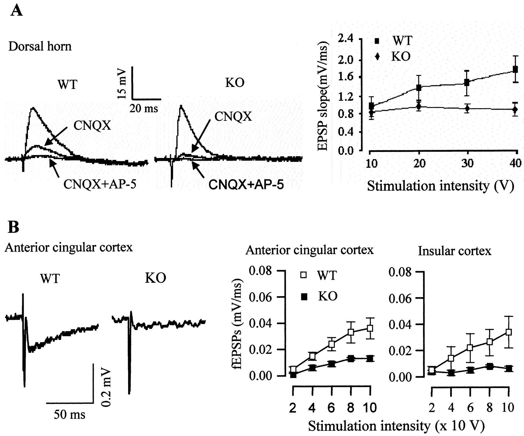
- Figure 5.
PSD-93 deletion attenuated NMDAR-mediated excitatory postsynaptic responses. A, Traces of EPSPs recorded in the presence or absence of CNQX (20 μm) or CNQX and AP-5 (100 μm) from the neurons of the superficial dorsal horn in adult wild-type (WT) and knock-out (KO) mice. Slow NMDAR-mediated synaptic responses were compared at four dorsal root stimulation intensities (10, 20, 30, and 40 V), revealing significant differences between wild-type (n = 6-8) and knock-out mice (n = 8-10; p < 0.05). B, Traces of NMDAR-mediated fEPSPs recorded in the presence of 20 μm CNQX from the anterior cingular cortex of forebrain in adult wild-type (WT) and knock-out (KO) mice. Similar traces also were found in insular cortex of forebrain (data not shown). NMDAR-mediated synaptic responses were compared at four stimulation intensities (40, 60, 80, and 100 V) in the anterior cingular cortex (WT, n = 8; KO, n = 10) and insular cortex (WT, n = 8; KO, n = 10), revealing significant differences between wild-type and knock-out mice (p < 0.05).
- Figure 6.
Role of PSD-93 in pain behavioral responses. A, Baseline response frequencies of paw withdrawal to mechanical stimulation that were elicited by various forces of von Frey monofilaments in intact mice. Withdrawal frequencies were similar in wild-type (+/+), heterozygous (+/-), and knock-out (-/-) mice (n = 15 for each group). B, Baseline withdrawal latency in response to thermal stimulation that was elicited by high-intensity radiant heat applied to the plantar sides of left and right hind paws in intact mice. Thermal thresholds of knock-out mice were not different from those of wild-type or heterozygous mice (n = 12 for each group). C, D, Effect of PSD-93 deletion on CFA-induced inflammatory pain. CFA produced a significant increase in paw withdrawal frequencies in response to 0.24 mN (low intensity) and 4.33 mN (moderate intensity) mechanical stimuli on the injured side in wild-type (WT) mice (repeated measures ANOVA, p < 0.001; n = 5), but not in knock-out (KO) mice (p > 0.05; n = 5). Asterisks indicate a significant difference on the injured side between wild-type and knock-out mice (*p < 0.05 and **p < 0.01, Bonferroni post-test). E, F, Effect of PSD-93 deletion on neuropathic pain. In wild-type mice (WT) the nerve injury produced a significant increase in paw withdrawal frequencies to 0.24 and 4.33 mN mechanical stimuli on the injured side (p < 0.0001; n = 14 for each group). This increase was significantly different from the paw withdrawal frequencies in the PSD-93 knock-out (KO) mice. Asterisks indicate a significant difference on the injured side between wild-type and knock-out mice (*p < 0.05 and **p < 0.01). The knock-out mice also exhibited a modest but significant mechanical hypersensitivity in response to low-intensity mechanical stimulation on days 8, 10, and 14 after surgery (E; p < 0.01), but not to moderate-intensity mechanical stimulation (F; p > 0.05).


