

Article Figures & Data
Figures
- Figure 1.
Ca 2+ responses elicited by the bitter stimuli cycloheximide (1–100 μm; A), quinine (10–5000μm; B), and denatonium (10–3000μm; C) were concentration dependent and had similar time courses. Shown are representative responses in three different taste cells at progressively higher stimulus concentrations. D, Concentration–response relationships show different potencies for cycloheximide (•), quinine (○), and denatonium (▴) in C57BL/6B mice. Data shown are means ± SEM of response amplitudes. Responses are from at least five cells. Detection thresholds for these compounds are similar to those found in behavioral studies in C57BL/6B mice (double-headed arrows above abscissa, cycloheximide ≪ quinine < denatonium) (for references, see Results).
- Figure 2.
Ca 2+ responses vary with cycloheximide sensitivity of the mouse strain. Concentration–response relationships for cycloheximide in C57BL/6B (•) and DBA/2J (○) mice show that taste cells are more sensitive to cycloheximide in DBA/2J mice. The corresponding behavioral thresholds show the same sequence (double-headed arrows above abscissa) (for references, see Results). Values shown are amplitudes of responses at varying concentrations. Responses are from seven (C57BL/6B mice) and 17 (DBA/2J) cells.
- Figure 3.
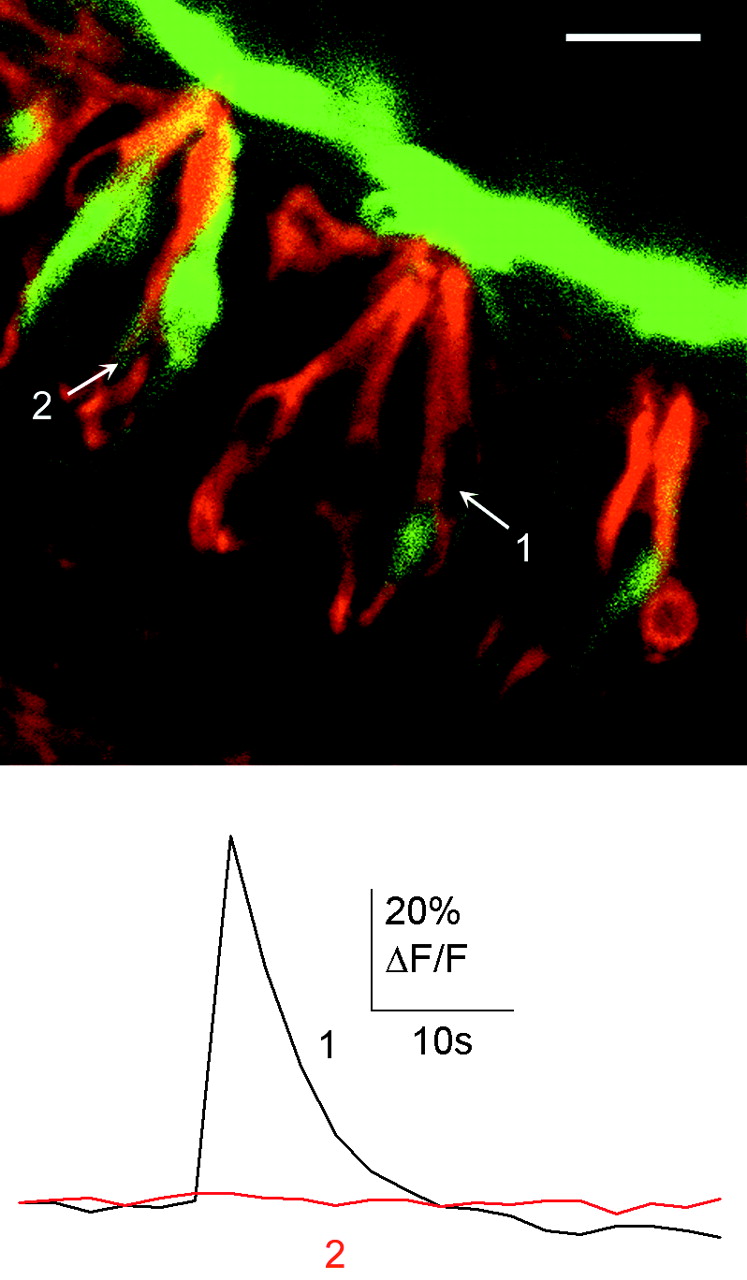
Bitter-sensitive cells express α-gustducin. A–C, α-Gustducin immunostaining (B) was present in many taste cells that were loaded with calcium green dextran (A). D, E, Stimulation with cycloheximide (100 μm) induced responses in two different cells in the same taste bud. Cells 1 and 2 responded to cycloheximide. Cell 1 was α-gustducin immunoreactive. Top image was acquired during calcium imaging (colors indicate pixel intensity mapping), and bottom image shows α-gustducin immunostaining (red) and calcium green dextran (green) after imaging. Scale bars: (in A) A–C, 20 μm; D, 10 μm.
- Figure 4.
Responses to cycloheximide (50 μm, bath applied) were similar in α-gustducin-immunopositive (gust +) and -immunonegative (gust –) taste cells. A, Superimposed Ca 2+ transients from four and five cells, respectively. B, Amplitudes of cycloheximide-elicited Ca 2+ transients ofα-gustducin-immunoreactive taste cells (n = 13) were not significantly different from those of taste cell lacking α-gustducin (n = 11; Student's t test; p = 0.21).
- Figure 5.
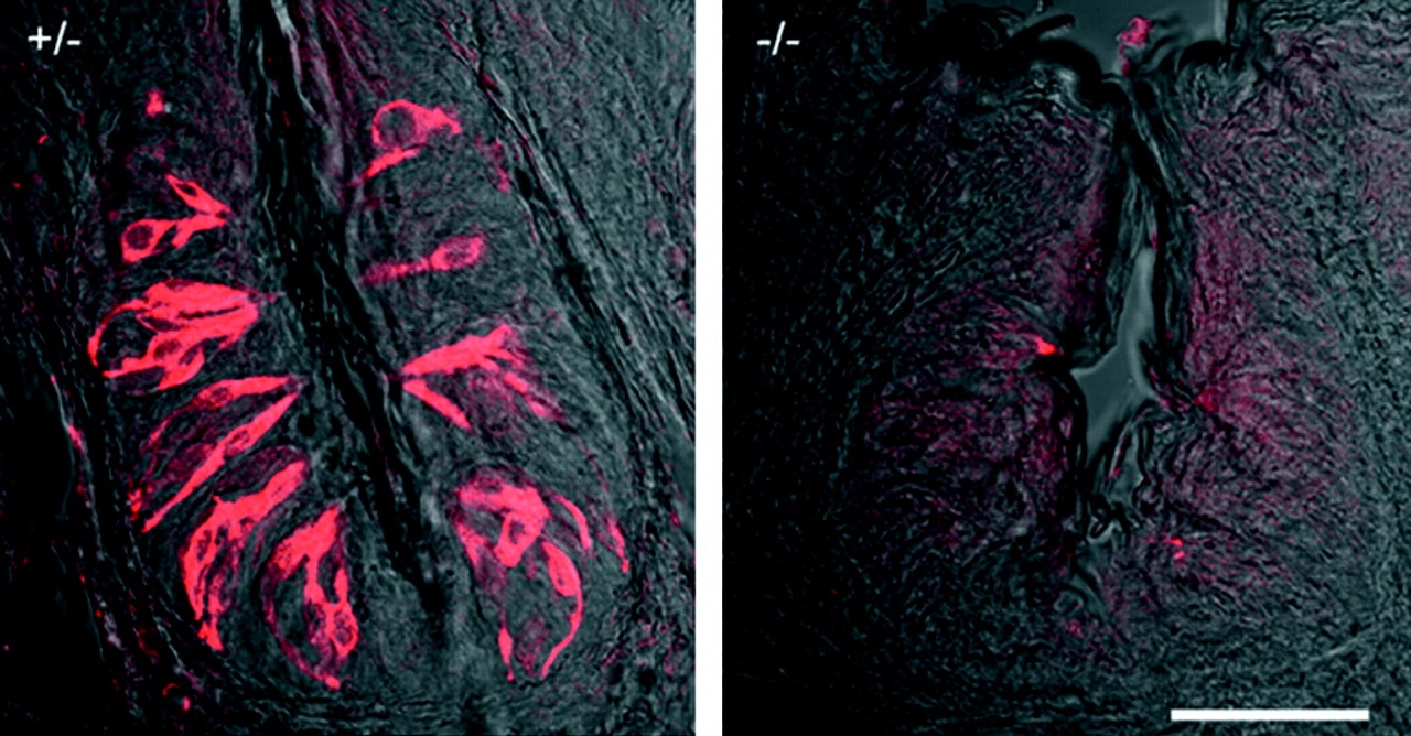
α-Gustducin immunostaining in mutant mice lacking α-gustducin (right, –/–) and their heterozygous littermates (left, +/–). Shown are sections (40 μm) of vallate papillae. Scale bar, 50 μm.
- Figure 6.
Bitter responses in taste cells of mice lacking α-gustducin. A, C, Ca 2+ transients elicited by cycloheximide (1–100 μm; A) and quinine (10–5000 μm; C) in two different taste cells from mice lacking α-gustducin were similar to those of wild-type mice (see Fig. 1). B, D, Taste cells inα-gustducin knock-out mice were less sensitive than those of heterozygous littermates. Note that α-gustducin knock-out mice (○) did not have taste cells sensitive to low concentrations of cycloheximide (< 10 μm) and quinine (< 500 μm).
- Figure 7.
The incidence of bitter-responsive cells was significantly reduced in α-gustducin knock-out mice (Student's t test; p < 0.01). Black bars, Heterozygous mice. White bars, Homozygous α-gustducin knock-out mice. Citric acid responses, which are not transduced throughα-gustducin, had the same incidence of occurrence in homozygous and heterozygous mice. Only taste buds with cells that showed responses to bitter stimuli and citric acid were included in this analysis. Results are from 12 taste buds from four homozygous mice and 10 taste buds from three heterozygous mice. Responses are from at least five taste cells. Proportion of cells = responsive cells/total cells tested. CX, Cycloheximide (100 μm); qui, quinine (5 mm); den, denatonium (5 mm); cit, citric acid (50 mm).
- Figure 8.
Bitter-sensitive cells expressed the G-proteinα subunit Gαi2. Gαi2 immunostaining was present in many taste cells that were loaded with calcium green dextran. Stimulation with cycloheximide (60 μm) induced a response in cell 1, which was Gαi2 immunoreactive. Cell 2 was Gαi2 immunoreactive but did not respond to cycloheximide. Scale bar, 20 μm.
- Figure 9.
Most bitter-sensitive cells express the G-protein α subunit Gαi2orboth Gαi2and Gα-gustducin.A, Cells loaded with CaGD. Cells 1–3 respond to cycloheximide (80μm). B, Immunoreactivity to α-gustducin. C, Immunoreactivity to Gαi2. D, Immunostaining and CaGD superimposed. E, Responses to cycloheximide (80 μm). Cycloheximide-sensitive cells (cells 1–3) were immunoreactive for Gαi2 alone (cell 2) or both Gαi2 andα-gustducin (cell 3). Cell 1 was not immunoreactive for either G-protein subunit. Cell 4 was Gαi2 immunoreactive but did not respond to cycloheximide. Scale bar, 20 μm.


Additional Files
HTML Page - index.htslp
Files in this Data Supplement:
- Supplemental Movie 1 - Ca2+ response to stimulation with the bitter compound cycloheximide in a taste bud that was subsequently immunostained for a-gustducin.� Confocal images of lingual slices showing 4 taste cells loaded with calcium green dextran (CaGD).� One cell (arrow) responds to cycloheximide with an increase in CaGD fluorescence intensity (shift from blue to green in the pseudocolor intensity scale).� This cell was immunoreactive for a-gustducin (red fluorescence).� a-Gustducin was also present in other cells.� Triangle at the level of the taste pore (top left) indicates the time of stimulus application.� Movie duration: 2.5 min; frame interval: 2.5 sec.� The immunostaining image is superimposed on the calcium-imaging recording at the beginning and at the end of the movie.
- Supplemental Movie 2 - Ca2+ response to stimulation with the bitter compound cycloheximide in taste buds that were subsequently immunostained for a-gustducin.� Confocal images of lingual slices showing 5 taste cells loaded with calcium green dextran (CaGD) in 2 different taste buds.� One cell (arrow) responds to cycloheximide with an increase in CaGD fluorescence intensity (shift from blue to green to red in the pseudocolor intensity scale).� This cell was immunoreactive for a-gustducin (red fluorescence).� a-Gustducin was also present in other cells.� Triangle (left) indicates the time of stimulus application.� Movie duration: 2.5 min; frame interval: 2.5 sec.� The immunostaining image is superimposed on the calcium-imaging recording at the beginning and at the end of the movie.
- Supplemental Movie 3 - Ca2+ response to stimulation with the bitter compound cycloheximide in a taste bud that was subsequently immunostained for a-gustducin.� Confocal images of lingual slices showing 3 taste cells loaded with calcium green dextran (CaGD).� Two cells (arrow and arrowhead) respond to cycloheximide with an increase in CaGD fluorescence intensity (shift from blue to green to red in the pseudocolor intensity scale), but only one cell (arrowhead) was immunoreactive for a-gustducin.� Triangle (top left) indicates the time of stimulus application.� Movie duration: 2.5 min; frame interval: 2.5 sec.� The immunostaining image is superimposed on the calcium-imaging recording at the beginning and at the end of the movie.


